Ever looked at a regular X-ray? It's a flat, two-dimensional picture. You see bones and tissues, but they're all layered on top of each other. It's like looking at the shadow of an object—you get the outline, but you lose all the depth.
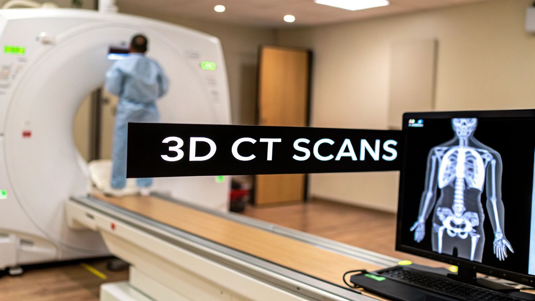
This is where 3D CT scans completely change the game. Instead of one flat picture, a CT scanner takes hundreds, sometimes thousands, of thin X-ray "slices" as it circles around the body. A powerful computer then stacks these slices together, almost like a digital deck of cards, to build a fully interactive 3D model.
Suddenly, doctors aren't just looking at a shadow. They're looking at a complete, detailed map of your anatomy.
Seeing in a New Dimension
This leap from flat slices to a spatial model is what makes 3D CT so incredibly valuable in medicine. It's the difference between looking at a road map and using a GPS with a satellite view.
Instead of mentally trying to reconstruct a 3D image from 2D slices, a physician can now:
- See Complex Anatomy Clearly: They can digitally rotate the model to see exactly how a tumor is connected to nearby blood vessels or understand the precise alignment of bone fragments in a severe fracture.
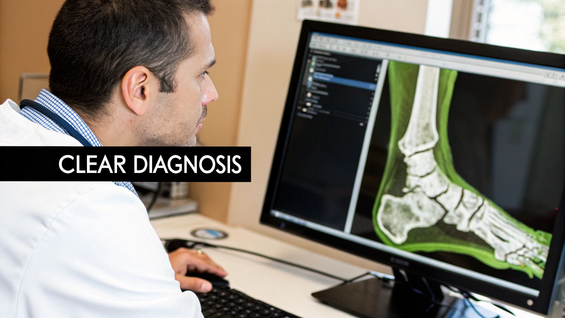
- Catch What's Hidden: Subtle abnormalities that might be missed or obscured in a flat image often become obvious when viewed in three dimensions.
- Plan Surgeries with Precision: Surgeons can essentially perform a "virtual fly-through" of a patient's body to map out the safest path for an operation before ever making an incision.
This technology provides a level of anatomical context that was once unimaginable. It's no wonder the global 3D imaging market is projected to hit around USD 115.98 billion by 2030, with this kind of healthcare innovation leading the charge.
The real power isn't just in creating a 3D picture. It's about giving clinicians the ability to interact with a patient's unique anatomy digitally, which leads to smarter decisions and, ultimately, better patient outcomes.
For those who want to keep a finger on the pulse of where this technology is headed, the discussions happening at ACR (American College of Radiology) conferences are a fantastic place to hear from the experts shaping the future of medical imaging.
How 2D Slices Become a 3D Model
So, how do we get from a stack of flat, black-and-white images to a fully interactive 3D model? The magic behind this is a process called 3D reconstruction. Think of it like a high-tech version of building a sculpture, but instead of using clay, the raw material is pure data from the CT scanner.
The computer takes each individual 2D slice—which is just a super-thin cross-section of the body—and computationally stacks them one on top of the other. This creates a complete "volumetric dataset," which is essentially a digital block containing every bit of anatomical information captured during the scan.
From there, specialized software uses sophisticated rendering techniques to bring this block of data to life, making it something doctors can actually see and manipulate. The jump from 2D slices to a functional 3D model hinges on these rendering processes, which are getting a massive boost from new AI-powered rendering techniques.
The workflow below shows this journey from raw scan data to the final 3D visualization.
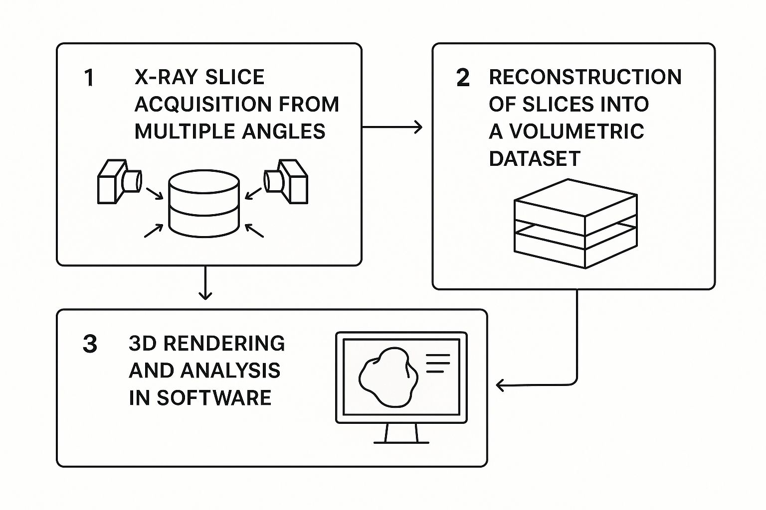
As you can see, it's a step-by-step pipeline that transforms abstract data points into a clear, tangible picture of a patient's anatomy.
Key Reconstruction Methods
Once we have that digital block of data, there are two main ways to visualize it. Each method gives a unique perspective, and the one a radiologist or surgeon chooses really depends on what they need to find out.
The first, and most common, is Volume Rendering.
Volume Rendering treats the entire dataset as a semi-transparent object. It assigns different colors and levels of opacity to different tissue densities. This lets a doctor peer right through skin and muscle to see the organs, bones, and blood vessels suspended inside.
Imagine a complex ship in a bottle, but you can make the bottle itself completely transparent. By tweaking the settings, clinicians can highlight or hide specific structures. For instance, they can dial down the visibility of all soft tissue to get a crystal-clear, unobstructed view of the skeleton—a game-changer for planning orthopedic surgery.
The second method is called Surface Rendering. Instead of showing everything at once, this technique is all about defining the edges of a particular structure, like the outer wall of the heart or a specific bone. It then generates a solid-looking, shaded 3D model of just that object's surface.
- When to use Volume Rendering: It’s perfect for seeing the big picture and understanding the spatial relationships between different parts, like figuring out exactly where a tumor is sitting in relation to a major blood vessel.
- When to use Surface Rendering: This is the go-to for examining the precise shape and contours of a single object, such as designing a custom implant for a complex bone fracture.
In the end, both techniques achieve the same goal: they turn raw scanner data into a powerful clinical tool. Doctors can spin, zoom, and even digitally slice through these models, giving them an incredibly deep understanding of a patient’s anatomy that informs everything from the initial diagnosis to the most delicate surgical procedures.
The Role of AI in Sharpening the Image
Artificial intelligence is taking the already powerful technology of 3D CT scans and making it profoundly smarter. Think of AI not just as a tool that captures images, but as an intelligent assistant for radiologists and clinicians, helping to refine every single step of the imaging process. Its work starts right at the reconstruction phase, where it can drastically speed up the creation of the final 3D model.
This acceleration is a game-changer. It means doctors get their hands on crucial diagnostic information much faster, which can be absolutely critical in an emergency. For example, AI can rapidly assemble a detailed 3D view of a patient's brain after a suspected stroke, giving physicians the intel they need to make split-second treatment decisions.
Enhancing Clarity and Reducing Noise
One of the biggest impacts of AI is in image enhancement. Raw CT data is often full of "noise" or artifacts—visual distortions that can easily obscure tiny, important details. AI algorithms are exceptionally good at identifying and scrubbing away this noise, almost like a highly specialized photo editor for medical images.
The result is a remarkably clear and crisp 3D model. What’s really significant here is that high-quality images can be produced even from lower-dose scans. By letting AI clean up the data, hospitals can reduce a patient's radiation exposure without compromising diagnostic accuracy. That’s a massive win for patient safety.
AI doesn't replace the expertise of a radiologist. Instead, it acts as a powerful amplifier, handling the heavy computational lifting so that human experts can focus on the critical task of interpretation and diagnosis.
Becoming a Diagnostic Partner
Beyond just cleaning up pictures, AI is quickly becoming a proactive partner in diagnostics. These models can be trained on immense libraries of scans, learning to recognize patterns that often point to a specific health issue. This lets them automatically analyze a 3D CT scan and flag potential areas of concern for the radiologist to review.
This is especially helpful for catching subtle or early-stage conditions that are easy to miss. For instance, an AI might flag:
- Tiny Pulmonary Nodules: It can spot small specks in the lungs that could be early signs of cancer, which are incredibly difficult for the human eye to catch consistently.
- Subtle Fractures: In complex trauma cases, AI can pinpoint hairline fractures in bones that might otherwise be overlooked during a first pass.
- Anomalies in Organs: It can highlight suspicious lesions or abnormalities in abdominal organs, immediately drawing the radiologist’s attention to where it’s needed most.
This automated pre-screening doesn't make the final call; it just serves up a well-organized and prioritized case for the physician. It’s this collaboration between human expertise and machine precision that is leading to faster, more accurate diagnoses and better outcomes for patients across the board.
How 3D CT Scans Are Changing Patient Care
The real magic of a 3D CT scan isn't just in the stunningly detailed images it produces. It's about what those images allow doctors to do. They get an interactive, true-to-life anatomical map that helps them plan safer, more effective treatments. This technology closes the gap between simply seeing a problem and knowing exactly how to fix it.
This shift towards more detailed imaging is driving huge demand. The global market for CT scanners was valued at USD 6.97 billion in 2023 and continues to grow. This isn't surprising, given the increasing need for better diagnostic tools to fight complex diseases like cancer and heart conditions. You can explore more on these market trends and what's fueling them.
A New Era for Surgical Planning
In oncology, the goal is always the same: remove the entire tumor while saving as much healthy tissue as possible. A 3D CT scan gives the surgical team a perfect digital copy of the patient’s anatomy. They can see exactly where the tumor is, how big it is, and how it’s tangled with nearby organs and blood vessels—all before making the first incision.
Think of it as a full dress rehearsal before the actual operation. Surgeons can spot potential roadblocks, map out the safest route, and walk into the operating room with a level of confidence that simply wasn't possible before. This translates directly to shorter surgeries, fewer complications, and better results for the patient.
Pinpoint Precision in Orthopedics and Cardiology
The impact is just as profound in other fields. An orthopedic surgeon dealing with a badly shattered bone can use a 3D model to see the fracture from every possible angle. This lets them meticulously plan a complex reconstruction or even design a custom implant that fits the patient’s unique anatomy like a glove.
Cardiologists rely on these scans, too. They get a clear, dynamic view of the heart and its web of arteries, allowing them to pinpoint blockages and check heart function without needing to perform invasive procedures. It’s a much safer way to get the answers they need.
Key Takeaway: 3D CT scans have taken medicine from a world of educated guesses to one of absolute precision. By giving doctors an exact digital model to work with, treatments can be tailored specifically to an individual's body.
Even in dentistry, 3D CT scans have become indispensable for placing dental implants with millimeter accuracy, which is crucial for a perfect, long-lasting fit. Whether it's fighting cancer or repairing a bone, the insights from these scans are fundamentally changing how doctors approach patient care.
The Pros and Cons of 3D CT Scans
3D CT scans offer a breathtaking view inside the human body, but like any powerful technology, they aren't a one-size-fits-all solution. Clinicians have to weigh the incredible advantages against some very real-world limitations. Getting a handle on this balance helps explain why these scans are ordered in specific situations where the benefits truly shine.
The biggest win, without a doubt, is the level of detail they provide. These rich 3D models give doctors a sense of depth and context that a flat, 2D image could never capture. This clarity translates directly into better surgical planning, which often means safer, more efficient operations and, ultimately, better outcomes for patients.
Unpacking the Primary Advantages
The power of a 3D CT scan goes far beyond just a pretty picture. It delivers actionable intelligence that fundamentally changes how patient care is delivered.
- See How Everything Connects: Surgeons can pinpoint the exact relationship between a tumor and the delicate blood vessels surrounding it. They can see how every bone fragment is positioned in a complex fracture.
- Spot Problems Sooner: Subtle issues that might be overlooked or misinterpreted on a standard 2D X-ray often become crystal clear when viewed from every angle in three dimensions.
- Tailor-Made Treatments: The detailed models are used to design custom surgical guides and patient-specific implants, taking the guesswork out of getting a perfect fit.
This technology allows clinicians to move from estimation to true precision. They can essentially perform a virtual dry run of a complex surgery, anticipating challenges long before they step into the operating room.
And the technology isn't standing still. The latest scanners use sophisticated algorithms to create even higher-resolution images, making it easier to spot conditions like cancer, stroke, and heart disease with greater accuracy. This constant improvement is a big reason why the global CT scan market is expected to hit USD 9.1 billion by 2033. You can read more about these market trends and the innovations behind them.
To truly appreciate the leap forward that 3D CT scans represent, it helps to see them side-by-side with traditional imaging.
Comparing 3D CT Scans with Traditional 2D Imaging
This table breaks down the key differences between the detailed, volumetric data from a 3D CT and the flat, two-dimensional view from something like an X-ray.
| Feature | 3D CT Scan | Traditional 2D Imaging (e.g., X-ray) |
|---|---|---|
| Dimensionality | Provides a full 3D volumetric view of structures. | Captures a flat, 2D projection of the body. |
| Anatomical Detail | Excellent visualization of soft tissues, bones, and blood vessels. | Primarily shows dense structures like bone; soft tissues are poorly defined. |
| Overlapping Structures | Eliminates the problem of overlapping anatomy, allowing clear views. | Structures are superimposed, which can obscure underlying issues. |
| Diagnostic Accuracy | High accuracy for detecting subtle fractures, tumors, and internal injuries. | Less sensitive; can miss small or non-displaced fractures and soft tissue problems. |
| Clinical Applications | Ideal for complex surgical planning, trauma assessment, and cancer staging. | Best for initial fracture diagnosis, chest screenings, and dental check-ups. |
| Radiation Dose | Involves a higher radiation dose. | Involves a much lower radiation dose. |
As you can see, each has its place. While an X-ray is perfect for a quick, low-dose look at a potential broken bone, a 3D CT scan is the tool you need when every last detail matters.
Considering the Practical Limitations
For all their power, 3D CT scans come with some important trade-offs. The most significant one is the radiation dose.
While a single scan is considered safe, it does expose a patient to more radiation than a standard X-ray. Medical teams are acutely aware of this and follow the ALARA principle (As Low As Reasonably Achievable), always using the minimum dose required to get the job done.
Cost is another major factor. The machinery behind a 3D CT scan is far more expensive to buy and maintain than a simple X-ray machine. Naturally, this higher overhead can mean a higher cost for the patient and the healthcare system.
Finally, there's the human element. Interpreting the sheer volume of data from a 3D scan is a specialized skill. Radiologists need extensive training to navigate these complex digital models and pull out the clinically important information. It's this mix of radiation, cost, and complexity that leads doctors to be selective, reserving 3D CT scans for cases where the profound diagnostic benefits clearly outweigh the drawbacks.
Where Diagnostic Imaging Is Headed
The story of the 3D CT scan is still being written. We're on the cusp of a new era where imaging isn't just about taking a static picture. It’s becoming a dynamic, highly personalized tool that’s completely reshaping how doctors plan and carry out treatments.
Think about this: surgeons are already using patient-specific anatomical models, 3D-printed straight from scan data, to rehearse complicated procedures before ever making an incision. This kind of preparation was science fiction not long ago. The next step is what many are calling “4D” CT, which introduces time into the equation. This will let us see organs as they move—a heart beating or lungs expanding and contracting in real-time.
Predictive Imaging is on the Horizon
Artificial intelligence is also playing a much bigger role than just cleaning up images. The real game-changer is training AI algorithms to spot subtle changes in scans over time and actually predict how a disease might progress.
For instance, an AI could be tasked with monitoring a patient's lung scans. It might flag tiny nodules that have the tell-tale signs of future growth, giving doctors a crucial heads-up for earlier intervention. This move toward predictive insight is going to be massive.
We’re moving toward a future where diagnostic imaging isn't just about seeing inside the body. It's about understanding and predicting its behavior in ways we're only just starting to grasp.
This kind of progress is shifting 3D CT scans from being a reactive tool used for diagnosis to a proactive one for managing a patient’s long-term health. It’s a fundamental change that promises a whole new standard of care.
Your Questions About 3D CT Scans, Answered
Even after getting the technical details, you probably still have a few practical questions. Let's walk through some of the most common things people ask about 3D CT scans.
Are They Safe? What About the Radiation?
This is usually the first question on everyone's mind, and for good reason. It’s true that 3D CT scans use more radiation than a simple X-ray, but radiologists and technicians are meticulous about safety.
They operate under a principle known as ALARA, which stands for "As Low As Reasonably Achievable." This means they use the absolute smallest dose of radiation necessary to get the high-quality images your doctor needs. The significant benefit of getting a precise diagnosis—whether for planning a major surgery or finding a critical issue early—almost always outweighs the very small risk from the radiation.
How Is a 3D CT Scan Different from an MRI?
It's easy to mix these two up since they both give doctors a detailed look inside the body. The real difference is how they create the images and what they’re best at revealing.
- 3D CT Scans: These use X-rays. The big advantages are speed (the scan itself can take just minutes) and its incredible ability to show bone, internal injuries, and blood vessels with amazing clarity. It's the go-to for trauma and emergencies.
- MRI Scans: These use strong magnets and radio waves—no radiation involved. An MRI takes much longer, but it provides unmatched detail of soft tissues. Think muscles, ligaments, brain tissue, and the spinal cord.
The bottom line: If speed is critical or your doctor needs to see bone, a CT scan is the tool for the job. If the focus is on getting an incredibly detailed picture of soft tissues, an MRI is usually the better choice.
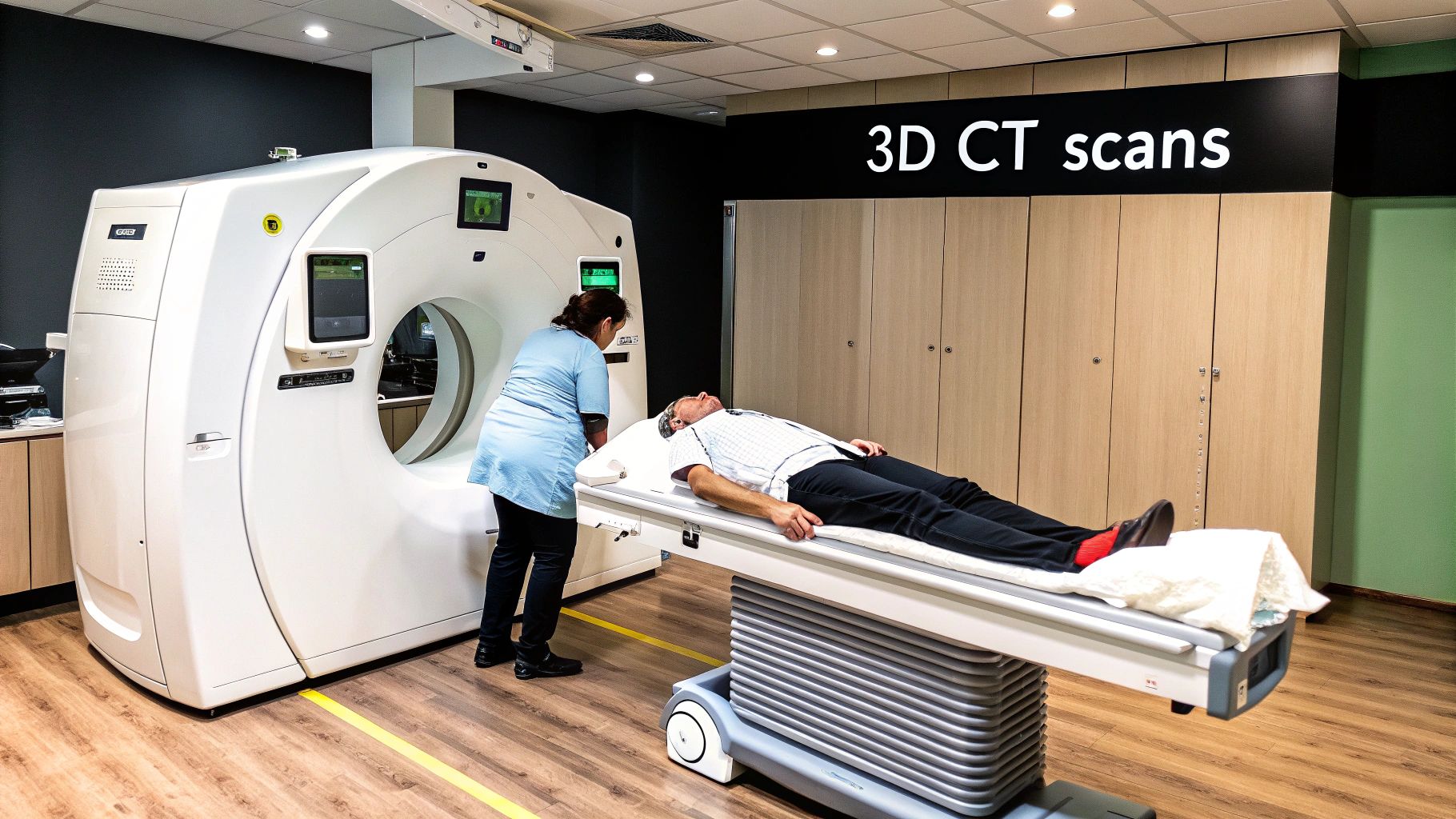
What Should I Expect During the Procedure?
The whole process is pretty straightforward and completely painless. You'll lie down on a comfortable, motorized table that gently moves into the center of a large, open ring—it looks like a big donut.
Depending on what your doctor is looking for, you might be given a contrast dye. You might drink it or have it injected, and it just helps certain organs or blood vessels stand out more clearly in the final image.
The scanner will quietly rotate around you, but you won't feel a thing. Your main job is to lie as still as you can, which helps ensure the pictures come out sharp. From start to finish, the entire scan is typically done in under 30 minutes.
At PYCAD, we're focused on advancing what's possible in medical imaging through smart AI. We partner with medical device companies to boost diagnostic accuracy and simplify workflows. See how we can help elevate your technology at https://pycad.co.


