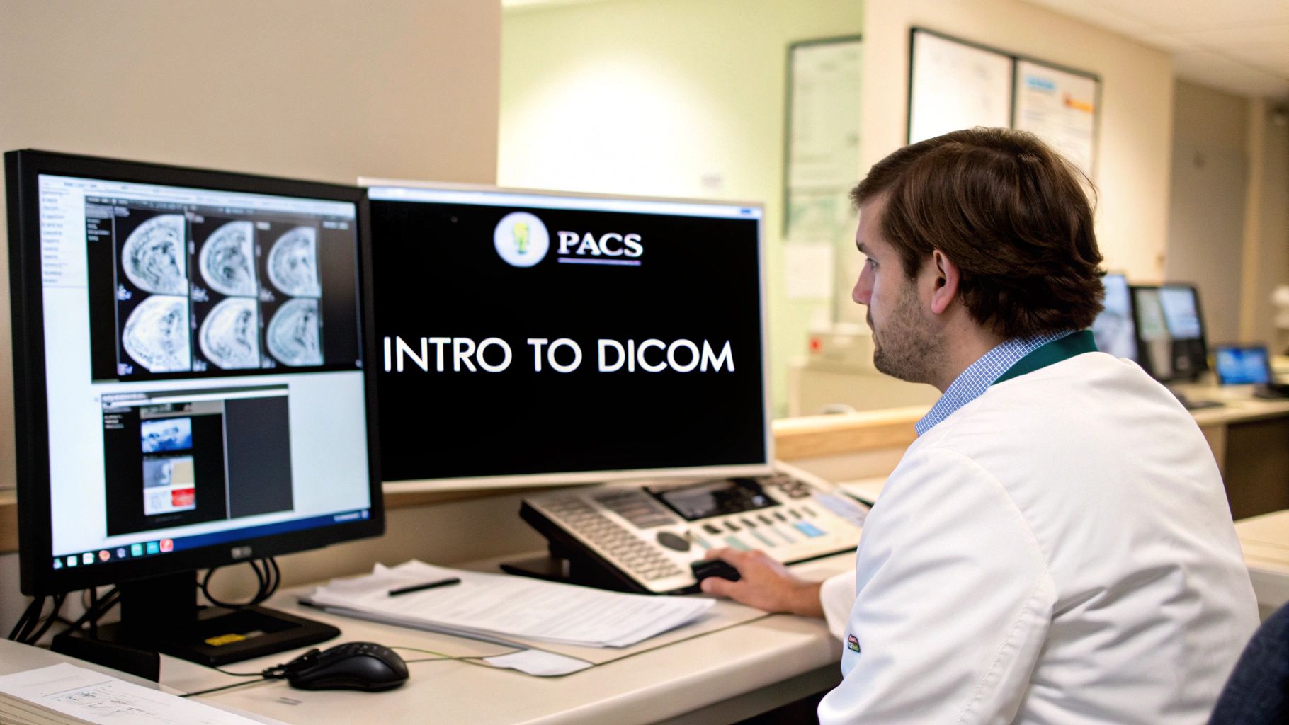
A DICOM file isn't just a picture; it's an entire data package. Think of it this way: a standard JPEG is like a simple photograph, but a DICOM file sample is that photograph with a full patient chart attached. This distinction is everything when you're working on a serious medical imaging project.
Frankly, without high-quality, realistic DICOM files, trying to develop an accurate AI algorithm or test new clinical software is a non-starter.

Why a DICOM File Sample Is Critical for Development
The real value of a DICOM file comes from its two-part structure. It bundles the pixel data—the visual image you see—with a comprehensive header full of metadata. This isn't just basic file info; it's a detailed record of everything from patient demographics (anonymized in sample files, of course) to the exact scanner settings used to capture the image. For any developer, this context is pure gold.
This kind of standardized format wasn't always a given. The journey began back in 1983 when the American College of Radiology (ACR) and the National Electrical Manufacturers Association (NEMA) joined forces. Before they created the standard, imaging devices from different manufacturers couldn't talk to each other, which created chaos and massive hurdles in the clinic. You can get a deeper dive into the standard's history on Wikipedia.
Fueling Accurate AI Models
When it comes to machine learning, this metadata isn't just a nice-to-have; it's absolutely essential. An AI model can't learn effectively from pixels alone. It needs the rich context that only a DICOM file can provide.
- Pixel Spacing and Slice Thickness: This tells the algorithm the physical size of each pixel and the distance between scan slices. Without it, making accurate anatomical measurements is impossible.
- Imaging Modality Details: Is it a CT, an MRI, or an X-ray? What was the exposure time? This information helps the model understand the inherent properties of the image it's analyzing.
- Study and Series Information: This data is crucial for organizing complex scans, making sure the model processes a sequence of related images in the correct order.
Lacking this data, an algorithm could easily misinterpret a benign cyst as a malignant tumor simply because it can't accurately gauge its size or density from the pixels alone.
Key Takeaway: A high-quality DICOM file sample provides the ground truth needed to train reliable and safe medical AI. It’s what elevates a project from a basic image recognition task to a clinically meaningful diagnostic tool.
At the end of the day, working with a proper DICOM file sample is about making sure your software or AI model is tested against the same kind of data it will face in a real-world clinical setting. That’s how you build a solid foundation for success.
Finding High-Quality DICOM Datasets
Knowing where to look is half the battle when you're hunting for the right DICOM file sample. Instead of just Googling and hoping for the best, it’s much more effective to go straight to the well-known public repositories. These archives are absolute gold mines for developers and researchers.
Many of the best datasets come from respected research and government institutions. Their whole purpose is to push medical science forward, which is great news for us. It means the data is usually well-organized, comes with solid documentation, and has been properly anonymized. This level of quality can save you a ton of headaches with data cleaning later on.
Think about it this way: if you're building an AI to spot lung nodules, you need a solid collection of chest CT scans. If you're working on a neuroscience project, you'll want a deep set of brain MRIs. Starting with the right repository makes all the difference.
Top Repositories for Your Project
One of the first places I always check is The Cancer Imaging Archive (TCIA). It’s a massive resource supported by the U.S. National Cancer Institute and is packed with cancer-related medical images. You can find just about everything—CT, MRI, digital pathology—and it’s often linked to clinical outcomes. For any AI project focused on oncology, TCIA is the gold standard.
Another fantastic source is the National Institutes of Health (NIH) Chest X-ray Dataset. With over 100,000 anonymized chest X-ray images, this collection is perfect for training models to classify diseases like pneumonia. The sheer volume of data here is a huge plus for building robust algorithms.
From my experience, the best dataset is always the one that closely matches the clinical problem you're trying to solve. A focused set of high-quality images from the right modality will always give you better results than a larger, more generic dataset.
As you explore these sources, keep a few things in mind:
- What’s the focus? Does the repository have a specialty, like a specific part of the body or a disease, that fits your project?
- Is it properly anonymized? You need to make sure the data has been de-identified according to standards like HIPAA. This is non-negotiable for ethical use.
- What are the rules? Always, always check the license. Most data is free for non-commercial research, but there might be strings attached for commercial projects.
Comparison of Public DICOM Sample Repositories
To help you get started, I've put together a quick comparison of some of the most popular repositories. This should give you a good sense of where to begin your search based on your specific needs.
| Repository Name | Primary Focus | Common Modalities | Anonymization Level | Access Requirements |
|---|---|---|---|---|
| The Cancer Imaging Archive (TCIA) | Oncology | CT, MRI, PET, Pathology | High | Free, registration may be needed |
| NIH Chest X-ray Dataset | Thoracic Diseases | X-ray (CXR) | High | Free, direct download |
| OpenNeuro | Neuroscience | MRI, fMRI, EEG | High | Free, open access |
| Medical Segmentation Decathlon | Various | CT, MRI | High | Free, challenge-based |
Each of these sources has its strengths, so take a moment to explore the one that seems like the best fit. Finding the right data is the foundational first step to a successful medical imaging project.
How to Inspect Your First DICOM File
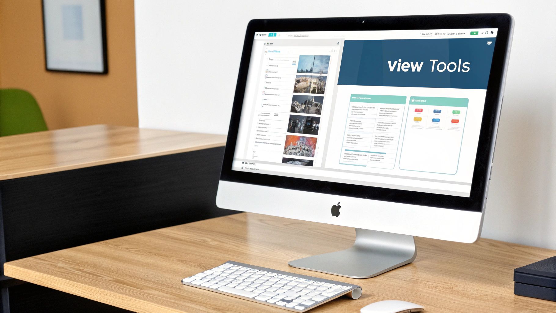
So, you've got a dataset that looks promising. Now for the fun part: popping the hood and seeing what's actually inside. This is where a good DICOM viewer becomes indispensable. These tools do more than just show you an image; they unlock all the rich metadata packed into every single file.
Plenty of fantastic, free viewers are out there. If you're on a Mac, Horos is a powerhouse open-source option and a big favorite in the community. For Windows folks, RadiAnt DICOM Viewer and MicroDicom are both solid choices with great features and intuitive interfaces. Your first real step is to download and install one of these to start exploring your dicom file sample.
Cracking Open the DICOM Header
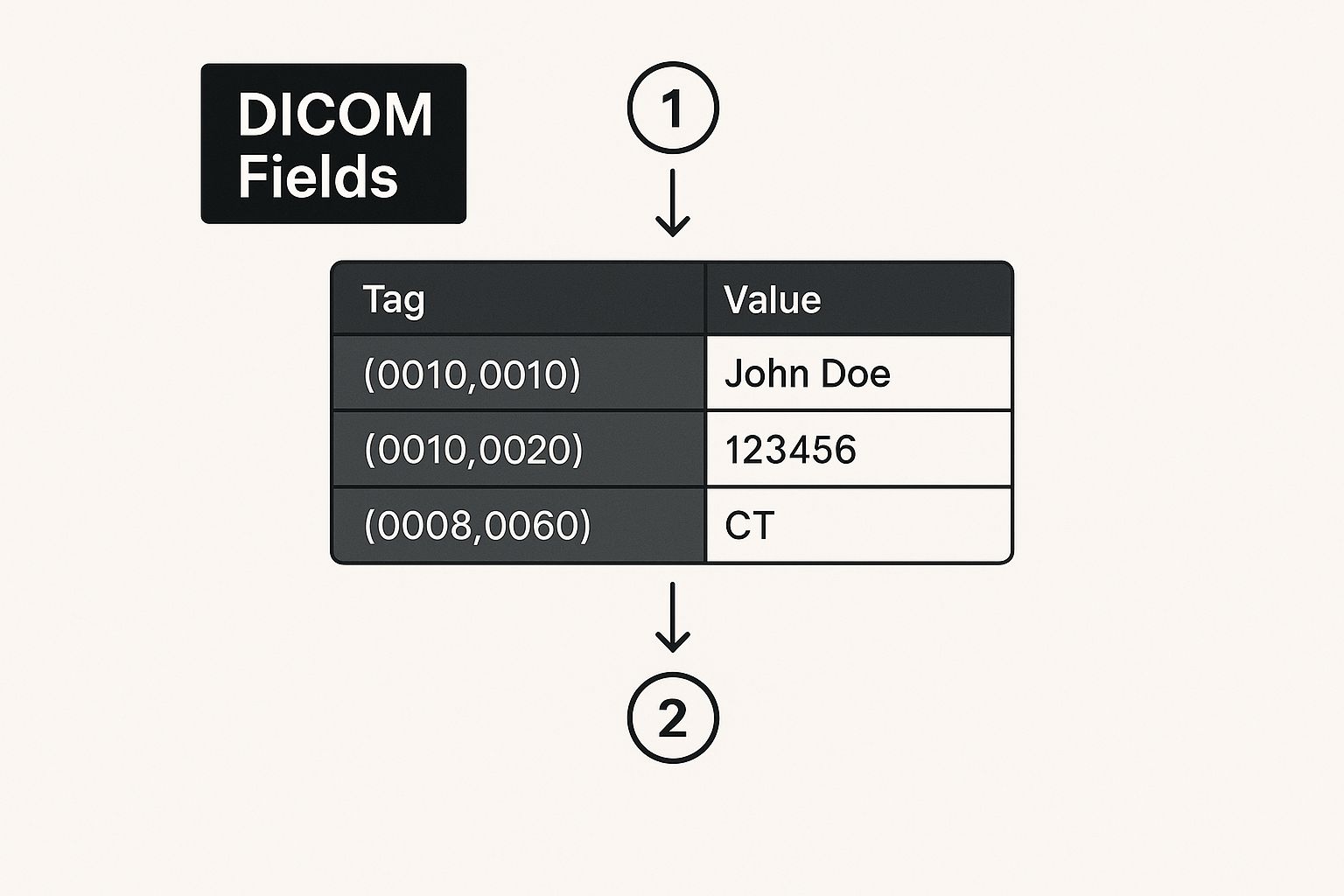
When you open a file for the first time, you'll immediately notice two key parts: the image itself and something called the DICOM header. The image is what we see, but the header is where the magic happens for any data science project. It contains all the contextual information, organized neatly by tags—unique codes that identify each piece of data.
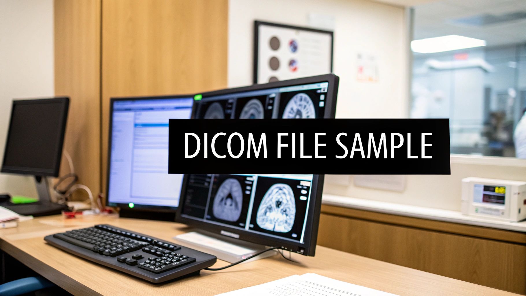
Think of it like this: the viewer shows you the medical image alongside all the crucial patient and study info, letting you connect the pixels to their clinical context.

A good viewer makes it easy to jump between analyzing the image and digging through the metadata that gives it meaning.
What to Look For in the Metadata
Digging into the metadata isn't just a box-ticking exercise. It's how you validate whether the data is actually good enough for what you want to do. The header holds everything from patient details to the exact settings used on the scanner. This tight integration is precisely what makes DICOM the standard in hospitals, as it allows for smooth communication between imaging devices and hospital-wide systems. If you want to go deeper, check out these insights on DICOM's role in healthcare.
Pro Tip: Immediately look for tags like (0028,0030) Pixel Spacing and (0018,0050) Slice Thickness. For any AI model that needs to make accurate measurements or build 3D reconstructions, these values are absolutely non-negotiable. If they're missing or inconsistent, you've got a problem.
Getting comfortable with this tag-based structure is the key. It's how you'll eventually write scripts to pull out the exact information you need to prepare your data for training a model. Once you start recognizing these essential tags on sight, you're no longer just looking at pictures—you're truly understanding your data.
Getting Your DICOM Sample Ready for AI Training
So, you've got your hands on a DICOM sample file. That’s a great start, but the real fun is just beginning. Raw medical images are almost never ready for an AI model right out of the gate. I like to think of it as prepping ingredients for a complex recipe—you have to wash, chop, and measure everything before you can even turn on the stove. This prep work, or preprocessing pipeline, is what turns that raw data into a high-quality meal for your model.
One of the very first things you'll need to tackle is normalizing the pixel intensity values. You'll quickly notice that images from different scanners, or even from the same scanner with different settings, can have wildly different levels of brightness and contrast. Normalization wrangles all these variations into a standard range, usually between 0 and 1. This step is absolutely critical; it prevents your model from getting confused by imaging quirks that have nothing to do with the actual anatomy.
This kind of consistency is the bedrock of reliable medical AI. It's what allows the entire healthcare imaging market—valued at around $35 billion back in 2020—to function, with DICOM ensuring devices from over 50 major manufacturers can talk to each other. If you're curious about how we got here, it's worth reading up on the history of the DICOM standard.
Fine-Tuning Your Dataset for Better Results
Once normalization is handled, you need to be absolutely certain your data is anonymized. Even if a dataset claims to be "de-identified," I always recommend running your own script to double-check and scrub any lingering Protected Health Information (PHI) from the DICOM headers. It’s a simple step that protects patient privacy and keeps you on the right side of compliance. Don't skip it.
Another incredibly useful technique, especially when you don't have a massive dataset, is data augmentation. This is where you artificially expand your collection of images by creating slightly modified versions of the ones you have.
A few common tricks of the trade include:
- Rotation: Tilting an image by a few degrees to mimic slight shifts in patient positioning.
- Flipping: Creating a mirror image of the scan.
- Zooming: Magnifying a random portion of the image to train the model to spot features at different scales.
These transformations effectively give you more data to train on, which helps build a much more resilient model—one that won't get tripped up by the small, real-world variations it's bound to see in a clinical setting.
Here’s a practical example: Imagine you're building a model to spot pneumonia in chest X-rays. By augmenting your DICOM samples with small rotations and brightness tweaks, you teach the algorithm to focus on the actual patterns in the lungs, not the specific angle of the X-ray or the machine's exposure settings.
By carefully working through these preprocessing steps—normalization, anonymization, and augmentation—you create a clean and powerful data pipeline. This isn't just busywork; it's the foundational effort that elevates a project from a simple experiment to an AI tool that could one day make a real difference.
Navigating the Inevitable Hurdles with DICOM Files

When you start gathering DICOM files from different sources, you'll quickly realize no two datasets are perfectly alike. You’re going to run into some bumps. It's just part of the process.
One of the biggest headaches I see time and again is wildly inconsistent metadata. You'll find crucial tags present in one set of scans and completely absent in another. If you're not prepared, this kind of variance can easily break your data processing pipelines or, worse, introduce bias into your AI model.
Another classic problem is wrestling with different Transfer Syntaxes. You'll get everything from massive, uncompressed image data to various compressed formats like JPEG 2000. Your software needs to be a jack-of-all-trades, ready to decode whatever comes its way. If it isn't, you'll be staring at errors and files that simply won't open, stopping your entire workflow in its tracks.
Keeping Your Data (and Your Project) Safe
Beyond the technical snags, you absolutely have to get the ethical and legal side right. This isn't optional. Every single DICOM file you work with must be properly anonymized to meet strict regulations like HIPAA. Using data that even might contain Protected Health Information (PHI) is a massive ethical breach and opens you up to serious legal trouble.
And while the DICOM protocol itself is solid, the ecosystem around it isn't a fortress. The tools we use can be a weak link.
I've seen security reports where malware was literally bundled with DICOM viewing software. It’s a stark reminder that while direct attacks on the protocol are rare, the tools and data sources we rely on are very much a target.
To steer clear of these pitfalls, you need a solid game plan. Here are a few practices that have become non-negotiable for my own projects:
- Scrub Your Metadata: Before you do anything else, run a script to standardize your metadata. Check for essential tags, fill in what you can, and flag any files that are too incomplete to be useful.
- Lean on Good Libraries: Use a battle-tested library that can handle the messy parts for you. In Python,
pydicomis the gold standard for a reason; it gracefully manages different Transfer Syntaxes so you don't have to. - Trust, But Verify Anonymization: Even if a dataset is labeled "anonymous," run your own de-identification script on it. Always. It’s a simple check that can save you from a world of hurt.
- Stick to Reputable Sources: Get your datasets and viewers from trusted places—think major academic institutions or government archives. This is your best defense against malware and questionable data.
Building these habits from the start ensures your project is built on a foundation of clean, consistent, and ethically sound data. It's the only way to do this work responsibly.
Got Questions About DICOM Samples?
Diving into the world of medical imaging can feel a little intimidating at first. You're bound to have some questions. Getting those sorted out early on lets you stop worrying about the basics and start focusing on what really matters—your project.
DICOM vs. JPEG: What's the Real Difference?
It’s a common starting point. A JPEG or PNG is just a picture—it’s the raw pixel data and nothing more. A DICOM file, on the other hand, is a whole different beast. Think of it as a smart container.
Inside, you don't just get the image; you get a treasure trove of metadata. This includes everything from anonymized patient details and the type of scanner used to the specific settings for that particular scan. That clinical context is everything. It's what allows a radiologist to make a diagnosis and, just as importantly, it's what you need to train an AI model that truly understands the data it's seeing.
Are These DICOM Samples Actually Free to Use?
For the most part, yes, especially when it comes to datasets from big research and government institutions like the NIH. They often make vast archives available for research and non-commercial projects.
But—and this is a big "but"—you always have to check the license. Some datasets prohibit commercial use, while others might just ask you to cite the original source. Make it a habit to read the terms before you download anything. A few minutes of due diligence upfront can save you a world of legal headaches later on.
How Do I Actually Open a DICOM File?
You can't just double-click a DICOM file and have it open in your default photo viewer. It won't work. You’ll need a dedicated DICOM viewer to properly interpret the file and its metadata.
Fortunately, there are some fantastic free options out there:
- For Mac users, Horos is pretty much the gold standard.
- If you're on Windows, both RadiAnt DICOM Viewer and MicroDicom are excellent choices.
For anyone working on the development side, the game changes. A graphical viewer is helpful, but programmatic access is essential. Libraries like
pydicomfor Python let you slice and dice DICOM files directly within your code. This is how you really get your hands on all that valuable metadata for building automated data pipelines.
This kind of direct access is the foundation of any serious machine learning project in medical imaging.
At PYCAD, building those exact kinds of pipelines is what we do. We turn raw, complex DICOM data into robust, AI-driven solutions. From data annotation to deploying a final model, we manage the entire technical journey. Learn more about our services at https://pycad.co.


