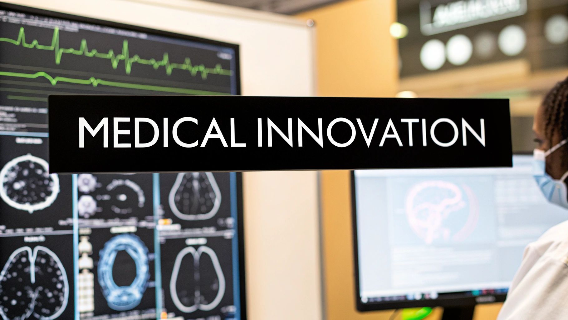
Medical Imaging Breakthroughs: Understanding Image Processing Algorithms
Image processing is playing an increasingly vital role in modern healthcare, allowing doctors to see inside the human body in remarkable detail. Through advanced computer vision and AI algorithms, medical professionals can now detect diseases earlier, make more accurate diagnoses, and create customized treatment plans for patients. From sharpening blurry X-rays to automatically spotting early signs of cancer, these tools are changing patient care.
The field has progressed significantly – from basic image enhancement to sophisticated AI systems that can analyze medical scans. Early algorithms focused on improving image quality to help radiologists interpret results better. Now, modern systems can automatically diagnose conditions, predict how patients will respond to treatments, and suggest personalized therapy approaches. The most effective algorithms combine speed, accuracy, reliability across different image types, and seamless integration into clinical workflows.
This progress builds on key concepts from signal processing, pattern recognition, and machine learning. The ability to analyze massive datasets of medical images has been crucial, allowing systems to spot subtle patterns that humans might miss. These algorithms keep improving as they learn from more examples.
We’ll explore eight powerful algorithms that are changing medical imaging, covering how they work and their real-world impact on healthcare. For anyone involved in medical technology – whether developing devices, researching new AI applications, or providing patient care – understanding these essential tools is key to delivering better health outcomes. Get ready for an eye-opening look at how image processing is improving diagnosis and treatment.
1. Convolutional Neural Networks (CNN)
Convolutional Neural Networks (CNNs) have become essential tools in medical image processing. These deep learning algorithms process image pixel data and learn patterns automatically, reducing the need for manual feature engineering that traditional methods require.
How CNNs Work
CNNs use multiple layers to analyze images. The convolutional layers scan images with filters to detect features like edges and textures. Pooling layers then reduce the data size while preserving important information. As data moves through the network, it identifies increasingly complex patterns and features directly from pixels – no manual feature extraction needed.
Key Features
- Multiple processing layers: Enable step-by-step pattern learning
- Automatic feature detection: Eliminates manual feature engineering
- Pattern recognition: Identifies complex visual elements
- Complete processing: Handles data from input to prediction
- Location flexibility: Spots patterns anywhere in images
Real-World Medical Applications
CNNs excel in several critical medical imaging tasks:
- Lung Cancer Detection: Analyzing CT scans to find early-stage nodules
- Brain Tumor Analysis: Precisely mapping tumor boundaries in MRI scans
- Eye Disease Screening: Examining retinal images for diabetic retinopathy
History and Development
While CNNs emerged in the 1980s through Yann LeCun’s work, they gained widespread use when computing power and large datasets became available. Success in image recognition competitions by teams from Google DeepMind and Stanford demonstrated their capabilities, leading to adoption across medical imaging.
Advantages and Limitations
Benefits:
- Strong pattern detection
- Minimal manual engineering needed
- Handles large image sets well
- Works with shifted/moved objects
Challenges:
- Needs extensive training data
- Heavy computing requirements
- Limited result explanations
- Risk of overtraining
Implementation Tips
- Use pre-trained models when data is limited
- Expand datasets with image modifications like rotations
- Add explanation tools like Grad-CAM for clarity
- Create balanced datasets to prevent biased results
CNNs provide powerful tools for medical imaging analysis. Understanding their strengths and limits helps healthcare organizations use them effectively to improve diagnoses and treatment planning.
2. Active Contour Models (Snakes)
Active Contour Models, also called “snakes,” are key tools for medical image segmentation. These algorithms use energy-minimizing splines guided by image forces to find object boundaries in medical images. Their ability to adapt dynamically makes them excellent at detecting complex anatomical structures.
The core principle of snakes is energy minimization. The snake is a curve that evolves to minimize both internal and external energy. Internal energy keeps the curve smooth and continuous, while external energy from the image data pulls the snake toward important features like edges and intensity changes. This balance helps the snake find accurate object boundaries.
Medical imaging professionals widely adopted snakes because they work well with noisy images and can detect smooth boundaries even when parts are missing. The dynamic nature of snakes lets them adjust to variations in shape and intensity – a major improvement over basic edge detection. This technique gained prominence through the work of Michael Kass, Andrew Witkin, and Demetri Terzopoulos.
Key features that make active contours effective:
- Dynamic adaptation: The curve evolves to match object shapes during processing
- Energy framework: Balances smoothness with image data for optimal results
- Force control: Allows fine-tuning based on image properties
- Continuous detection: Produces smooth, connected boundary lines
Common medical applications include:
- Heart chamber analysis: Finding chamber boundaries in ultrasound and MRI
- Tumor measurement: Outlining tumors in CT and MRI for treatment planning
- Blood vessel mapping: Tracing vessels in angiograms
Benefits of Active Contours:
- Noise resistant: Handles typical medical image artifacts well
- Smooth results: Creates continuous boundary curves
- User control: Allows manual adjustments when needed
- Gap filling: Can bridge missing boundary segments
Limitations to consider:
- Starting position matters: Initial placement must be near the target
- Local minima issues: May settle on suboptimal solutions
- Processing demands: Can be slow, especially for 3D images
- Shape limitations: Basic snakes struggle with holes and branches
Implementation tips:
- Start close: Place initial contour near the target boundary
- Adjust parameters: Test different internal/external force settings
- Use GVF: Gradient Vector Flow helps avoid local minima
- Consider variants: Try topology-adaptive methods for complex shapes
Active Contour Models remain essential in medical image analysis because they handle challenging real-world scenarios well. While initialization and speed can be tricky, their flexibility makes them invaluable for many medical imaging tasks. Ongoing research keeps expanding what these algorithms can do.
3. Watershed Segmentation
Watershed segmentation uses the concept of topographic mapping to segment images. Picture a grayscale image as a 3D landscape where pixel intensity values represent elevation. When rain falls on this landscape, it flows downhill and collects in basins. The ridges between these basins, called watershed lines, mark the boundaries between image segments. This approach works especially well for separating touching or overlapping objects in medical images.
How it Works:
The watershed algorithm detects local minima (basins) in the image gradient and simulates flooding from these points. As the water level rises, different basins begin to merge. The points where basins meet become watershed lines, defining the boundaries between segments.
Key Features:
- Edge detection: Uses image gradients to find object boundaries
- Region growing: Expands from seed points to segment touching objects
- Morphological basis: Employs operations like dilation and erosion
- Marker control: Allows manual markers to guide segmentation
- Boundary accuracy: Precisely detects object edges
- Processing speed: Faster than many other methods
- Complete boundaries: Creates fully closed object contours
Advantages and Limitations:
| Advantages | Limitations |
|---|---|
| Precise edge detection | Can over-segment images |
| Quick processing time | Affected by image noise |
| Works with defined edges | Needs image smoothing |
| Creates closed boundaries | Marker selection takes experience |
Managing Common Issues:
Over-segmentation happens when too many local minima are detected, creating excess regions. This can be addressed through:
- Marker-based approach: Using predefined markers to guide segmentation
- Image smoothing: Applying filters to reduce noise before processing
- Region merging: Combining segments after initial segmentation
Medical Applications:
The watershed method helps analyze:
- Cell microscopy: Finding individual cells in crowded images
- Brain MRI: Separating brain structures and regions
- Bone analysis: Examining bone architecture in CT scans
Background:
Serge Beucher and Christian Lantuéjoul developed the watershed transform for image processing in the late 1970s. Their work made the technique a standard tool in medical imaging.
Implementation Tips:
- Choose markers carefully to prevent over-segmentation
- Test different pre-processing steps to optimize results
- Use metrics like Dice coefficient to evaluate accuracy
- Consider combining watershed with other segmentation methods
The watershed method’s ability to separate touching objects and process images quickly makes it vital for medical image analysis. Through proper pre-processing and marker selection, it can extract meaningful data from complex medical images.
4. SURF (Speeded Up Robust Features)
SURF (Speeded Up Robust Features) is a patented system that detects and describes local features in images. Its exceptional speed and reliability make it valuable for medical image analysis, particularly when dealing with varying scales and rotations across different images. This makes it ideal for time-critical applications like image-guided surgery and quick diagnoses.
The algorithm gets its speed from two key components: integral images and box filters. Integral images enable quick calculations of Haar wavelet responses at multiple scales. Box filters act as approximations of Gaussian derivatives, making computations much faster without major accuracy loss. This approach gives SURF a significant speed advantage over its predecessor SIFT.
SURF finds unique image features that remain consistent despite changes in scale or rotation. It creates compact vector descriptions of these features for efficient image matching. Some key medical applications include:
- Medical image registration: Matching images from different scanning methods (CT, MRI) or time periods to track changes and plan treatments
- Ultrasound feature tracking: Monitoring anatomical structures in real-time during ultrasound procedures
- Anatomical landmark detection: Finding specific points on bones and organs to guide surgery and diagnosis
Herbert Bay at ETH Zurich developed SURF to be faster than SIFT while maintaining similar accuracy. This speed advantage is crucial for medical imaging where large datasets and real-time processing are common.
Main Benefits:
- Superior speed: Processes images much faster than SIFT through efficient calculations
- Scale adaptation: Successfully matches features across different image sizes
- Noise handling: Works well with the image noise common in medical scans
- Reliable matching: Creates distinct feature descriptions for accurate image alignment
Key Limitations:
- Patent restrictions: May require licensing for commercial use
- Slightly less precise: Speed optimizations can reduce accuracy compared to SIFT
- Memory usage: Requires significant memory when processing large images
- Limited transformation handling: Works best with scale and rotation changes, less so with other image distortions
Implementation Advice:
- Use scale layers: Process at multiple scales to improve results and efficiency
- Customize thresholds: Adjust detection settings to control feature quantity
- Choose matching methods: Select appropriate algorithms like FLANN for optimal feature pairing
- Enable parallel processing: Use multi-core systems or GPUs to speed up large-scale processing
For companies and researchers in medical technology, understanding these characteristics helps determine if SURF fits their needs. While licensing costs matter, its combination of speed and reliability makes it valuable for many medical imaging tasks requiring both accuracy and quick results.
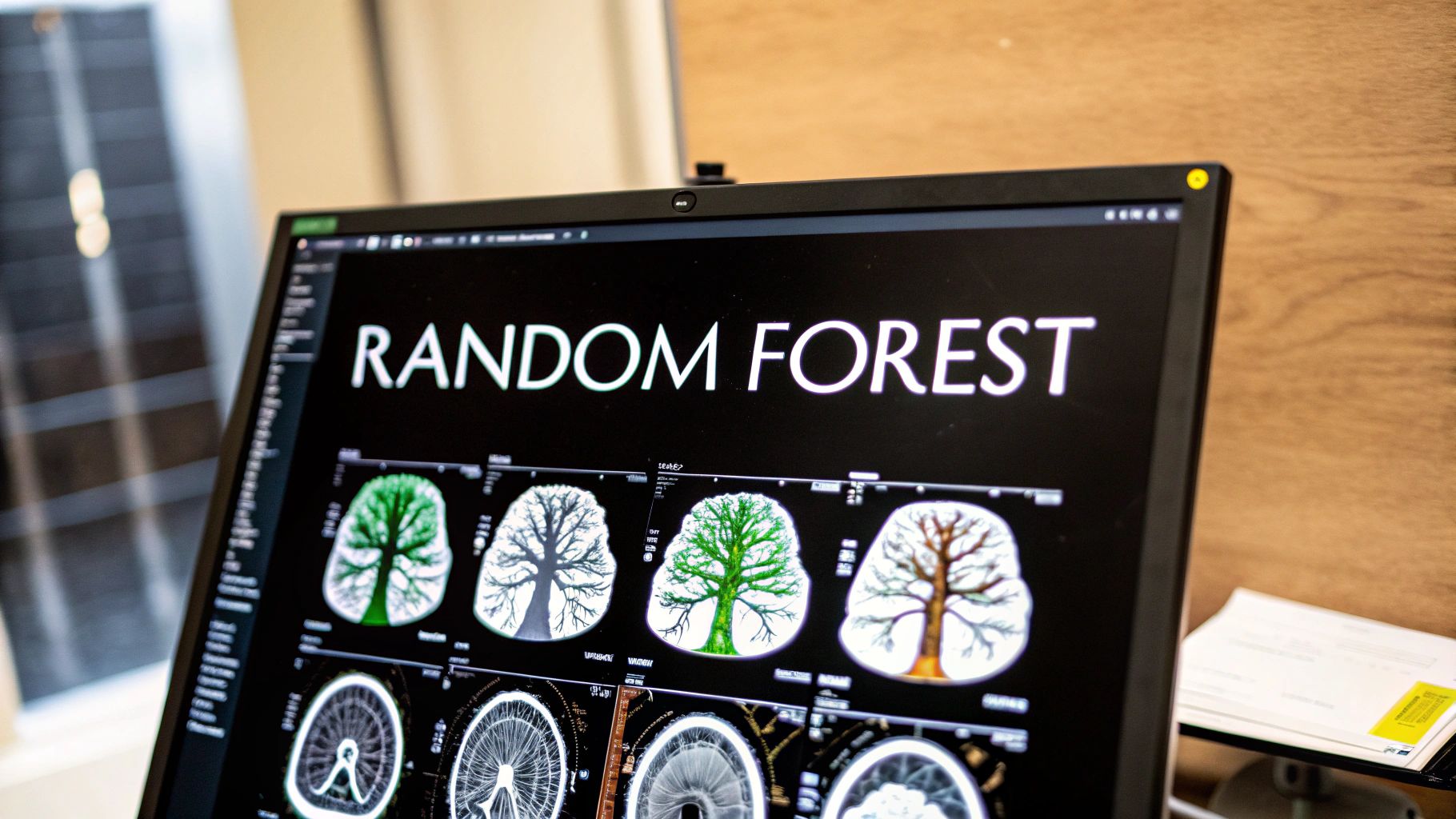
5. Random Forest Segmentation
Random Forest segmentation is a type of machine learning that divides medical images into distinct regions representing different tissues, organs, or abnormalities. This method combines multiple decision trees to create reliable and precise segmentation results. Its effectiveness with complex medical data has made it a common choice for analyzing medical images.
The strength of Random Forests comes from having many decision trees work together. Rather than using just one tree, which can be unreliable, it pools the results from numerous trees – each trained on different data samples. This group approach helps handle noise and unusual data points that often appear in medical images.
How it Works
Random Forest segmentation builds many decision trees. Each tree learns from a randomly selected subset of the training data, with some data points appearing multiple times while others are left out. At each decision point in a tree, only some features are considered for making splits. This randomness helps create diverse trees that work well together. For a new image pixel, each tree “votes” on what type of tissue it is. The final label comes from the majority vote, while the vote distribution shows how certain the system is about its decision.
Features and Benefits
- Multiple Tree Approach: Using many trees reduces overfitting and helps handle new data better
- Feature Analysis: Uses image properties like brightness, texture and shape to classify pixels
- Shows Uncertainty: Provides confidence levels for classifications
- Fast Processing: Trees can work in parallel to speed up training and predictions
Pros
- Handles Complex Images: Works well with varied tissue types and structures
- Good with New Data: Less likely to overfit compared to single trees
- Shows Confidence Levels: Indicates how sure the system is about each classification
- Resists Overfitting: Group approach and random features help prevent this common issue
Cons
- Needs Good Features: Success depends on choosing the right image characteristics
- Uses Lots of Memory: Storing many trees requires significant computer memory
- Can Take Time: Training a large forest can be slow
- Hard to Optimize: Finding the best settings for trees and features takes work
Real-World Examples
- Brain MRI Analysis: Used to identify white matter, gray matter, and fluid in brain scans
- Organ Finding in CT: Helps locate organs like liver, kidneys, and lungs automatically
- Finding Problems: Detects issues like tumors or inflammation in medical images
Implementation Tips
- Balance Tree Numbers: Add trees until performance levels off
- Choose Features Carefully: Pick image properties that matter for your task
- Test Thoroughly: Use cross-validation to check performance
- Study Important Features: Look at which image aspects help most with segmentation
Historical Note
Leo Breiman and Adele Cutler developed and promoted Random Forests.
Random Forest segmentation has become essential in medical imaging because it works reliably, handles complex data well, and shows how confident it is in its results. Its proven success across many medical applications makes it valuable for researchers, doctors, and medical technology companies.
6. Histogram Equalization
Histogram equalization is a key image processing method that improves image contrast in medical imaging. This technique works by redistributing pixel intensity values to reveal hidden details in low-contrast images.

Key Features:
- Distribution normalization: Evens out the spread of intensity values across the image
- Automatic contrast: Optimizes contrast based on the image histogram without manual input
- Histogram analysis: Uses the image’s intensity distribution data
- Global and local options: Can be applied to entire images or specific regions using CLAHE for better local detail
Advantages:
- Easy to implement: Simple integration into existing image processing workflows
- Strong contrast boost: Significantly improves visibility in poor contrast images
- Minimal user input: Works automatically with few parameters needed
- Fast processing: Quick enough for real-time applications
Limitations:
- Noise amplification: Can make image noise more apparent
- Artificial appearance: May create unnatural-looking results
- Detail loss: Some image details may be reduced
- Limited applicability: Works better with some image types than others
Clinical Applications:
- X-ray enhancement: Better visualization of bones and soft tissues
- MRI contrast: Clearer distinction between different tissue types
- Mammogram analysis: Improved detection of subtle breast tissue changes
Technical Evolution:
The core principles have remained stable for decades, but modern computing power has enabled wider adoption in medical imaging. Research institutions and medical device companies continue refining the technique for clinical use.
Implementation Tips:
- Consider CLAHE: Use this adaptive method to preserve local contrast while minimizing noise
- Monitor noise levels: Check processed images for unwanted noise amplification
- Visual verification: Always review enhanced images to confirm appropriate results
This established technique remains vital for improving medical image quality and supporting accurate diagnosis. Understanding its capabilities and constraints helps medical professionals apply it effectively.
7. U-Net Architecture
The U-Net network has become a critical tool for biomedical image analysis. Named for its “U” shaped design, this architecture has proven highly effective for medical image segmentation tasks, even with small training datasets. Its success comes from handling the specific needs of medical imaging – from detailed tissue boundaries to subtle anatomical variations.
Medical images present unique challenges that traditional segmentation methods often failed to address. These include complex structural details, minor variations between healthy and diseased tissue, and the need for precise boundary detection, especially when working with limited data.
How U-Net Works
At its core, U-Net uses a symmetric encoder-decoder structure. The encoder path compresses the input image through multiple layers, capturing key features at different scales. The decoder path then rebuilds the image by gradually increasing resolution to create the final segmentation mask. What makes U-Net special are its skip connections – direct paths that carry detailed information from encoder layers to matching decoder layers. This preserves important fine details that would otherwise be lost during compression, leading to more accurate results. The combination of broad context analysis and precise detail preservation is what drives U-Net’s strong performance.
Key Features and Benefits
- Symmetric design: Enables both broad feature extraction and precise reconstruction
- Skip connections: Keep fine details intact for better boundary detection
- Deep feature analysis: Captures comprehensive contextual information
- Multi-scale processing: Handles objects of varying sizes effectively
- Small dataset training: Works well even with limited training examples
- High accuracy: Produces detailed, precise segmentations
- Size flexibility: Successfully segments both microscopic and large anatomical structures
- Quick processing: Delivers results efficiently for time-sensitive cases
Pros and Cons
| Pros | Cons |
|---|---|
| Works with small datasets | Complex architecture |
| Highly accurate results | High GPU memory needs |
| Handles different sizes | Training can be unstable |
| Fast processing speed | Complex data preparation |
Real-World Applications
U-Net has shown excellent results across many medical use cases:
- Cell analysis: Finding and outlining individual cells in microscope images
- Organ segmentation: Mapping organs like the liver and heart in CT/MRI scans
- Cancer detection: Identifying and measuring tumors across different imaging types
Implementation Tips
- Use data augmentation: Expand your training set with rotations, flips and scaling
- Add batch normalization: For more stable and faster training
- Tune learning rate: Carefully adjust to optimize performance
- Consider variants: Look into 3D U-Net for volume data or attention U-Net for focused analysis
Origins and Impact
U-Net was created by Olaf Ronneberger, Philipp Fischer, and Thomas Brox at the University of Freiburg for biomedical image segmentation. Their work quickly gained widespread adoption in medical imaging and inspired many variations.
Thanks to its reliability, precision, and ability to work with limited training data, U-Net has become essential for medical device companies, healthcare tech firms, researchers and doctors. It continues to drive advances in medical image analysis and patient care.
8. Gabor Filter
The Gabor filter is a specialized filter named after Dennis Gabor that has become essential in medical image processing for analyzing textures. Its main strength lies in detecting specific patterns and extracting detailed texture information from medical images. What makes it particularly useful is its ability to examine both spatial details and frequency patterns at the same time.
At its core, a Gabor filter combines a sine wave with a Gaussian curve. This structure allows it to pick up specific patterns, directions and scales within an image. Here are the four key characteristics that define Gabor filters:
- Directional Analysis: The filters can detect features pointing in specific directions, like edges and lines
- Multi-Scale Processing: Adjustable Gaussian settings enable analysis at different levels of detail
- Frequency Detection: The sine wave component helps identify specific frequency patterns
- Precise Localization: The Gaussian curve ensures accurate positioning of detected features
Key Benefits:
- Detailed Texture Analysis: Provides thorough information about texture patterns
- Scale Flexibility: Can examine features at both fine and broad levels
- Directional Detection: Effectively finds structures oriented in specific ways
- Biologically-Based: Mirrors how visual cells in the brain process information
Main Limitations:
- Complex Setup: Choosing the right filter settings requires careful testing
- Heavy Processing: Running multiple filters demands significant computing power
- Large Filter Sets: Complete analysis often needs many different filters
- Memory Usage: Storing filter data can use substantial computer memory
Medical Applications:
- Blood Vessel Analysis: The filters help find and outline blood vessels in eye scans by detecting their linear structure, aiding diagnosis of conditions like diabetic retinopathy
- Tissue Examination: Different tissues show distinct texture patterns that Gabor filters can detect, helping distinguish healthy from diseased tissue in mammograms and ultrasounds
- Cancer Detection: The texture analysis capabilities help automated systems spot suspicious patterns that could indicate breast cancer
Implementation Tips:
- Test Filter Settings: Carefully adjust parameters through testing to get optimal results for your specific use case
- Create Filter Collections: Build sets of filters covering relevant angles and scales
- Use Multiple Scales: Analyze both fine details and broader patterns
- Speed Up Processing: Use parallel computing when possible to handle intensive calculations
Originally created for signal processing, Gabor filters have proven invaluable in medical imaging by reliably capturing subtle texture differences that can indicate various medical conditions. Their effectiveness at finding important patterns, combined with their similarity to human visual processing, has made them a vital tool in medical image analysis. Research continues to find new ways to apply these filters to improve medical diagnosis and treatment.
Medical Image Processing: 8-Algorithm Side-by-Side Comparison
| Algorithm | 🔄 Complexity | ⚡ Resources | 📊 Use Cases | ⭐ Advantages | 💡 Tips |
|---|---|---|---|---|---|
| Convolutional Neural Networks (CNN) | High complexity with deep layered structure | Requires large datasets & high computational power | Medical image segmentation, classification & pattern recognition | Automated feature extraction & translation invariance | Use transfer learning & data augmentation |
| Active Contour Models (Snakes) | Moderate; sensitive to initialization & local minima | Moderate load with iterative energy minimization | Boundary detection in noisy or incomplete images | Effective smooth boundary detection & interactive adjustments | Initialize contour near target & tune parameters |
| Watershed Segmentation | Moderate; involves pre‑processing & marker selection | Low to moderate; benefits from fast computation | Region‐based segmentation for cells, tissues & bone structures | Fast processing with clear edge detection & closed contours | Use markers to control segmentation & reduce noise |
| SURF (Speeded Up Robust Features) | Low to moderate; simpler than SIFT | Memory intensive but benefits from efficient algorithms | Feature extraction in image registration, tracking & landmark detection | Fast, scale & rotation invariant with robust matching | Adjust threshold parameters & use octave pyramids |
| Random Forest Segmentation | Moderate; requires ensemble tuning & feature engineering | Memory heavy with longer training durations | Tissue classification, organ segmentation & lesion detection | Good generalization with uncertainty measures and resistance to overfitting | Balance the number of trees & use cross‑validation |
| Histogram Equalization | Low; straightforward contrast enhancement | Computationally efficient | Enhancing contrast in X‑rays, MRI & mammograms | Simple, effective global normalization of intensity distribution | Consider adaptive variants and monitor noise amplification |
| U‑Net Architecture | High; complex encoder‑decoder with skip connections | Demands significant GPU memory & extensive dataset prep | Precise biomedical segmentation for cells, organs & tumors | Accurate segmentation even with few training images | Employ extensive data augmentation & batch normalization |
| Gabor Filter | Moderate to high; requires careful parameter selection | Computationally intensive due to large filter bank | Texture analysis in retinal imaging, tissue characterization & pattern recognition | Accurate detection of texture and orientation features | Optimize filter parameters & consider multi‑scale processing |
The Future of Medical Imaging: Algorithms at the Forefront
Medical image processing algorithms are driving major advances in diagnostics, treatment planning, and disease understanding. Advanced techniques like Histogram Equalization and Gabor Filters improve image quality, while segmentation methods such as Active Contour Models, Watershed, and Random Forest algorithms help identify anatomical structures. The rise of deep learning, especially architectures like U-Net and Convolutional Neural Networks (CNNs), has enabled computers to detect and classify medical conditions with remarkable accuracy rates above 90%.
Implementing these tools requires deep technical knowledge and careful clinical consideration. Selecting algorithms depends heavily on the specific medical task – whether enhancing image clarity, isolating organs, or finding abnormalities. Success demands thorough validation and testing to ensure the results are reliable and accurate in real medical settings.
This field demands continuous education and adaptation. Medical imaging professionals must:
- Stay current with new research findings
- Participate in key conferences and forums
- Connect with peers in the medical imaging community
- Test and refine existing methods
- Help develop new approaches
Current developments focus on:
- More automated analysis workflows
- Integration of different imaging types
- Transparent AI systems that can explain their decisions
- Real-time processing capabilities
- Personalized diagnostic approaches
Key Takeaways:
- Medical image processing algorithms play a vital role in modern healthcare
- Choosing the right algorithm requires matching it to specific clinical needs
- Ongoing learning keeps professionals current in this fast-changing field
- Future advances will focus on automation, data integration, and clear AI reasoning
Looking to bring AI capabilities to your medical imaging work? PYCAD provides complete medical AI services – from data preparation to model deployment. We help optimize medical devices for better diagnostic precision and clinical workflows. Our solutions cover data annotation, privacy protection, API deployment, and user interface creation. Visit us to learn how we can strengthen your medical imaging applications through proven AI methods.


