Think about how a radiologist traditionally views an MRI. They’re looking at a series of flat, two-dimensional images—a stack of digital slices. While each slice holds important data, trying to mentally piece them together to understand a complex anatomical structure is incredibly difficult and often imprecise. This is where the magic of 3D MRI reconstruction comes in.
From 2D Slices to 3D Models
Instead of just looking at the individual blueprints, 3D reconstruction acts like a digital construction crew. It takes that entire stack of 2D images and assembles them into a single, fully interactive three-dimensional model.
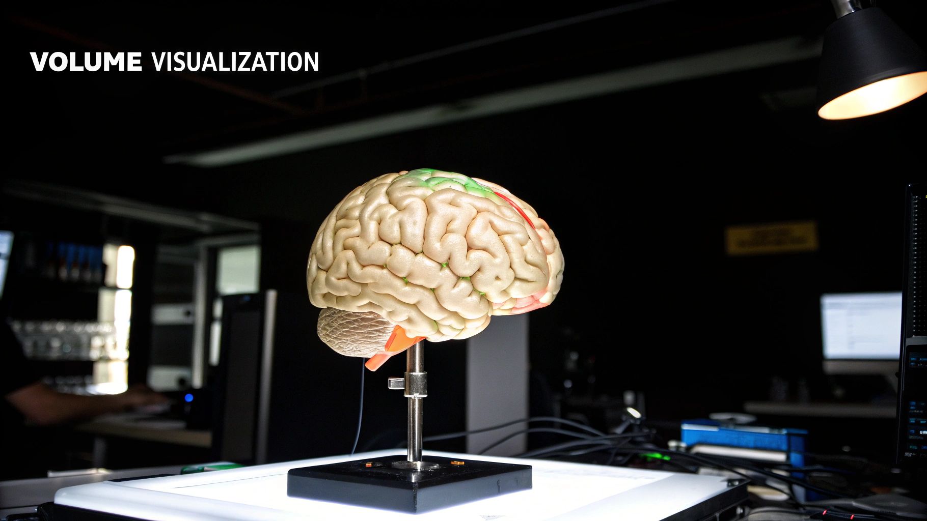
Suddenly, clinicians aren't just scrolling through static images. They can grab the 3D model, rotate it, view it from any angle, and even digitally peel back layers to see what's happening underneath. This is a monumental shift from static slices to a dynamic, volumetric view, giving doctors a much deeper understanding of the patient's anatomy.
A Brief History of Volumetric Imaging
This capability didn't just appear out of nowhere. It's the result of decades of hard work and innovation in medical imaging. The real breakthroughs in MRI started in the late 1970s, leading to the first full-body scan of a person in 1977.
By the mid-1980s, commercial MRI systems had become powerful enough to produce the high-resolution data that makes accurate 3D reconstruction possible. Seeing the timeline of these developments really puts into perspective how far the technology has advanced.
Why It Matters in Modern Medicine
This isn't just a fancy visual trick; it has a profound impact on patient care across many different medical fields. A clear, comprehensive view of a patient's unique anatomy directly leads to better outcomes.
- Diagnostic Accuracy: Clinicians can more easily identify subtle issues, like tiny tumors or complex ligament tears, that are often hidden or ambiguous in a series of 2D images.
- Surgical Planning: Surgeons can essentially perform a "dry run" of a complex operation on the 3D model, planning the safest route to avoid critical nerves and blood vessels before ever making an incision.
- Patient Communication: It's one thing to describe a medical condition, but it's another to show it. A 3D model helps doctors explain a diagnosis or a planned procedure to patients in a way that’s intuitive and easy to grasp.
To truly grasp the depth of topics like 3D MRI reconstruction, from its foundational principles to cutting-edge advancements, learning how to read scientific papers effectively is an invaluable skill.
This guide will walk you through the whole process, from how the raw data is captured inside the MRI machine to the sophisticated algorithms that build the final model. We'll also dive into how AI is making scans faster and images sharper, and look at the real-world applications in neurology, orthopedics, and cardiology that are changing patient's lives.
The Core Principles of MRI Data Acquisition
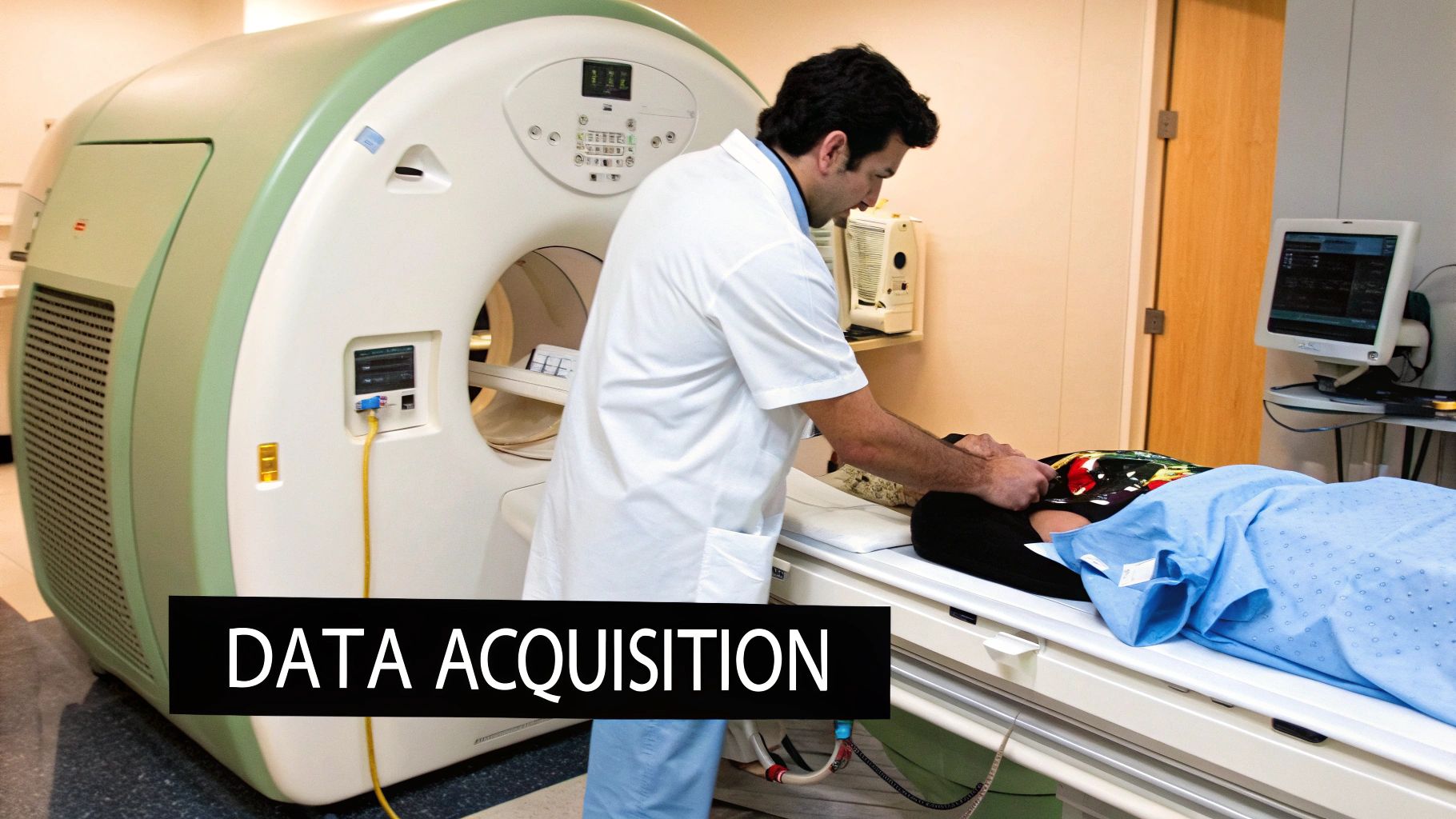
To really get a handle on 3D MRI reconstruction, you first have to understand where the data comes from. An MRI machine isn't like a camera that just snaps a picture. Instead, it meticulously gathers a massive amount of raw signal data, which a computer then has to translate into the detailed anatomical images we see in the clinic.

It all starts when a patient is placed inside a powerful magnetic field. This strong field gets the protons in the body's water molecules to all align in the same direction. Then, a series of radiofrequency (RF) pulses are beamed in, knocking these protons out of that alignment. When the RF pulse stops, the protons relax and snap back into place, releasing their own faint radio signals in the process. These are the signals the MRI scanner’s coils are designed to detect.
This raw data isn't an image yet. It's collected in a complex data matrix called k-space. It’s more helpful to think of k-space as the blueprint for an image rather than the image itself.
Think of it this way: The final 3D image is a beautiful symphony. K-space is the sheet music containing all the individual notes—the frequency and phase information—that the computer will later "play" to produce that symphony. Every data point is a crucial note in the composition.
The center of k-space holds the low-frequency data, which dictates the image's overall contrast and brightness—the broad strokes. The outer edges of k-space contain the high-frequency data, which is responsible for all the fine details and sharp edges. How this k-space "sheet music" gets filled in depends entirely on the acquisition technique chosen.
Choosing the Right Acquisition Technique
The radiographer’s choice of acquisition technique has a huge impact on the final image quality, how long the scan takes, and the characteristics of the raw data itself. There are two main ways to go about it: a 2D multi-slice approach or a true 3D volumetric approach. Each has its own strengths and weaknesses.
To better understand these differences, let's compare the two primary methods for acquiring the data needed for a 3D view.
Comparison of 2D and 3D MRI Acquisition Techniques
| Characteristic | 2D Multi-Slice Acquisition | 3D Volumetric Acquisition |
|---|---|---|
| Data Collection | Acquires data slice by slice, stacking them to form a volume. | Excites and captures data from an entire volume (a "slab") at once. |
| Scan Time | Often faster for scans covering a very large area with lower resolution. | Can be slower, but often more efficient for high-resolution imaging of a specific area. |
| Voxel Shape | Typically anisotropic (rectangular), with slice thickness greater than in-plane resolution. | Produces isotropic voxels (perfect cubes), ideal for multi-planar reformatting. |
| Slice Gaps | Prone to small gaps between slices, which can miss small pathologies. | No inter-slice gaps, providing continuous data throughout the volume. |
| Signal-to-Noise Ratio (SNR) | Generally lower SNR compared to 3D for a given scan time. | Higher SNR because the entire volume is excited repeatedly. |
| Best For | Routine scans, fast surveys, situations where motion is a major concern. | Detailed anatomical studies (e.g., neuro, musculoskeletal), MRA, where reformatting is key. |
As the table shows, the choice between 2D and 3D acquisition is a balancing act. While 2D is a reliable workhorse, the seamless, high-resolution data from a 3D volumetric scan provides the best possible raw material for a high-quality 3D model.
The specific type of pulse sequence, like a Spin Echo (SE) or Gradient Echo (GRE), adds another layer of control. SE sequences are known for being robust and producing excellent image quality, but they take longer. GRE sequences are much faster, which is why they're a great fit for 3D volumetric scans, though they can be more sensitive to certain image artifacts.
Ultimately, this initial choice is critical. The data acquisition method sets the absolute ceiling on the quality of the final 3D MRI reconstruction. A 3D volumetric acquisition, for instance, provides those perfect, cube-shaped isotropic voxels that allow for clear, distortion-free reconstructions from any angle. Understanding these foundational decisions makes it clear why the algorithms that come next are so essential for turning raw signals into a clinically powerful 3D model.
How Algorithms Build a 3D MRI Model
Once the MRI scanner has gathered all the raw data—what we call k-space data—the real magic of 3D MRI reconstruction begins. This is where powerful algorithms step in. Think of them as computational translators, converting the abstract language of frequency signals into a clear, detailed anatomical model. It's a fascinating journey from a chaotic collection of raw data to a crystal-clear diagnostic tool.
At the very core of this process is a mathematical workhorse called the Fourier Transform. It’s the master decoder. K-space data, filled with information about signal frequencies and phases, is like an encrypted message. The Fourier Transform is the key that unlocks it, rearranging the data from the frequency domain into the spatial domain. In simpler terms, it turns raw signal information into a recognizable image slice.
This is the foundation for almost every MRI reconstruction. But to create truly exceptional 3D models and sidestep common imaging problems, we often need to bring in more advanced techniques.
Advanced Reconstruction Techniques
While the Fourier Transform is the standard, modern imaging demands faster scans and higher-quality results. This push has led to some incredibly sophisticated algorithms that can build better images, even when working with less-than-perfect data. Two of the most important are Iterative Reconstruction and Compressed Sensing.
-
Iterative Reconstruction: Picture an artist sketching a portrait. They start with a rough outline, look at their work, spot the flaws, and make corrections. They repeat this cycle—sketch, review, refine—until the portrait is just right. Iterative algorithms work the same way. They generate an initial image, compare it to the original raw data, identify errors like noise or artifacts, and then refine the image over and over. Each loop, or "iteration," produces a cleaner, more accurate picture.
-
Compressed Sensing: This technique has been a game-changer for cutting down scan times. It's built on a clever principle: if you already have a good idea of what a final medical image should look like (meaning, it's not just random static), you don't actually need to collect all the data. Compressed Sensing algorithms can take a dramatically undersampled k-space—from a scan done in a fraction of the time—and intelligently fill in the missing pieces to build a complete, high-quality image. It’s a bit like solving a giant Sudoku puzzle; even with many empty squares, you can figure out the missing numbers based on the rules and the numbers you already have.
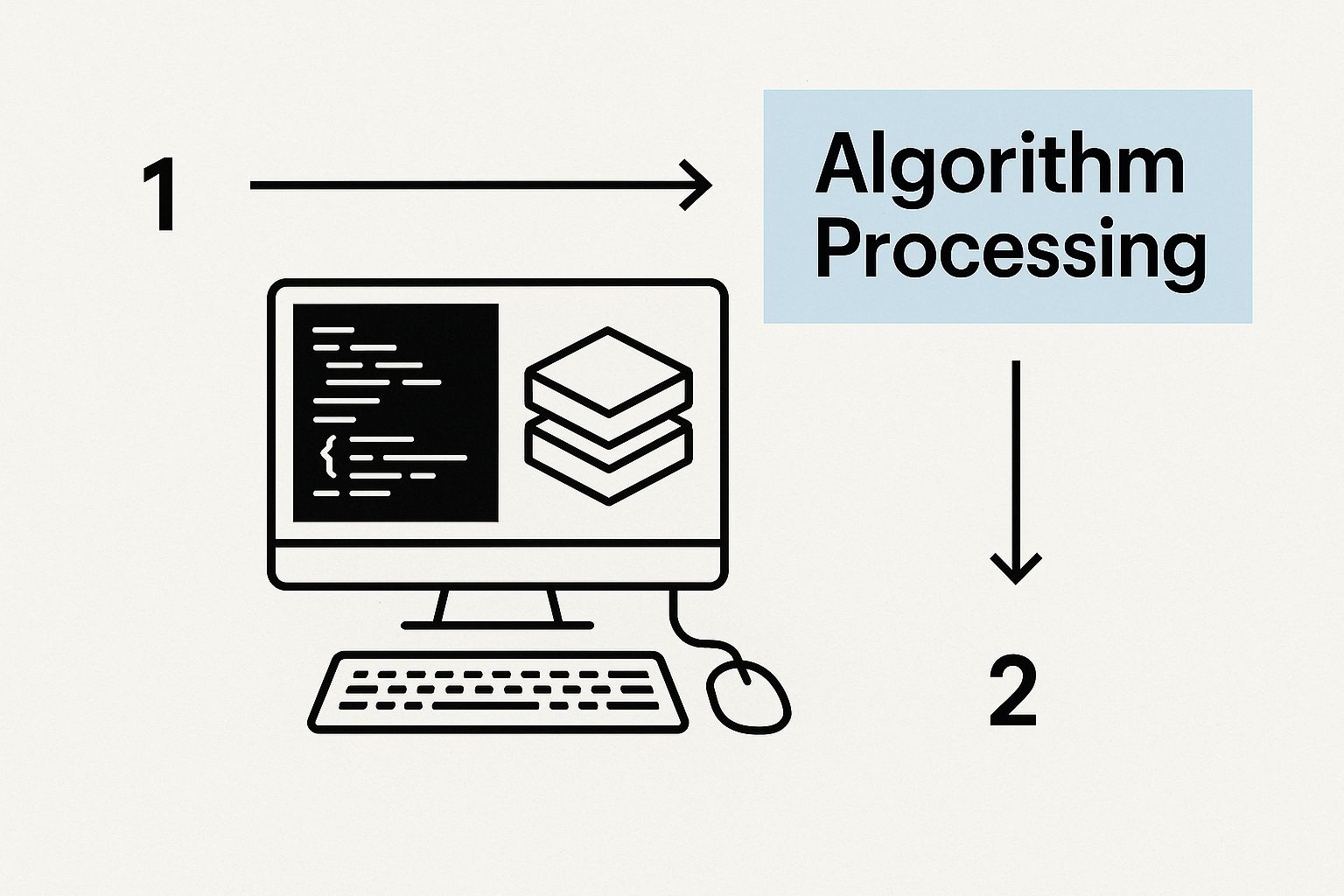
The image below gives you a great visual of how these complex algorithms turn raw data into a final 3D model.

You can see the journey here, from the raw k-space signals passing through the algorithmic engine to produce the final, coherent 3D slices.
Overcoming Common Imaging Hurdles
A major reason we rely on these advanced algorithms is to tackle real-world problems that can ruin the quality of a final model. Two of the biggest culprits are motion artifacts and image noise.
Motion Artifacts
Even the slightest patient movement during a scan, like breathing or a small twitch, can blur the image and create frustrating ghost-like artifacts. This happens because any movement corrupts the k-space data while it's being collected.
Motion-correction algorithms are specifically designed to detect and compensate for this corrupted data. Some track the patient's movement and adjust the reconstruction on the fly, while others work after the fact, identifying and fixing inconsistencies in the raw data before the final image is ever created.
Image Noise
That random, grainy pattern in an image, known as noise, can easily obscure fine anatomical details. It's a common problem, especially with faster scans where the signal-to-noise ratio (SNR) is naturally lower. Iterative reconstruction methods are fantastic at cutting through this noise, as each refinement step helps smooth out those random fluctuations while carefully preserving the true anatomical structures underneath.
This kind of algorithmic power has a rich history. A huge milestone came back in 1983 when Michael Vannier, a radiologist at the Mallinckrodt Institute of Radiology (MIR), published the first-ever 3D reconstruction from single CT slices of a human head. This pioneering work in the early 1980s, done with what we'd now consider ancient hardware, really paved the way for the volumetric reconstruction techniques that were eventually adapted for MRI. You can learn more about these historical breakthroughs and see just how much they influenced modern imaging.
At the end of the day, the choice of algorithm has a direct impact on how useful the 3D MRI reconstruction is in a clinical setting. By using these intelligent methods, we can get faster scans, clearer images, and ultimately, more confident diagnoses.
The Role of AI in Modern 3D MRI Reconstruction
If advanced algorithms like Compressed Sensing are skilled artists, think of Artificial Intelligence as their brilliant, lightning-fast assistant. AI, and specifically deep learning, is fundamentally reshaping the world of 3D MRI reconstruction. It's doing more than just speeding up old methods; it’s opening the door to capabilities we once thought were out of reach, turning raw scan data into clinically rich 3D models with incredible speed and accuracy.

This influence is felt at every point in the reconstruction pipeline. AI models, most often Convolutional Neural Networks (CNNs), are put through rigorous training using massive datasets of medical images. This process teaches them to recognize the incredibly complex patterns of human anatomy and, just as importantly, the tell-tale signs of noise and imaging artifacts.
With this deep-seated knowledge, AI can perform complex reconstruction tasks with a precision that’s hard to match. It's like having an expert who can glance at a blurry, incomplete picture, instantly clean it up, and then perfectly trace every important object. That's what AI brings to MRI—it acts as an accelerator, a quality expert, and a detail-obsessed analyst all at once.
Slashing Scan Times
One of AI's most celebrated contributions is its power to dramatically shorten MRI scan times. Anyone who's had an MRI knows that long scans are uncomfortable, and for hospitals, they are expensive. AI-powered reconstruction tackles this head-on by enabling aggressive undersampling techniques.
By deliberately capturing less k-space data, we can cut scan times down significantly. The catch? Traditionally, this sparse data would result in a noisy, artifact-filled image that's clinically useless. But this is exactly where AI shines.
A deep learning model, trained on thousands of paired high-quality and undersampled scans, learns to intelligently "fill in the blanks." It can reconstruct a high-resolution image from this incomplete data, providing clinicians with the diagnostic detail they need in a fraction of the time. The result is a better experience for the patient and higher throughput for the clinic.
Boosting Image Quality and Sharpness
Beyond just speed, AI is also a master of image cleanup. Every MRI scan is prone to random noise and artifacts, whether from slight patient movements or tiny hardware imperfections. These glitches can easily obscure critical details and complicate a diagnosis.
AI-driven denoising algorithms are trained to tell the difference between a true anatomical signal and unwanted noise. Because they've learned what a "perfect" scan should look like, they can intelligently strip away the noise and correct artifacts without blurring the important underlying structures.
This process yields images that are not only cleaner but noticeably sharper. For a radiologist, that added clarity can mean the difference between confidently spotting a tiny lesion and missing it completely. It provides a more reliable foundation for building the final 3D model.
The ability of AI to derive meaningful insights from complex information is transforming industries far beyond medicine. The core principles, like those discussed in building competitive advantage with AI for decision making, show a clear parallel in how sophisticated algorithms create actionable intelligence.
Automating Anatomical Segmentation
To create a truly useful 3D model, you first need to perform segmentation—the process of outlining specific structures, like a tumor, a ventricle in the brain, or a specific muscle group. Doing this by hand, slice by 2D slice, is an incredibly tedious task that's notoriously slow and subject to human inconsistency.
AI steps in to automate this entire process. A properly trained model can instantly identify and delineate specific organs or tissues throughout the entire 3D volume. The advantages here are game-changing:
- Speed: It turns a task that could take a radiologist hours into a job that takes just minutes.
- Consistency: The AI applies the exact same criteria every single time, removing the natural variability between different human operators.
- Precision: In many cases, it can detect and trace boundaries with a level of detail that surpasses what's even possible by hand.
This automated segmentation is crucial for advanced applications like planning surgery, where a surgeon needs precise volumetric measurements of a tumor, or in large-scale research analyzing brain structure changes across hundreds of patients. By offloading these labor-intensive tasks, AI frees up clinicians to focus on what matters most—diagnosis and treatment planning—making the entire 3D MRI reconstruction workflow more powerful than ever before.
How 3D MRI Is Making a Difference in Real-World Medicine
The real test of any medical technology isn't how clever the algorithms are, but how much it helps patients. When it comes to 3D MRI reconstruction, the leap from the lab to the clinic is fundamentally changing how doctors diagnose diseases, plan surgeries, and even talk to patients. Creating these interactive, detailed models of the human body gives them a level of clarity that was once unimaginable.
https://www.youtube.com/embed/aNAZxV8yQHM
This isn't just a minor upgrade from flat, black-and-white images. It's an entirely new way of looking inside the body. Let’s dive into how this technology is making a tangible difference in neurology, orthopedics, and cardiology.
A New Level of Precision in Neurosurgery and Brain Mapping
Nowhere is anatomical detail more critical than in the brain. For a neurosurgeon, a 3D MRI reconstruction is like having a high-resolution GPS for the most intricate organ in the body.
Think about the immense challenge of removing a brain tumor. With traditional 2D scans, it's tough to get a true feel for the tumor's size, shape, and—most importantly—its relationship to the critical structures surrounding it, like blood vessels or neural pathways. A 3D model changes the game completely.
- Smarter Surgical Planning: Surgeons can digitally spin the model around, examining it from every angle to find the safest possible entry point. This helps them plan a route that minimizes any damage to healthy tissue and accurately measure the tumor's volume for a complete resection.
- Guidance in the Operating Room: These 3D maps can be loaded directly into neuronavigation systems, giving the surgeon real-time guidance during the operation. This helps ensure every bit of the tumor is removed while protecting vital brain functions.
- Clearer Patient Communication: Being able to show a patient a 3D model of their own brain and walk them through the planned surgery is incredibly powerful. It makes a scary, abstract procedure feel more understandable, which is crucial for building trust and getting informed consent.
Seeing the Full Picture of Complex Joint Injuries in Orthopedics
Orthopedics is another field that has been transformed by 3D MRI. The complex web of ligaments, tendons, and cartilage in joints like the knee or shoulder is notoriously difficult to assess fully with 2D images.
Imagine an athlete with a major knee injury, potentially with several torn ligaments. A 3D model gives the orthopedic surgeon a complete, 360-degree view of the entire joint.
This comprehensive view is essential. It lets the surgeon see not just which ligaments are torn, but the precise location and severity of those tears. That level of detail is a prerequisite for planning a complex reconstruction surgery.
The 3D model helps answer critical questions long before the first incision is made: What's the best angle for placing a new graft? How will the repaired ligaments work together once they heal? This kind of preoperative insight leads to more precise surgeries and better long-term outcomes for patients eager to get back on their feet. Afterward, these detailed models are invaluable for monitoring how the soft tissues are healing.
A Deeper Look into Diagnosing and Treating Heart Conditions
The heart isn't a static image; it's a dynamic, three-dimensional pump. Understanding its unique structure is the key to treating cardiac disease effectively. For cardiologists and cardiac surgeons, 3D MRI reconstruction delivers stunningly detailed models of the heart's chambers, valves, and major vessels.
This is especially critical in pediatric cardiology, where doctors diagnose complex congenital heart defects. These are structural issues present from birth that can be incredibly intricate. A 3D model lets the medical team:
- Pinpoint the Defect: They can clearly see exactly how a child’s heart deviates from a normal structure, like a hole between chambers or a malformed valve.
- Plan the Correction: Surgeons can use the model to "rehearse" the procedure, planning the delicate repairs needed to restore proper blood flow.
- Track Post-Surgical Healing: After the operation, follow-up 3D models help doctors monitor how the heart is healing and adapting over time.
By providing such a clear, intuitive view of a patient’s unique anatomy, 3D reconstruction gives doctors the confidence to make better diagnoses and create highly personalized surgical plans, directly improving outcomes for some of their most vulnerable patients.
While today’s 3D MRI reconstruction is already impressive, what we have now is just the tip of the iceberg. The field is moving incredibly fast, and researchers are pushing hard to transform those static 3D models into something far more dynamic and insightful.
The next giant leap forward is all about adding the fourth dimension: time. This brings us to 4D MRI, which is essentially a movie of a 3D model, showing anatomy as it moves and changes. Think about watching a high-fidelity 3D rendering of a heart beating in real-time or seeing lungs expand and contract with every single breath.
This isn’t just a visual gimmick. It offers a direct window into how organs actually work, helping doctors spot functional issues, not just structural abnormalities. A 4D model could instantly reveal an irregular heart valve flutter or a subtle contraction problem that a series of static images would completely miss. We're moving from asking "What does it look like?" to "How does it behave?"
Adding Function to Form
Another major evolution is weaving quantitative MRI (qMRI) data directly into our 3D anatomical models. A standard MRI is great at showing you the "what" and "where" of anatomy. But qMRI goes a step further by measuring the actual physical properties of tissue—things like water content, fat fraction, or blood flow.
When you overlay this quantitative data onto a high-resolution 3D model, you get a much richer, more complete story. Imagine a 3D brain model where different colors instantly show areas with dangerously low blood flow, pointing directly to tissue at risk of a stroke. This fusion of structure and function gives clinicians a comprehensive diagnostic tool in one intuitive visualization.
The ultimate goal is to move from a simple anatomical map to a fully functional, personalized model. This sets the stage for creating a "digital twin" of a patient.
The Rise of the Digital Twin
The idea of a digital twin is easily the most exciting frontier on the horizon. This isn't just a static model; it's a highly accurate, virtual replica of a patient's organ or even their entire body that can be used as a dynamic simulation tool.
What does that look like in practice?
- Treatment Simulation: A surgeon could perform a complex procedure on a patient's digital twin first, testing different approaches to see which one delivers the best outcome—all before making the first incision.
- Personalized Medicine: Oncologists could simulate how a specific patient’s tumor might react to various chemotherapy drugs, tailoring a treatment plan for maximum impact with minimal side effects.
This kind of technology paves the way for a future where medical care is planned, tested, and fine-tuned in a risk-free virtual space. As powerful computing becomes more widely available, these advanced 3D MRI reconstruction techniques will give us a deeper understanding of human health than we ever thought possible.
Frequently Asked Questions
Still have a few questions floating around about 3D MRI reconstruction? You're not alone. Let's tackle some of the most common ones to clear things up and give you a solid grasp of how this technology works.
What Is the Main Difference Between 2D and 3D MRI?
The real difference comes down to how the data is gathered and what you can do with it afterward. A conventional 2D MRI captures individual, distinct slices of tissue. Think of it like taking separate photos of each page in a book. A radiologist then has to mentally piece those pages together to understand the full picture.
A 3D MRI reconstruction, on the other hand, captures an entire volume of tissue—a complete "slab"—all at once. This gives you a continuous, gap-free block of data. From there, powerful software assembles it into a cohesive 3D model that you can turn, slice, and inspect from any angle. It’s like graduating from a flip-book of static images to holding a detailed sculpture in your hands.
How Long Does a 3D MRI Reconstruction Scan Take?
This is a classic "it depends" question, but the answer is probably faster than you'd guess. The exact time hinges on what's being scanned and how much detail is needed. While some 3D sequences can take a bit longer than their 2D counterparts, modern advancements have dramatically sped things up.
Thanks to techniques like Compressed Sensing and AI-driven reconstruction, many high-resolution 3D scans can be wrapped up in just 5 to 10 minutes. That's a game-changer for patient comfort and clinic throughput.
Is 3D MRI Reconstruction Safe?
Absolutely. The safety profile is one of MRI's biggest strengths. Whether it's 2D or 3D, the technology relies on strong magnetic fields and radio waves, not ionizing radiation. This means there is no radiation exposure—a key advantage over CT scans or X-rays.
The "reconstruction" part of the process is purely a software task. It all happens on a computer after you've left the scanner, posing no additional risk or procedure for the patient.
Can Any MRI Scan Be Turned into a 3D Model?
Not quite. For a truly high-quality 3D MRI reconstruction, you need to start with the right ingredients. The best results come from scans that were intended for 3D from the get-go, using volumetric acquisition sequences that capture seamless, high-resolution data.
You can technically try to stack old 2D slices and have a computer fill in the blanks, but the resulting model is often a rough approximation. It's prone to gaps, stair-step artifacts, and misalignments that make it unsuitable for precise clinical work. For a 3D model you can trust, you have to acquire the data correctly from the start.
At PYCAD, our expertise lies in applying sophisticated AI to unlock the full potential of medical imaging data. If you're looking to improve your reconstruction workflows and boost diagnostic precision, we can help. See how our custom solutions work and what they can do for you.


