The Evolution of 3D Medical Visualization
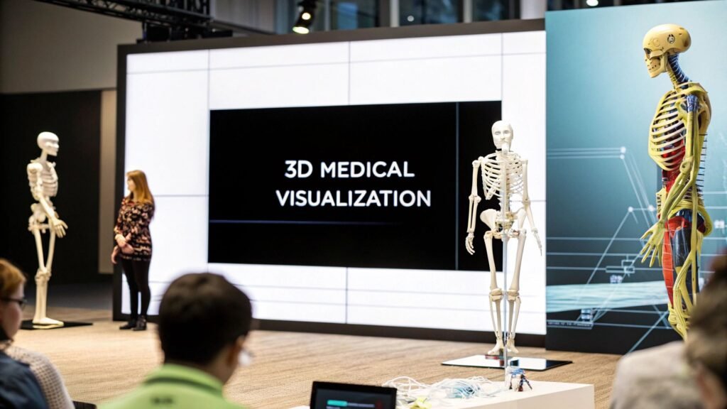
Medical imaging has undergone a dramatic transformation. Early X-rays provided a limited, two-dimensional glimpse inside the human body. Now, 3D medical visualization offers detailed, interactive models that have revolutionized diagnosis and treatment. This progress allows medical professionals to visualize structures previously unseen, providing unparalleled clarity.
From Grainy Images to Interactive Models
Initially, 3D medical visualization was an expensive and specialized tool primarily used in research. The technology relied on costly hardware and complex software, limiting its availability for most healthcare providers. The resulting images were often grainy and challenging to interpret, requiring extensive training and expertise. This restricted the practical use of 3D visualization in routine clinical practice.
However, advancements in computer processing power and software algorithms have significantly enhanced the quality and accessibility of 3D medical visualization. Images are now sharper, more detailed, and easier to manipulate. Physicians can explore anatomical structures from different angles and perspectives. For instance, examining complex bone fractures with greater clarity allows for more precise surgical planning. This detail improves diagnostic accuracy and enables personalized treatment plans.
Democratizing Access to Advanced Imaging
The cost of 3D imaging hardware has also decreased significantly. In the 1980s, acquiring cutting-edge 3D imaging hardware required a substantial investment, often between $250,000 and $350,000. Today, comparable technology is available for under $10,000. This drastic price reduction, coupled with improved software and networking, has democratized access to 3D medical visualization, making it a vital tool in various healthcare settings. This evolution is driven by progress in both computer hardware and software. Find more detailed statistics here
Increased processing power has also led to a surge in the amount of data that can be captured and processed. Modern CT scanners can produce datasets of 3,000 to 5,000 slices, compared to less than 100 slices in earlier models. The thickness of each slice has also decreased from 3-4 mm to under 1 mm, providing significantly more detail. This increased precision allows medical professionals to identify subtle anomalies that older technology might have missed.
The Future of 3D Medical Visualization
The ongoing development of 3D medical visualization promises further advancements. Integrating with technologies like artificial intelligence (AI) and virtual reality (VR) will enhance its capabilities and applications. Artificial intelligence can automate image analysis, aiding in the detection of abnormalities and streamlining diagnostic workflows. Virtual reality can create immersive environments for surgical planning and medical training, allowing surgeons to "practice" procedures before operating on a real patient. These innovations will continue to transform healthcare, improving diagnostic accuracy, personalizing treatments, and ultimately leading to better patient outcomes.
Real-World Impact of 3D Medical Visualization
3D medical visualization is rapidly changing how medical professionals approach patient care. This technology provides interactive, detailed anatomical models that empower more accurate diagnoses and more effective treatment strategies. Its influence spans various medical specialties, ultimately improving patient outcomes and reshaping clinical practices.
Transforming Surgical Planning and Execution
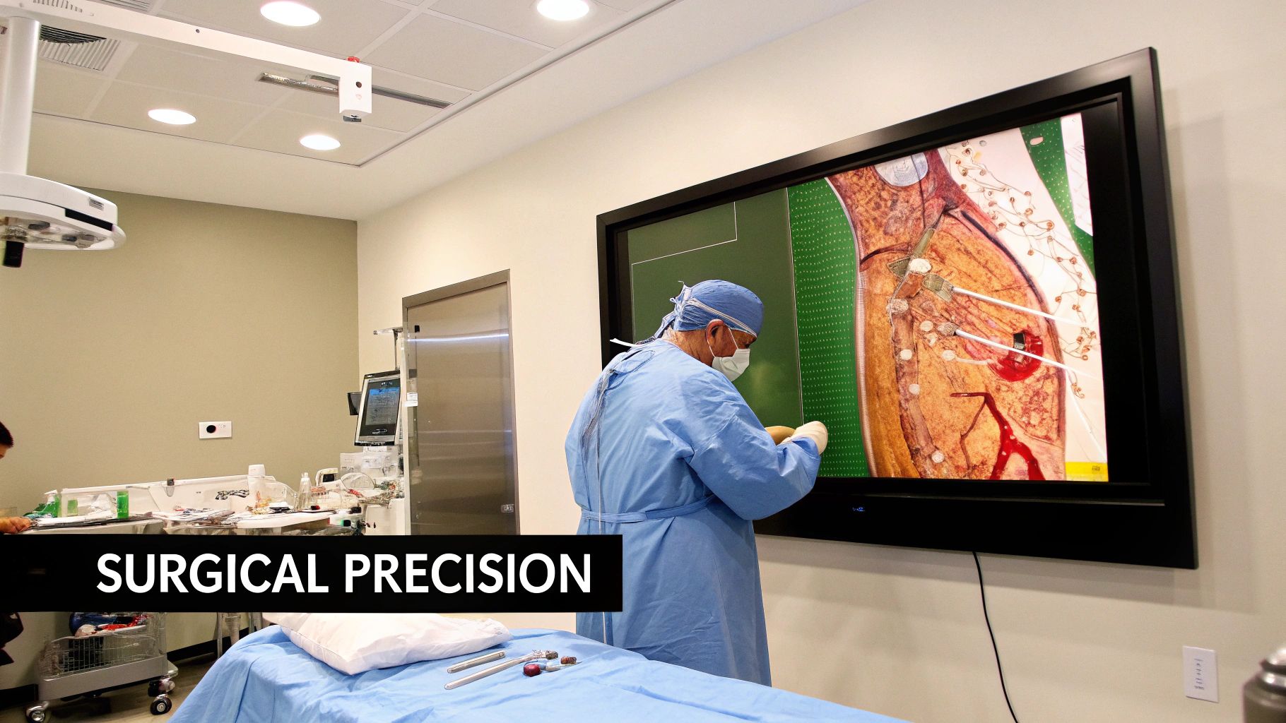
One of the most profound benefits of 3D medical visualization is its impact on surgical planning. Using 3D models generated from patient scans, surgeons can now virtually rehearse complex procedures. This "virtual surgery" allows them to anticipate challenges, refine their surgical approach, and minimize invasiveness.
This pre-operative planning also allows for precise placement of implants and prosthetics, leading to better functional outcomes. In specialties like orthopedics, where precise alignment is essential for joint replacements and fracture repairs, this level of detail is crucial. The ability to virtually "see" the procedure beforehand contributes significantly to surgical success.
Enhancing Diagnostic Accuracy in Radiology
3D medical visualization has become indispensable in radiology. Radiologists can utilize 3D imaging to uncover subtle anomalies that might be overlooked in traditional 2D images. This enhanced accuracy is paramount for early disease detection and prompts timely intervention.
From the discovery of X-rays in 1895, medical imaging has evolved through ultrasound, CT scans, and MRI. 3D imaging, particularly with CT scans, provides highly detailed visualizations of internal structures, aiding in the diagnosis of complex conditions like cancer and heart disease. Techniques like volumetric rendering, which preserves all CT scan data, enable the visualization of object thickness and internal contours, proving invaluable in specialties such as orthopedic surgery.
This three-dimensional perspective allows radiologists to better understand the spatial relationships between anatomical structures, ultimately improving their ability to characterize lesions and assess the extent of diseases. In challenging diagnostic scenarios, 3D medical visualization offers a more comprehensive understanding of the patient's condition.
Improving Patient Communication and Education
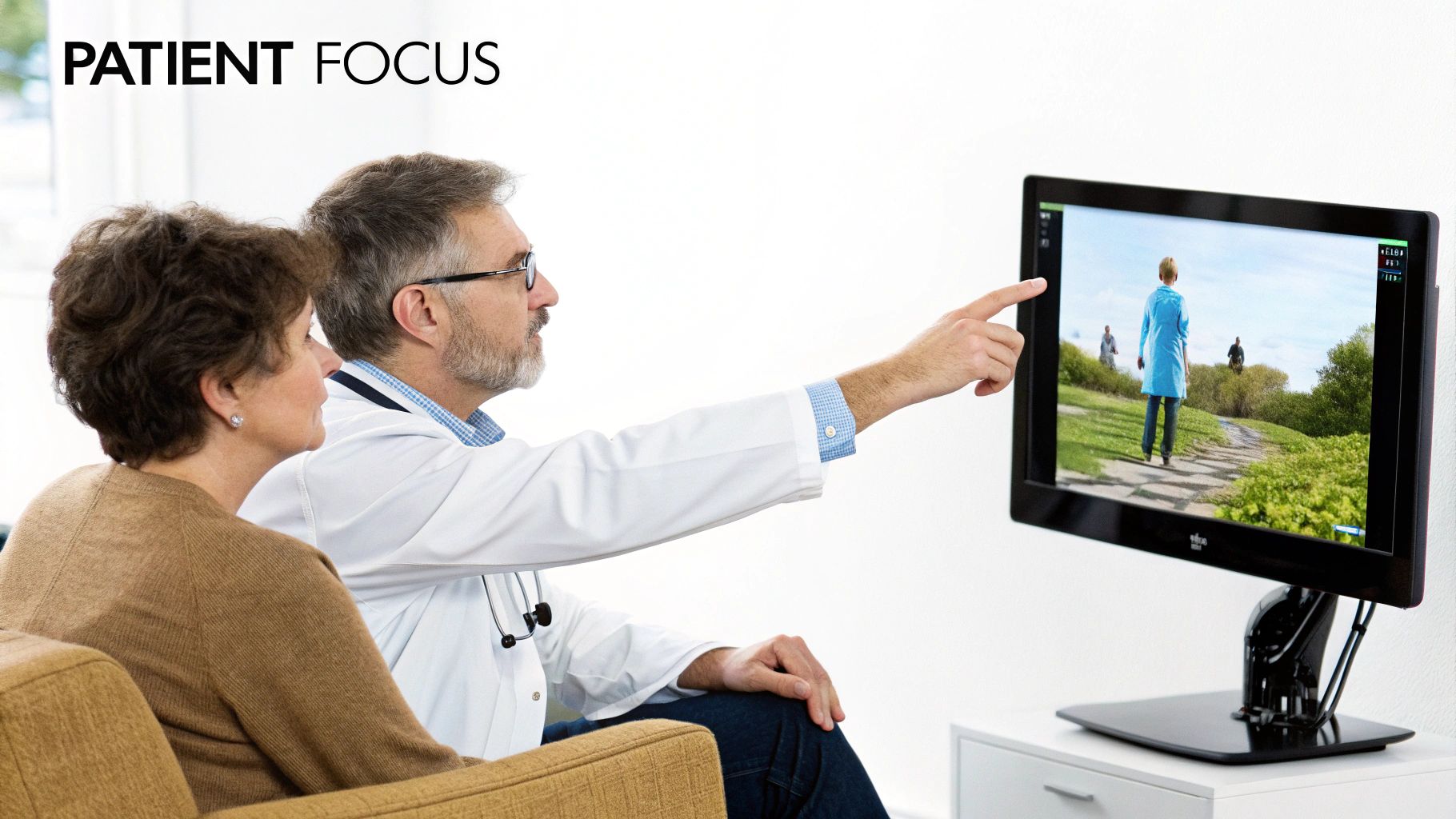
Beyond diagnostics and surgical planning, 3D medical visualization also strengthens patient communication and education. These visual aids help patients better understand their diagnosis, treatment plan, and potential risks and benefits.
By using 3D models, doctors can effectively explain complex medical concepts, making them easier for patients to grasp. This improved understanding can significantly reduce patient anxiety and empower them to make more informed decisions about their care. Visualizing a procedure beforehand can greatly alleviate stress and uncertainty.
Specialty-Specific Applications and Benefits
To further illustrate the breadth of impact, let's examine how 3D medical visualization is applied across various medical specialties:
To understand the breadth of impact, the table below outlines how 3D visualization is transforming various medical specialties:
3D Medical Visualization Applications by Specialty
This table compares how different medical specialties utilize 3D visualization technology and the specific benefits gained in each field.
| Medical Specialty | Primary 3D Visualization Applications | Key Clinical Benefits |
|---|---|---|
| Orthopedics | Pre-operative planning for joint replacements and fracture repairs | Precise implant placement, improved surgical outcomes |
| Cardiology | Visualization of heart structures and blood flow | Accurate diagnosis and treatment of heart disease |
| Neurology | Enhanced visualization of the brain and spinal cord | Improved diagnosis and treatment of neurological disorders |
| Oncology | Improved tumor detection and characterization | More targeted and effective cancer treatments |
This table demonstrates the impressive versatility of 3D medical visualization and how its tailored application benefits different medical fields. The ability to visualize complex anatomical structures and processes in three dimensions is leading to more accurate diagnoses, more effective treatments, and ultimately, better patient care. As technology continues to advance, 3D medical visualization will undoubtedly play an even greater role in shaping the future of healthcare.
Beyond Screens: 3D Printing and Physical Visualization
3D medical visualization is no longer limited to computer screens. Through the power of 3D printing, tangible models are transforming medical practice, patient care, and education. This shift from digital images to physical objects opens exciting possibilities for healthcare.
From Visualization to Fabrication: A New Dimension in Healthcare
3D printing allows the creation of patient-specific anatomical models based on medical imaging data. Surgeons can physically hold and examine these models from all angles, even practicing complex procedures on them. This hands-on approach enhances surgical planning, leading to improved outcomes. For instance, in complex heart surgery, a 3D printed model of the patient's heart allows surgeons to visualize specific anatomical challenges and preemptively plan the most effective surgical approach.
3D printing is also revolutionizing prosthetics and implants. Custom-designed prosthetics, created with precision from 3D visualizations, provide a perfect anatomical fit for individual patients. This personalized approach improves the fit, function, and comfort of prosthetics, restoring mobility and independence. Patient-specific implants follow a similar trajectory, offering tailored solutions for diverse medical needs. This movement toward personalized medicine showcases the power of combining 3D medical visualization and 3D printing.
Not only is the integration of 3D printing with 3D medical visualization transforming patient care, but it’s also revolutionizing medical education. Students can now interact directly with 3D printed models, gaining a deeper understanding of complex anatomy and surgical techniques. These tangible models provide realistic representations of the conditions encountered in real-world procedures, better preparing students for clinical practice.
The use of 3D printing in medical visualization has grown significantly, especially for creating physical models and prosthetics. In 2008, the first 3D printed prosthetic leg was created, marking a major milestone. Explore this topic further. This technology enables the creation of personalized prosthetics and implants tailored to each patient's unique anatomy, improving both fit and function. 3D printing also creates detailed organ and tissue models, invaluable for surgical planning and medical education. This allows surgeons to practice intricate procedures, minimizing risks and improving patient outcomes.
The Future of Hands-On Healthcare
As 3D printing technology continues to advance, the potential applications in healthcare are extensive. We can expect even more sophisticated models using varied materials and textures to accurately simulate real tissues and organs. Bioprinting, the printing of living cells and tissues, holds immense potential for regenerative medicine and organ transplantation. The ability to create functional organs and tissues from a patient's cells could eliminate the need for organ donors and drastically change the treatment of organ failure.
These advancements point toward a future where 3D medical visualization and 3D printing will further transform diagnosis, treatment, and education in the medical field. The potential for creating personalized, highly accurate representations of the human body holds great promise for improving patient outcomes and advancing the healthcare industry.
Measuring the True Impact of 3D Medical Visualization
3D medical visualization offers exciting possibilities. Its true value, however, lies in measurable improvements to patient care and healthcare systems. This means going beyond the visual appeal and examining the tangible benefits that justify the investment. This section explores how leading institutions quantify the return on their investment in 3D medical visualization.
Quantifying the ROI of 3D Visualization
Hospital administrators and health economists are increasingly focusing on data-driven approaches to evaluate the effectiveness of new technologies. For 3D medical visualization, this involves tracking key performance indicators (KPIs) that demonstrate its clinical and operational impact. These metrics provide concrete data to support continued investment, moving beyond anecdotal evidence.
Diagnostic accuracy is one such crucial metric. By comparing diagnostic accuracy with and without 3D visualization, institutions can measure how this technology improves the identification of subtle abnormalities. This can lead to earlier diagnoses and more effective treatment plans. Additionally, reductions in surgical complications are a key indicator of improved surgical planning and execution.
Another important area to consider is the impact on hospital efficiency. 3D visualization can streamline surgical workflows, potentially leading to shorter hospital stays and reduced overall costs. These factors contribute not only to improved patient outcomes but also to the overall financial health of the healthcare system.
Evaluating these metrics requires robust statistical analysis. For example, understanding the difference between percent change and percentage-point change is essential. If 3D imaging adoption rises from 10% to 20%, it's a 10 percentage-point increase, not a 100% increase. Learn more about proper statistical analysis here: https://journalistsresource.org/home/percent-change-math-for-journalists/
Key Metrics in 3D Medical Visualization Evaluation
The following table summarizes key metrics used to assess the clinical and operational impact of implementing 3D medical visualization technologies.
Key Metrics in 3D Medical Visualization Evaluation
| Metric Category | Specific Measurements | Clinical Significance |
|---|---|---|
| Diagnostic Accuracy | Sensitivity, specificity, positive predictive value | Improved early disease detection, reduced misdiagnosis |
| Surgical Outcomes | Complication rates, reoperation rates, surgical time | Enhanced surgical planning, reduced patient risk |
| Hospital Efficiency | Length of stay, readmission rates, cost per case | Streamlined workflows, optimized resource utilization |
| Patient Satisfaction | Pain scores, functional outcomes, quality of life | Improved patient experience and recovery |
These metrics provide a framework for evaluating the effectiveness of 3D visualization and its contribution to improved patient care. Implementing and interpreting these metrics, however, can present challenges.
Addressing Implementation Challenges
Integrating 3D medical visualization into existing workflows can be challenging despite its potential benefits. Factors such as staff training, data integration, and workflow adjustments require careful planning and execution. Furthermore, interpreting the collected data requires expertise in statistical analysis to ensure accurate and meaningful conclusions. Honest discussions about these challenges are essential for successful implementation.
Despite these challenges, the potential benefits of 3D medical visualization make it a valuable investment. By focusing on measurable outcomes and rigorously evaluating the impact on patient care and operational efficiency, hospitals can demonstrate the true value of this powerful technology. This data-driven approach helps justify continued investment and paves the way for broader adoption of 3D medical visualization in the future.
AI and 3D Medical Visualization: The Perfect Partnership
3D medical visualization has significantly improved healthcare. Now, integrating Artificial Intelligence (AI) is pushing the boundaries even further. This powerful synergy transforms medical imaging, allowing for automated image processing and highlighting crucial details, ultimately impacting diagnostic workflows and patient care.
Automating the Tedious, Highlighting the Critical
AI algorithms excel at automating repetitive tasks. In 3D medical visualization, this means automating the often tedious image processing, freeing medical professionals to concentrate on diagnosis and treatment planning. For example, AI can segment organs and tissues in 3D images, a task that previously demanded significant manual effort.
AI algorithms can also be trained to detect subtle abnormalities that might be missed by the human eye. By analyzing large datasets of medical images, AI identifies patterns indicating disease, highlighting areas of concern for radiologists to examine more closely. This increases diagnostic accuracy and allows for earlier detection of critical conditions. Think of it as having a highly trained assistant pre-screening images and flagging potential problems for expert review.
Transforming Diagnostic Workflows
This potent combination of AI and 3D medical visualization is changing diagnostic workflows across various medical specialties. AI-powered tumor identification software can automatically detect and outline tumors in 3D images, offering crucial information for oncologists. This accelerates the diagnostic process, enabling faster treatment decisions.
Patient-specific treatment simulations are another area of impact. Using 3D visualizations and AI, physicians can simulate surgical procedures or radiation treatments, optimizing plans for individual patients. This personalized approach minimizes risk and improves outcomes, truly individualizing care.
Real-World Implementations and Remarkable Outcomes
Leading medical centers are implementing AI-powered 3D medical visualization technologies with impressive results. By incorporating AI into diagnostic processes, these institutions are seeing improved diagnostic accuracy, faster turnaround times, and better patient outcomes.
Some hospitals, for instance, use AI-powered software to analyze 3D mammograms, increasing the detection rate of breast cancer. Others are using AI to assist in diagnosing neurological disorders based on 3D brain scans. These real-world applications demonstrate the practical value of this technology in clinical settings.
PYCAD: Leading the Integration of AI in Medical Imaging
PYCAD is at the forefront of integrating AI into medical imaging, specializing in AI services that span data handling, model training, and deployment. Since 2023, PYCAD has completed over 10 successful projects, demonstrating its expertise in this evolving field.
PYCAD's solutions support medical device optimization by enhancing diagnostic accuracy and operational efficiency. Their service offering includes data annotation and anonymization, deploying trained models as APIs, and creating MVP UIs. They work closely with clients to tailor solutions that meet specific needs, ultimately contributing to improved healthcare outcomes.
The Future of 3D Medical Visualization
3D medical visualization has already made a substantial impact on healthcare. Looking ahead, we can anticipate even more significant changes, driven by emerging technologies set to reshape patient care. These developments will not only refine current techniques but also unlock entirely new possibilities for diagnosis, treatment, and medical training.
Immersive Visualization With AR/VR
Augmented Reality (AR) and Virtual Reality (VR) are poised to revolutionize how medical professionals interact with 3D medical visualizations. Imagine surgeons stepping inside a patient's virtual anatomy before making an incision. AR and VR are making this a reality.
These technologies create immersive experiences, enabling surgeons to examine organs and tissues from all angles. They can plan surgical approaches with exceptional precision and practice complex procedures in a risk-free virtual setting.
This direct interaction with patient-specific 3D models has the potential to drastically improve surgical outcomes and minimize complications.
Real-Time 3D Visualization During Procedures
The future of 3D medical visualization goes beyond pre-operative planning. Real-time 3D visualization during procedures is on the horizon.
This means surgeons will have access to live, dynamic 3D models during an operation. This technology will facilitate more precise navigation within the body, decreasing the chances of damaging vital structures, and boosting the effectiveness of minimally invasive procedures.
For instance, real-time 3D visualization could guide the precise placement of catheters or instruments during complex cardiovascular procedures.
Democratizing Access Through Cloud Computing
While 3D medical visualization is becoming increasingly accessible, obstacles remain, particularly for smaller hospitals and clinics. Cloud computing offers a solution by providing scalable and cost-effective access to sophisticated visualization software and robust data storage.
This means advanced 3D visualization capabilities will no longer be confined to large medical centers. Community hospitals globally can utilize cloud-based solutions to offer their patients the same level of cutting-edge care.
This widespread availability of vital diagnostic tools has the potential to transform healthcare delivery for millions.
Multi-Modal Imaging Integration
Different imaging modalities such as CT, MRI, and PET scans each provide unique insights into the human body. The future of 3D medical visualization lies in integrating these distinct data sources into a single, comprehensive 3D model.
This multi-modal approach fosters a more complete understanding of a patient's condition. For example, merging anatomical data from a CT scan with metabolic data from a PET scan can provide a more accurate assessment of tumor activity, leading to more effective treatments. This data integration will improve diagnostic accuracy and personalize patient care.
AI-Powered Automation and Analysis
Artificial Intelligence (AI) is playing a pivotal role in shaping the future of 3D medical visualization. AI algorithms can automate image processing tasks, analyze extensive datasets to pinpoint patterns and anomalies, and generate predictive models to anticipate disease progression.
This AI-driven automation and analysis will free up medical professionals' time, enabling them to concentrate on patient care. It will also significantly improve the precision and speed of diagnoses. This powerful combination will continue to optimize patient outcomes and the overall efficiency of healthcare systems.
PYCAD: Powering the Future of Medical Imaging
PYCAD is spearheading the integration of AI into medical imaging, offering expertise in data handling, model training, and deployment. With a successful track record since 2023, PYCAD empowers medical device manufacturers and healthcare technology companies to enhance diagnostic accuracy and operational efficiency. Their solutions encompass data annotation, anonymization, API deployment of trained models, and creation of MVP UIs. Partner with PYCAD to fully realize the potential of AI in 3D medical visualization and transform your approach to healthcare.


