Demystifying DCM Files: What They Are and Why They Matter
Navigating medical imaging can be complex. A key element in this field is the DCM file, essential for storing and sharing medical images. Understanding its purpose and importance is crucial for everyone in healthcare, from doctors and researchers to patients.
What Is a DCM File?
A DCM file, short for Digital Imaging and Communications in Medicine (DICOM), is more than just an image. It's a complete package containing the image and vital related data. This metadata includes patient information, scan type, date and time, and equipment used. Unlike JPEGs or PNGs, which primarily focus on visual data, DCM files offer a more comprehensive record.
The Importance of the DICOM Standard
The DICOM standard is the foundation of modern medical imaging. It ensures seamless sharing and interpretation of images and data across different systems. Imagine every hospital using a unique image format – sharing would be incredibly difficult, potentially delaying diagnoses and treatment. DICOM solves this problem.
This standardization is crucial for:
- Interoperability: DICOM enables communication between medical devices from various manufacturers, simplifying image transfer between facilities.
- Patient Care: The detailed metadata in DCM files provides clinicians with critical context for accurate diagnoses and treatment plans.
- Research: Standardized data enables large-scale research, driving advancements in medical imaging technology and diagnostic techniques.
Created in 1985, the DICOM standard has evolved into the universal language of medical imaging. Its journey highlights its significance in modern healthcare. Learn more: Explore the history of DICOM.
Why DCM Files Matter to You
Whether you're a healthcare professional, researcher, or patient, DCM files are important. For providers, they are the primary way to view and analyze images. Researchers use them for large-scale data analysis and developing new imaging techniques. For patients, access to your DCM files empowers you to manage your healthcare journey.
Understanding how to open and view these files is essential for informed decisions. With AI companies like PYCAD integrating AI into medical imaging, the importance of DCM files continues to grow, promising advanced diagnostic capabilities and better patient care.
Finding Your Perfect DCM Viewer: Software That Actually Works
Choosing the right DICOM (Digital Imaging and Communications in Medicine) viewer can be daunting, given the sheer number of options. This section helps you navigate the available software solutions that healthcare professionals rely on. We'll explore which viewers are best for quick image reviews versus detailed analysis, highlighting the strengths of both free and paid software.
Free vs. Paid: Choosing the Right Tool for the Job
Free DCM viewers are often sufficient for basic image viewing needs. They're particularly useful for medical students or those requiring only occasional access. However, for advanced functionality like 3D rendering, measurement tools, or integration with hospital systems, investing in paid software is generally recommended. This investment can significantly streamline workflows and enhance diagnostic capabilities.
Platform Compatibility: Finding the Right Fit for Your System
Your operating system will influence your viewer choices. Windows users have a wide selection of free and paid options, such as MicroDicom and RadiAnt DICOM Viewer. Mac users might prefer OsiriX or Horos. Linux users can explore software like Ginkgo CADx. Cloud-based and mobile viewers offer additional flexibility, allowing access to images from anywhere.
Key Features to Consider: Beyond Just Opening the File
Certain features have a major impact on user experience and overall efficiency:
- Image Manipulation: Basic tools like zoom, pan, windowing, and leveling are essential.
- Measurement Tools: Accurate length, angle, and area measurements are crucial for diagnosis.
- 3D Rendering: Creating 3D reconstructions offers a comprehensive perspective on complex anatomical structures.
- Annotation and Reporting: Adding notes and generating reports facilitates clear communication.
- Integration Capabilities: Seamless integration with PACS (Picture Archiving and Communication System) and other hospital systems is vital for streamlined workflows.
The following infographic illustrates key differences between standard image formats and DCM files:
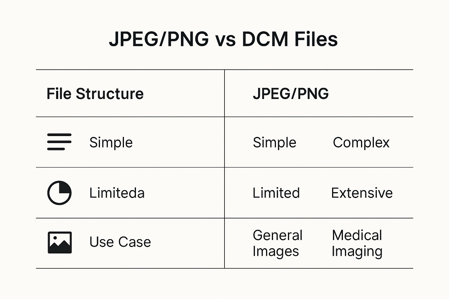
As the infographic demonstrates, DCM files, unlike JPEGs or PNGs, contain a wealth of metadata essential for medical interpretation. This complexity necessitates specialized software. Opening a .dcm file, adhering to the DICOM Standard, requires specific software because of its rich metadata. This metadata includes patient demographics, image acquisition parameters, and the image data itself. Specialized viewers decode this information, making it accessible to medical professionals. Approximately 95% of hospitals worldwide use DICOM-compliant systems.
To help you select the right viewer, we've compiled a comparison table outlining the features and benefits of some popular options:
Top DCM Viewers Compared: Finding Your Perfect Match
This comparison reveals which DCM viewers excel for different needs, from quick viewing to professional analysis.
| Software Name | Platforms | Free/Paid | Key Features | Best For |
|---|---|---|---|---|
| MicroDicom | Windows | Free | Basic image viewing, manipulation | Quick viewing |
| RadiAnt DICOM Viewer | Windows | Paid | Advanced visualization, 3D rendering, measurements | Professional use |
| OsiriX | Mac | Paid | Comprehensive features, research capabilities | Students, professionals |
| Horos | Mac | Free/Paid | Powerful image processing, plugin support | Research, advanced analysis |
| Ginkgo CADx | Linux | Free | Image analysis, CAD features | Research, specialized tasks |
| ImageJ | Cross-platform | Free | Basic image viewing, analysis | Quick viewing, educational purposes |
| 3D Slicer | Cross-platform | Free | Advanced 3D visualization, segmentation | Research, advanced analysis |
The table above summarizes key features and ideal use cases for each viewer. Consider factors like platform compatibility, required features, and budget when making your decision.
Recommendations Based on Your Needs:
- For Quick Viewing: Free viewers like MicroDicom (Windows) or ImageJ (cross-platform) are ideal.
- For Students and Educational Purposes: OsiriX MD (Mac) or RadiAnt DICOM Viewer (Windows) strike a good balance between features and cost.
- For Professional Use and Advanced Analysis: Consider premium options like Horos (Mac) or 3D Slicer (cross-platform), which offer robust features and advanced analysis capabilities.
By carefully considering your needs, platform, and budget, you can select a DCM viewer that enhances both efficiency and diagnostic accuracy. The right software can dramatically improve how you work with and interpret these vital medical images.
Opening DCM Files: A Straightforward Approach That Works
Now that you understand DCM files and have selected the right viewer, let's explore how to open them. This section provides clear, step-by-step instructions, making sure you can view your DCM files regardless of your technical expertise. We'll cover everything from opening a single file to the advanced techniques used by medical professionals.
Opening a Single DCM File: The Basics
Opening a single DCM file, like one from a recent medical scan, is usually a simple process. Here's a breakdown:
- Locate the File: Find the DCM file on your computer. It typically has a
.dcmextension. - Open Your Viewer: Start your chosen DICOM viewer software.
- Import the File: Most viewers have an "Import" or "Open" feature. Select this and navigate to your DCM file’s location.
- View the Image: The image, along with its metadata, will then be displayed in the viewer.
This easy process provides quick access to the image and the important patient information within the DCM file.
Managing Multiple DCM Files: Working With Entire Studies
Medical scans often consist of multiple images, which together form a complete study. Handling these multiple DCM files requires a slightly different approach:
- Organize Your Files: Keeping all DCM files for a single study in a dedicated folder is recommended. This simplifies access and prevents files from different studies getting mixed up.
- Import the Series: Many viewers allow you to import an entire folder, or "series," of DCM files simultaneously. This is far more efficient than opening individual files.
- Navigate the Images: Use the viewer's navigation tools to move between images within the study. Many viewers provide specialized features like cine mode for viewing image sequences.
This streamlined workflow is essential for efficient review and analysis of complex medical imaging studies.
Customizing Your View: Techniques From the Pros
Medical professionals customize their viewing settings depending on the imaging modality (CT, MRI, X-ray) and their diagnostic needs. Here are a few common techniques:
- Windowing and Leveling: This important technique adjusts image contrast and brightness to highlight specific structures or tissues. For example, different windowing settings are used to visualize bones versus soft tissue in CT scans.
- Measurement Tools: Viewers often offer tools to measure distances, angles, and areas within the images. These are essential for precise diagnoses.
- 3D Reconstructions: Advanced viewers can generate 3D models from a series of 2D slices, providing a more complete view of complex anatomy. If you are a developer and need to document how to interact with DICOM image viewing, API documentation generator tools can be helpful.
These advanced features enable in-depth analysis and a more complete understanding of medical images.
Enhancing Image Quality and Navigation: Hidden Settings and Shortcuts
Beyond the basics, viewers often have less obvious settings and shortcuts that can significantly improve your experience:
- Image Filters: Some viewers offer image filters that enhance details or reduce noise, ultimately improving diagnostic clarity.
- Navigation Shortcuts: Keyboard shortcuts significantly speed up image navigation, particularly helpful when reviewing large studies.
- Custom Layouts: Viewers often allow you to personalize the display layout, arranging images and tools in a way that suits your specific workflow.
These hidden features can greatly enhance efficiency and image quality. Whether for casual viewing or professional use, exploring these settings can optimize your viewing environment. Mastering these techniques can significantly improve your interaction with and understanding of DCM files.
Protecting Patient Privacy When Working With Medical Images
Medical images, particularly those stored as DCM files, contain highly sensitive patient information. Understanding how to open and handle these files securely is paramount. It's not simply a matter of technical proficiency; it's about respecting patient privacy and adhering to ethical and legal standards.
Understanding The Risks: Why Casual Handling Is Dangerous
DCM files contain a wealth of information beyond just the image itself. This metadata often includes patient identifiers such as names, birth dates, medical record numbers, and even specific health conditions. Casual handling, like sharing files without proper security or viewing them on unsecured devices, can expose this sensitive data to unauthorized access and potential breaches.
Real-World Breaches: Lessons Learned
Real-world incidents demonstrate the severe consequences of mishandling DCM files. In one instance, a hospital employee mistakenly uploaded a DCM file to a public cloud storage service, exposing hundreds of patients' data. Another case involved a researcher losing an unencrypted USB drive containing thousands of DCM files, resulting in a significant data breach. These examples highlight the need for vigilance and proactive security measures.
From a cybersecurity perspective, opening a .dcm file requires careful consideration. DICOM files often contain personally identifiable information, making secure handling crucial. While the DICOM standard itself offers security features like encryption, reported vulnerabilities highlight the ongoing need for strong security practices. The risk of data breaches has led to the development and implementation of stricter measures, including anonymization tools.
Practical Steps For Protecting Patient Data
Fortunately, preventing such breaches often involves straightforward yet highly effective practices. Protecting patient privacy is achievable with the right approach. Here are some key recommendations:
- Use Strong Passwords: Secure all devices and accounts with strong, unique passwords.
- Encrypt Your Data: Encrypt DCM files, particularly during electronic storage or transmission. This adds a vital layer of security, rendering data inaccessible even if a device is lost or stolen.
- Restrict Access: Limit access to DCM files to authorized personnel only, implementing strict access control measures.
- Anonymize Data When Possible: When sharing files for research or educational purposes, anonymize the data by removing or replacing identifying information.
- Stay Updated: Regularly update software with the latest security patches to protect against known vulnerabilities.
Compliance With Regulations: HIPAA and GDPR
Protecting patient privacy isn't simply a best practice; it's a legal requirement. Regulations like HIPAA in the U.S. and GDPR in Europe establish stringent guidelines for handling sensitive patient data. Compliance is not only ethical but also essential for avoiding legal repercussions. Understanding these regulations is crucial for responsible DCM file handling, even without becoming a legal expert. This often involves implementing the security measures outlined above and ensuring all personnel handling DCM files are trained on proper procedures.
By adopting these practices, individuals and organizations can handle DCM files responsibly and uphold patient confidentiality. This diligence builds trust, protects patients, and contributes to a more secure and ethical healthcare environment.
Solving Common DCM File Problems Before They Frustrate You
Opening DCM files can be tricky. Even seasoned professionals sometimes struggle. This guide tackles the most common obstacles, offering practical solutions vetted by medical imaging specialists and PACS administrators. Knowing these issues and their fixes can save you time and reduce frustration.
Why Won't My DCM File Open?
Several things can prevent a DCM file from opening correctly. A common culprit is an incompatible viewer. DCM files need special software like DICOM viewers designed for their unique format. A standard image viewer won't work and might even corrupt the file. File corruption is another frequent issue, often arising during transfer or storage on damaged media. When working with DCM files, remember patient privacy is paramount. For more on this, check out these data privacy best practices.
Troubleshooting Common DCM File Errors
Incomplete datasets can also cause problems. A DCM file might belong to a larger study with multiple images. If some are missing or corrupted, the whole study might not load. Incompatibility between software versions can also create issues. Older DCM files from legacy systems may not work with newer viewers, and vice-versa.
Before diving into specific solutions, let's take a look at a helpful table that outlines common DCM file problems, their potential causes, and recommended solutions. This table serves as a quick reference guide when you encounter issues.
DCM File Errors Solved: Your Troubleshooting Roadmap
When your DCM files won't cooperate, this guide provides specific solutions for each frustrating scenario.
| Error Message/Issue | Possible Causes | Recommended Solutions |
|---|---|---|
| File Not Found | Incorrect file path, missing file | Double-check the file location, search for the file, contact the source if the file is missing |
| Incompatible Viewer | Using a standard image viewer, outdated viewer software | Use a DICOM-compatible viewer, update your software to the latest version |
| Corrupted File | Damaged media, incomplete transfer | Attempt file recovery using recovery software, request a new copy from the source |
| Incomplete Dataset | Missing DCM files, corrupted files within the series | Locate missing files, replace or repair corrupted files within the series |
| Software Incompatibility | Older DCM file format, newer viewer version, or vice versa | Use a viewer compatible with the file format, convert the file (if possible) to a compatible format |
| Unable to Render 3D Image | Insufficient image slices, incompatible viewer | Verify 3D rendering support in the viewer, ensure enough images for 3D reconstruction |
This table offers a starting point for troubleshooting. These straightforward solutions are frequently used by imaging professionals and often resolve common problems quickly.
Retrieving Images From Outdated Media
Retrieving images from older media like CDs or DVDs can be difficult. These media degrade, leading to file corruption. Specialized data recovery tools and techniques may be needed. Having the right software to view older DCM file formats is also essential.
By understanding these common DCM file problems and their solutions, you can navigate the complexities of medical imaging data more effectively. This knowledge gives you practical steps to ensure smooth access to critical images when needed.
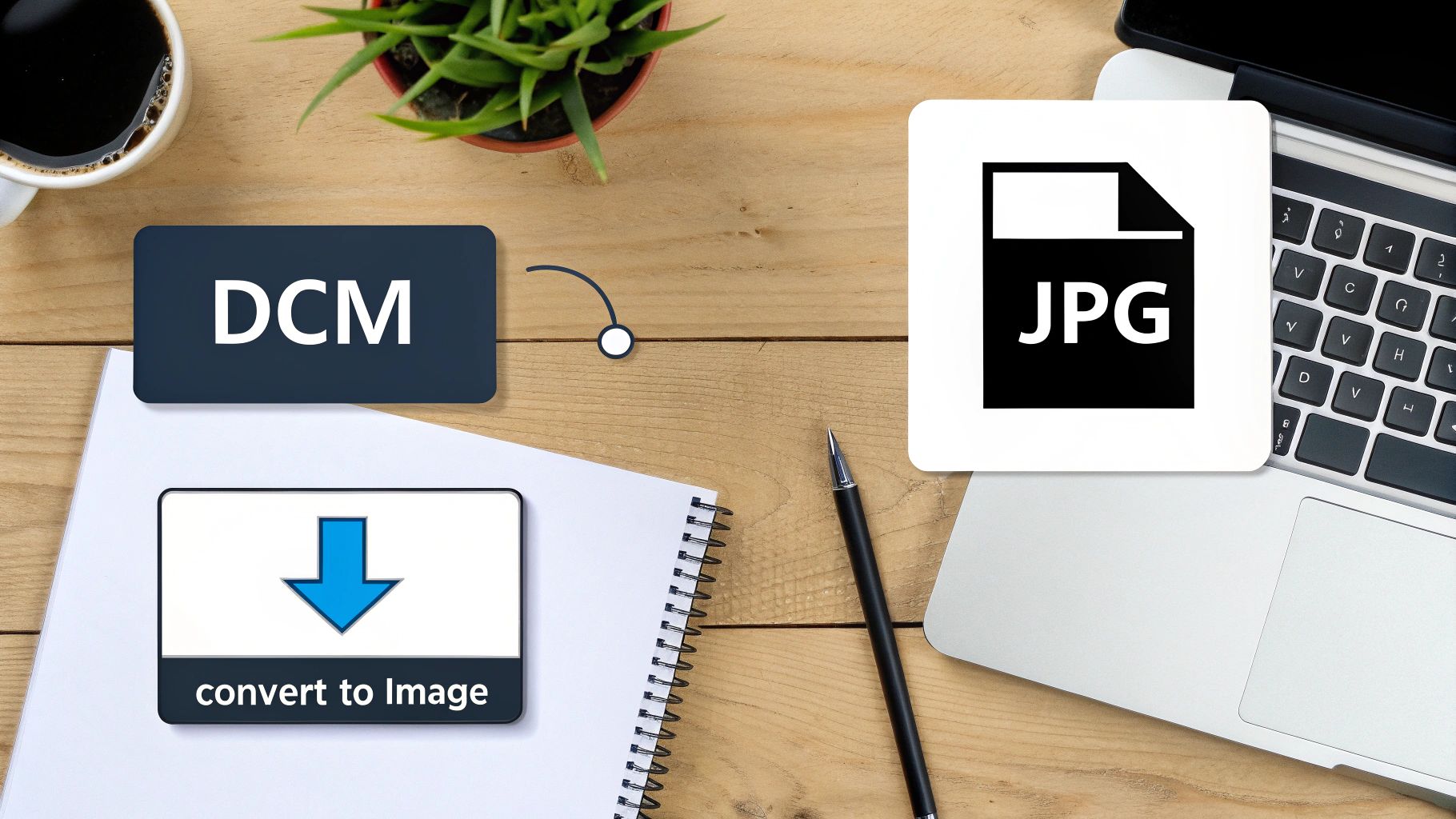
Converting DCM Files: When, Why, and How To Do It Right
Converting Digital Imaging and Communications in Medicine (DICOM) files, commonly known as DCM files, requires a strategic approach. While DICOM is the gold standard for medical imaging, sharing these images in more accessible formats is sometimes necessary. This section explains the nuances of DCM file conversion, guiding you through the process and emphasizing the importance of preserving critical image data.
Why Convert DCM Files? Balancing Accessibility and Diagnostic Quality
The primary reason for converting DCM files is accessibility. Many individuals do not have access to specialized DICOM viewers. Converting to standard formats like JPEG, PNG, or TIFF allows for easy sharing with patients, colleagues without specialized software, and for use in presentations or publications. However, conversion can sometimes lead to data loss, especially if not done carefully.
Understanding which formats preserve diagnostic quality and which don't is crucial. For example, JPEG, while widely compatible, uses lossy compression. This can degrade image details. Formats like PNG and TIFF offer lossless compression, maintaining image integrity.
When Conversion Makes Sense: Real-World Applications
Several scenarios justify DCM file conversion:
- Patient Education: Sharing images with patients in easily viewable formats improves their understanding and engagement in their care.
- Collaboration and Consultation: Sharing images with colleagues who lack DICOM viewers facilitates quicker consultations and second opinions.
- Educational Materials: Converted images can be integrated into presentations, publications, and educational resources.
- Legal and Insurance Purposes: Providing images in accessible formats simplifies communication in legal or insurance contexts.
How To Convert DCM Files: Step-by-Step Instructions
You can convert DCM files through various methods:
- Specialized DICOM Software: Many DICOM viewers have built-in export functionalities for converting to various formats while preserving scaling information.
- Online Converters: Several online tools facilitate DCM conversion, offering convenience but requiring careful consideration of privacy and security.
- Image Editing Software: Some image editing software, such as GIMP or Adobe Photoshop, can open and convert DCM files, though they might not preserve all metadata.
Here's a general process using specialized DICOM software:
- Open the DCM File: Load the DCM file into your chosen DICOM viewer.
- Select Export: Find the "Export" or "Save As" function.
- Choose the Format: Select your desired output format (JPEG, PNG, TIFF).
- Adjust Settings: Configure options such as compression level and resolution.
- Save the File: Save the converted file to your desired location.
Batch Processing: Handling Multiple Files Efficiently
When working with large studies containing numerous images, batch processing is invaluable. Most DICOM software offers batch export options for converting multiple files simultaneously. Maintaining organization during batch processing is essential; ensure files are appropriately named and structured to avoid confusion.
Maintaining Image Integrity: Preserving Crucial Details
During conversion, prioritizing the preservation of critical details is crucial. Scaling information, in particular, is essential for accurate measurements and interpretations. Choose formats and settings that maintain this information. Be mindful that conversion, even with lossless compression, might still discard some metadata.
Potential Pitfalls: When To Avoid Conversion
While conversion offers several benefits, some situations require extra caution:
- Diagnostic Interpretation: Never use converted images for primary diagnosis, as they may not contain all the details present in the original DCM file.
- Medical-Legal Cases: In these scenarios, always keep the original DCM files as the primary source of information.
By carefully considering the advantages and disadvantages, and following best practices, you can ensure successful and responsible DCM file conversion. Balancing accessibility and preserving information is essential for effective image sharing and communication in medical and educational settings.
Ready to elevate your medical imaging analysis with AI-powered solutions? PYCAD offers comprehensive services tailored for medical device manufacturers, healthcare technology companies, and researchers. From data handling to model deployment, PYCAD empowers you to unlock the full potential of your medical images. Visit PYCAD today to learn more.
Keep Reading
Related Articles
Explore the full DICOM HubAll services, tools, and guides on this topic — in one place.
Visit Hub →