Understanding DCM Files and Why They Matter
Let's start with the basics. DCM files, short for DICOM (Digital Imaging and Communications in Medicine), aren't your typical image files. They're specialized containers specifically designed for medical images, holding much more than just the picture itself. Think of a DCM file as a comprehensive medical record. It encapsulates not only the image but also vital patient information, technical scan parameters, and even details about the equipment used. This wealth of information is crucial for accurate diagnoses and effective treatment planning.
This inherent complexity distinguishes DCM files from standard image formats like JPEG or PNG. Regular image viewers simply can't interpret the extensive metadata embedded within a DCM file. Attempting to open one with a typical photo viewer will likely result in an error or display only a fraction of the data.
Why Regular Image Viewers Can't Handle DCM Files
Standard image formats primarily focus on visual data. They store the image pixels and basic information like color and resolution. DCM files, however, go much deeper. They incorporate structured data elements that adhere to the DICOM standard. This standard ensures interoperability between different medical imaging systems. This allows seamless sharing of information between hospitals, clinics, and research institutions. For instance, a radiologist can access and interpret a CT scan taken at another facility without compatibility problems.
The Importance of DCM Files in Healthcare
Since its introduction in 1993, the DICOM standard has transformed medical imaging. By 2025, billions of DICOM (.dcm) images are estimated to be actively used worldwide for clinical care. Discover more insights about DICOM This widespread adoption highlights the essential role DCM files play in modern healthcare. They are fundamental for:
-
Accurate Diagnoses: The rich metadata within DCM files offers crucial context for accurate interpretation by medical professionals.
-
Treatment Planning: Detailed information enables precise treatment planning, particularly in fields like radiation therapy and surgery.
-
Research and Education: DCM files are indispensable for medical research and education, allowing the study and analysis of vast medical image datasets.
-
Long-Term Archiving: The standardized format ensures the long-term accessibility of medical images, vital for patient follow-up and research.
Understanding the unique nature of DCM files and their importance in healthcare is the first step towards selecting the right tools and methods for opening and using them effectively. Whether you're a medical professional, a researcher, or simply accessing your own medical records, knowing how to handle these files is essential.
Top Free DICOM Viewers That Actually Work
You don't need expensive software to open DCM files. This section reveals reliable, free DICOM viewers trusted by medical professionals and researchers. We'll explore options like RadiAnt DICOM Viewer, known for its user-friendly interface, Horos for Mac users needing professional-grade features, and 3D Slicer for advanced analysis. Each viewer has unique strengths, from basic image viewing to sophisticated 3D reconstruction.
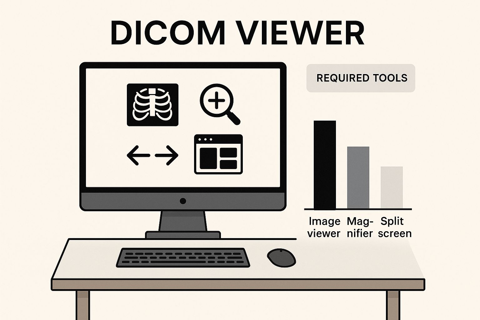
The infographic above depicts a workspace highlighting essential DICOM viewing tools. It emphasizes how accessible viewing these specialized files can be with readily available software. These free viewers empower users to interact with DCM files, laying the groundwork for deeper exploration with more advanced tools. Exploring AI-based image content retrieval systems like the one described in this article, An AI-Based Image Content Retrieval System, offers further insights into how medical images are analyzed and accessed.
To help you choose the best viewer for your needs, we’ve compiled a comparison table of the viewers discussed in this section.
Free DICOM Viewer Comparison: Comparison of popular free DICOM viewers showing features, compatibility, and best use cases.
| Viewer Name | Operating System | Key Features | Best For | Download Size |
|---|---|---|---|---|
| RadiAnt DICOM Viewer | Windows, macOS, Linux | User-friendly interface, supports various modalities, basic measurement tools | Users seeking a straightforward and easy-to-use viewer for basic image review | Varies by OS |
| Horos | macOS | Advanced visualization, MPR capabilities, open-source | Mac users requiring professional-grade features for detailed image analysis | ~500 MB |
| 3D Slicer | Windows, macOS, Linux | 3D reconstruction, advanced analysis tools, open-source | Researchers, clinicians, and those needing sophisticated 3D visualization and analysis | ~700 MB |
This table provides a quick overview of each viewer's strengths and target users. The download size is approximate and may vary depending on the version and operating system. Remember to download software only from official and trusted sources.
RadiAnt DICOM Viewer: A Simple and Effective Solution
RadiAnt is an excellent starting point for those new to viewing DCM files. Its intuitive interface makes navigating complex medical image data surprisingly easy. This viewer excels at displaying a wide range of medical image modalities, including X-rays, CT scans, and MRI images.
RadiAnt also offers basic measurement tools, allowing users to assess distances and areas within the images. This makes it a practical choice for quick reviews and basic analysis.
- Key Features: User-friendly interface, supports various modalities, basic measurement tools
- Best For: Users seeking a straightforward and easy-to-use viewer for basic image review
Horos: Professional-Grade Features for Mac Users
Horos is a powerful, free, open-source DICOM viewer designed specifically for macOS. It provides a more extensive feature set than RadiAnt, including advanced visualization tools and image manipulation capabilities.
Horos allows for multi-planar reconstruction (MPR), enabling users to view images in different anatomical planes. This feature is particularly useful for surgical planning and complex anatomical assessments.
- Key Features: Advanced visualization, MPR capabilities, open-source
- Best For: Mac users requiring professional-grade features for detailed image analysis
3D Slicer: Advanced Analysis and 3D Reconstruction
3D Slicer stands out for its robust 3D reconstruction features. This free, open-source software platform is used extensively in medical research and surgical planning. It allows users to create 3D models from a series of 2D DICOM images.
This functionality is invaluable for understanding complex anatomical structures and planning interventions. 3D Slicer offers a wide array of analysis tools, making it a powerful resource for researchers and clinicians.
- Key Features: 3D reconstruction, advanced analysis tools, open-source
- Best For: Researchers, clinicians, and those needing sophisticated 3D visualization and analysis
Choosing the right DICOM viewer depends on your specific needs. Whether you need a simple viewer for quick checks like RadiAnt, advanced features like those in Horos, or robust 3D capabilities like 3D Slicer, free options exist to open and effectively use DCM files. Each provides a valuable pathway to accessing and understanding the information within these medical files.
Professional Medical Software for Advanced DCM Handling
Free DICOM viewers are a great starting point if you need to open and view DCM files. But when basic viewing isn't enough, healthcare institutions require the advanced functionality of professional medical software. This section explores enterprise-grade solutions, examining their clinical-grade features, integration capabilities, and cost considerations.
Enterprise-Grade DICOM Viewers: OsiriX MD, Synapse, and Carestream Vue
Platforms like OsiriX MD, Synapse, and Carestream Vue go far beyond simply viewing files. They provide sophisticated tools essential for medical professionals. These include:
- Multi-Planar Reconstruction (MPR): This feature allows viewing images in different anatomical planes, crucial for surgical planning and complex diagnoses.
- Precise Measurement Capabilities: Professional software offers accurate tools for measuring distances, angles, and volumes within medical images, assisting in quantitative analysis.
- Advanced 3D Rendering: Creating detailed 3D models from 2D image slices is invaluable for visualizing complex structures and planning interventions.
- Comprehensive Annotation Systems: These platforms allow detailed annotations and markups directly on images, which facilitates collaboration and communication among medical professionals.
These advanced features transform how medical professionals interact with DICOM data, enabling more accurate diagnoses and effective treatment planning.
Integration and Compliance: Seamless Workflows and HIPAA Adherence
Beyond advanced features, professional solutions seamlessly integrate with existing hospital systems. Integration with PACS (Picture Archiving and Communication Systems) streamlines clinical workflows, ensuring efficient access to patient data.
Critically, these platforms maintain strict HIPAA compliance, safeguarding sensitive patient information. They adhere to rigorous security standards, ensuring data privacy and integrity. This commitment to security is paramount in healthcare.
Investment Considerations: Licensing, Training, and Return on Investment
Implementing professional DICOM software involves significant investment considerations. Licensing costs vary based on features, the number of users, and the size of the institution.
Training requirements are another crucial factor. While some platforms offer intuitive interfaces, mastering advanced features might require specialized training. This investment in training ensures medical professionals can effectively use the software's full potential.
However, these investments are often justified by the enhanced capabilities, improved workflows, and, ultimately, better patient outcomes. The increased efficiency and accuracy in diagnosis and treatment planning contribute significantly to a positive return on investment. Whether you are part of a small clinic or a large hospital system, evaluating these factors helps determine the right solution for your clinical needs and budget.
Imagine a complex medical case requiring detailed analysis of a CT scan. A free viewer might allow you to open the DCM file and view the images. However, professional software like OsiriX MD empowers radiologists to perform MPR, create 3D reconstructions, and take precise measurements, leading to a more accurate and informed diagnosis. This enhanced capability justifies the investment, especially considering the potential impact on patient care. Carefully considering these factors allows healthcare institutions to make informed decisions about which professional DICOM software best suits their specific needs.
Step-by-Step Guide: Opening Your First DCM File
Ready to view your first DCM file? This guide provides a practical walkthrough using proven methods. We'll cover everything from choosing the right viewer to navigating the image data.

Choosing the Right DICOM Viewer
The first step is selecting a suitable DICOM viewer. Your options range from free viewers like RadiAnt and Horos to professional software like OsiriX MD. The best choice depends on your individual needs and technical capabilities. This guide uses the free and user-friendly RadiAnt DICOM Viewer. The general principles, however, apply to most viewers.
Downloading and Installing the Viewer
- Download RadiAnt: Visit the official RadiAnt website and download the correct version for your operating system.
- Installation: Run the installer and follow the on-screen prompts. This typically involves accepting license agreements and choosing an installation location.
Opening Your First DCM File with RadiAnt
With RadiAnt installed, you can now open your DCM file:
- Launch RadiAnt: Open the application. You'll initially see a blank interface.
- Import the DCM File: Click the "Load" button or use the File menu to open your DCM file. You may need to browse your computer to find the file.
- Viewing the Image: The DCM image will then appear in the viewer window.
Navigating and Interpreting the Image
DICOM viewers provide various tools for navigating and interpreting medical images. Here are some key features in RadiAnt:
- Window and Level: Adjust these settings (often with sliders or your mouse wheel) to change the image contrast, similar to adjusting brightness and contrast on a standard image.
- Zoom and Pan: Use these tools to move around the image and examine specific areas in detail.
- Image Series: If your DCM file is part of a series (like a CT or MRI scan), navigate through the slices using arrow keys or a dedicated navigation tool.
- Metadata: Access the metadata linked to the DCM file. This usually includes patient information, scan parameters, and other vital details.
Handling Different DCM File Types
DCM files come in various formats:
- Single Images: These represent a single snapshot, such as an X-ray.
- Multi-Frame Studies: These contain multiple images, such as those from a CT or MRI scan.
- Compressed DICOM Formats: These use compression to reduce file size. Make sure your viewer supports these formats.
RadiAnt handles these different types effectively. Use the navigation controls to move between slices in multi-frame studies. Compressed formats may need extra decoding steps based on your viewer.
Troubleshooting Common Issues
You might occasionally encounter these issues:
- Format Compatibility: Ensure your viewer supports the specific DICOM format of your file.
- Display Problems: Adjust the window and level settings or update your graphics card drivers.
- File Corruption: Corrupted files may not open. Try getting a fresh copy of the file.
By following these steps and learning your viewer's features, you can confidently open and explore any DCM file. This allows you to access important insights from medical imaging data. Whether you're a healthcare professional, researcher, or viewing your own medical images, this guide provides a strong foundation for understanding and using this important file format.
Web-Based DCM Viewers and Mobile Solutions
Accessing DICOM (DCM) files doesn't always require dedicated software. Sometimes, quick access is needed on devices where installations aren't an option. Web-based DICOM viewers offer a practical solution, allowing you to open DCM files directly in your browser. This eliminates the need for downloads and installations.
Online DICOM Viewing Platforms: Advantages and Limitations
Several secure online platforms offer DCM viewing capabilities. These viewers are particularly useful for:
- Quick Consultations: Share medical images rapidly with colleagues for a second opinion.
- Remote Access: View images from anywhere with an internet connection.
- Limited Installations: Access DCM files on devices where software installation isn't feasible.
However, web-based viewers have limitations compared to desktop applications. Functionality may be restricted, and some advanced features like 3D reconstruction might not be available.
Additionally, internet connection speed significantly impacts the viewing experience, especially with larger files. This can become a bottleneck when working with complex image series. Some platforms may also have file size limits.
Privacy and Security in Web-Based Viewers: Protecting Patient Data
When uploading medical images to web services, privacy and security are paramount. Choose platforms that prioritize data protection and comply with relevant regulations like HIPAA. Look for features like:
- Secure Encryption: Ensures data is protected during transmission and storage.
- Access Controls: Allows you to manage who can view and access uploaded images.
- Data Retention Policies: Clearly outlines how long data is stored and how it is disposed of.
Carefully evaluating these features is essential for safeguarding sensitive patient information.
Mobile DICOM Viewers: On-The-Go Access to Medical Images
Mobile apps bring DCM viewing to smartphones and tablets, providing invaluable portability for healthcare professionals. However, screen size can affect the review of complex medical images.
Consider whether the mobile solution provides sufficient image quality for clinical review, especially for detailed diagnoses. Mobile apps are ideal for quick image checks or consultations, but might not be suitable for in-depth analysis or complex cases.
Choose an app that balances portability with the necessary features and image quality for your specific needs. Remember that some apps may have limited offline functionality.
Choosing between a web-based viewer and a mobile app depends on your specific requirements and the context in which you’ll use them. While convenient, these solutions are not always replacements for dedicated desktop software, especially for tasks requiring advanced features, high image quality, and robust security. Selecting the right tools for opening and managing DCM files is vital for optimal workflows, accurate interpretations, and proper handling of sensitive medical data.
Solving Common DCM File Problems
Working with Digital Imaging and Communications in Medicine (DCM) files can be straightforward, but sometimes issues arise that prevent them from opening correctly. This troubleshooting guide addresses the most frequent problems users encounter and offers practical solutions.
Identifying and Resolving Compatibility Issues
One common hurdle is compatibility between the DCM file and the viewer software. Not all DICOM viewers are created equal. Some viewers excel at displaying specific medical imaging types, like 2D X-rays, while others are designed for complex 3D models from CT or MRI scans. If a viewer struggles with a particular file, it might simply be a software limitation, not a file problem. Trying a different viewer often resolves this.
RadiAnt is a versatile viewer suitable for many DCM files. For advanced 3D rendering, consider 3D Slicer. Also, check if your viewer supports the compression format used in the DCM file, as compatibility can vary.
Deciphering Error Messages and Troubleshooting Corrupted Files
Error messages, while frustrating, offer valuable clues. "File not found" usually indicates an incorrect file path or a missing file. Double-check the file location. "Invalid DICOM format" often suggests file corruption. Try obtaining a fresh copy of the file if this occurs.
Sometimes, a DCM file might open, but the image appears distorted or patient information is missing. This could point to corruption or incorrect display settings within the viewer. Experiment with the window and level controls, which adjust image brightness and contrast, to improve visibility.
Handling Password-Protected and Encrypted Files
Security measures, like password protection or encryption, are common with DCM files. If prompted for a password, you'll need the correct credentials from the healthcare provider. This protection safeguards patient privacy and accessing the content without the password is impossible.
Alternative Approaches and Conversion Considerations
If your usual DICOM viewer isn't working, consider web-based viewers or mobile DICOM viewer apps. These offer convenient access without software installation, but they may have limitations in functionality.
Converting DCM files to JPEG or PNG is possible but comes with risks. Vital metadata can be lost, and image quality might suffer. Also, legal and ethical considerations surround medical file conversion, particularly concerning patient confidentiality. Stick to dedicated DCM viewers whenever possible.
To help you quickly diagnose and resolve common DCM file issues, refer to the following table:
Common DCM File Problems and Solutions
Quick reference guide for resolving typical DCM file opening issues
| Problem | Likely Cause | Solution | Prevention Tip |
|---|---|---|---|
| File won't open | Incompatible viewer | Try a different viewer like RadiAnt or 3D Slicer | Use a versatile DICOM viewer |
| Error: "Invalid DICOM format" | Corrupted file | Obtain a fresh copy of the file | Verify file integrity after transfer |
| Image displays incorrectly | Incorrect window/level settings | Adjust window/level in the viewer | Familiarize yourself with viewer settings |
| Missing patient data | File corruption or conversion error | Obtain original DCM file | Avoid unnecessary file conversions |
| Password prompt | File is encrypted | Obtain password from provider | Store passwords securely |
This table offers a handy overview of common DCM file problems and their solutions. By understanding these troubleshooting techniques, you'll be better prepared to handle and resolve issues that may occur when working with DCM files.
Best Practices for DCM File Management
Successfully opening a DCM file is just the first step. Proper DCM file management ensures long-term accessibility, maintains file integrity, and upholds crucial patient privacy standards. This involves implementing effective strategies for storing, organizing, and backing up these vital medical images.
File Naming and Folder Structure: Organization for Efficiency
A well-defined file naming convention is crucial for easily locating specific DCM files. Incorporate essential information like patient ID, date of the scan, and modality (e.g., CT, MRI). For example, a filename like PatientID_20241026_MRI_Brain.dcm is far more informative than a generic name like Image1.dcm.
A logical folder structure simplifies navigation and management. Organize files by patient, modality, or date. This structured approach significantly streamlines workflows, especially when dealing with large numbers of files.
Backup Strategies: Preventing Data Loss
Regular backups are essential to protect against data loss. Implement a robust backup strategy using multiple methods:
- External Hard Drives: Regularly back up DCM files to an external drive kept in a secure location.
- Cloud Storage: Cloud services like Dropbox offer convenient and automated backups, ensuring data redundancy.
- Onsite Server Backups: For larger institutions, dedicated server backups provide additional security and accessibility.
Troubleshooting DCM files can be challenging. If you encounter issues, think of it like diagnosing a car problem with service engine light diagnostics. Understanding the structure and common problems of DCM files helps identify solutions.
Legal and Ethical Considerations: Patient Privacy and Data Retention
Handling medical images requires strict adherence to legal and ethical guidelines. Prioritize patient privacy by implementing strong security measures:
- Data Encryption: Encrypt DCM files both in storage and during transmission.
- Access Controls: Restrict access to authorized personnel only.
- De-identification: When sharing files for research or education, remove all identifying patient information.
Adhere to data retention policies mandated by regulations like HIPAA. These policies dictate how long medical records, including DCM files, must be kept and how they should be disposed of securely.
Optimizing Storage and Maintaining File Integrity
Large DCM files can consume significant storage space. Consider using lossless compression techniques to reduce file size without compromising image quality. Regularly verify file integrity to ensure data remains accurate and usable over time. This involves checking for file corruption and implementing measures to prevent data degradation.
Preparing for Secure Transmission: Sharing with Specialists
Sharing DCM files with specialists requires secure transmission methods. Utilize encrypted email services, secure file transfer platforms, or dedicated medical image sharing networks. These methods protect patient privacy and ensure the integrity of the transmitted data.
Version Control and Audit Trails: Documentation for Clinical Decision-Making
Implement version control for DCM files, especially with multiple revisions or annotations. This allows tracking changes and reverting to previous versions if needed. Maintain detailed audit trails to document who accessed, modified, or transmitted the files. These practices support clinical decision-making, facilitate collaboration, and ensure compliance with regulatory requirements.
By following these best practices, healthcare professionals can ensure the secure, efficient, and ethical management of DCM files throughout their lifecycle. Proper management protects valuable medical data and supports optimal clinical workflows and enhances patient care.
Ready to elevate your medical imaging workflows with AI solutions? Explore PYCAD's advanced capabilities at https://pycad.co. PYCAD empowers medical device manufacturers and healthcare providers to enhance diagnostic accuracy and operational efficiency through innovative AI integrations.