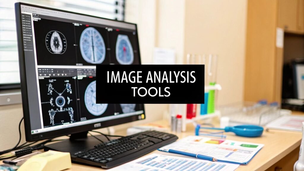
From microscopic cellular structures to complex MRI scans, extracting meaningful data from visual information is a cornerstone of modern science and medicine. The right image analysis software is crucial, transforming raw pixels into quantifiable insights that drive diagnostics, research, and innovation. However, the market is crowded with options, ranging from open-source academic tools to powerful commercial platforms, making the selection process a significant challenge for researchers, developers, and IT departments alike.
This guide simplifies that decision. We've compiled a comprehensive list of the leading image analysis platforms, offering a deep dive into their core features, ideal use cases, and practical limitations. Innovations in this field are often accelerated by dedicated funding, such as recent grants for collaborative research in computational neuroscience, which pushes the boundaries of what these tools can achieve.
Here, you will find a detailed breakdown of each solution, complete with screenshots, direct links, and an honest assessment to help you identify the perfect fit for your specific project needs. We will explore everything from DICOM compatibility and 3D rendering to advanced algorithmic capabilities. We also evaluate how each tool compares to our own specialized solution, PYCAD, providing a clear benchmark for performance in medical imaging contexts. Let's explore the tools that can sharpen your analytical edge.
1. Pycad Convert
Pycad Convert stands out as a premier choice in image analysis software, specifically engineered for the rigorous demands of medical imaging. It is a powerful, yet remarkably intuitive, platform that excels at streamlining data preparation, a critical first step in any sophisticated analysis workflow. This tool addresses the fundamental challenge of format incompatibility by providing seamless, high-fidelity conversion between essential medical imaging formats like DICOM, NIfTI, PNG, and JPEG.
Its primary strength lies in its ability to do more than just convert files. Pycad Convert integrates essential processing capabilities directly into the workflow, enabling users to perform 2D segmentation and generate detailed 3D reconstructions from their datasets. This consolidation of features makes it an indispensable asset for preparing data for advanced AI modeling and in-depth clinical review, significantly reducing the time and technical expertise required. The user-friendly interface is designed for efficiency, ensuring that researchers and clinicians can focus on analysis rather than on cumbersome data manipulation.
Key Strengths & Use Cases
- Broad Format Support: Its core function is flawless conversion between formats like DICOM and NIfTI, crucial for multi-modal studies or preparing data for machine learning frameworks that require standardized inputs.
- Integrated Advanced Processing: The platform includes built-in tools for 2D segmentation and 3D reconstruction, allowing users to extract regions of interest and visualize anatomical structures without needing separate software. This is particularly valuable for surgical planning and volumetric analysis.
- Streamlined AI/ML Data Prep: Pycad Convert is ideal for preparing large imaging datasets for AI model training. It simplifies the process of standardizing, annotating, and structuring data, accelerating the entire research and development lifecycle.
- Part of a Larger Ecosystem: As a component of the broader PYCAD ecosystem, it offers a clear pathway to more advanced AI-driven tools for model deployment and analysis, ensuring scalability for growing projects.
Considerations and Access
While its specialization in medical imaging is a significant advantage for its target audience, this focus naturally limits its utility outside of healthcare and life sciences research. To fully harness its advanced features, some foundational knowledge of medical imaging concepts is beneficial.
Access: Pycad Convert is available as a free, web-based tool. Users can access its full functionality directly through their browser without installation.
Our Takeaway: For professionals in medical imaging who need a reliable, efficient, and powerful tool for data conversion and preparation, Pycad Convert is a definitive choice. It masterfully bridges the gap between raw data acquisition and meaningful analysis.
Pros:
- Supports multiple key medical image formats for broad compatibility.
- Features advanced 2D segmentation and 3D reconstruction capabilities.
- User-friendly interface simplifies complex image processing tasks.
- Integrates smoothly with PYCAD’s larger AI-powered ecosystem.
Cons:
- Primarily focused on medical imaging, limiting its scope for other industries.
- Advanced features are best utilized with some background in medical imaging.
Website: https://pycad.co/pycad-convert/
2. Media Cybernetics – Image-Pro Software
Media Cybernetics' Image-Pro is a powerful and highly versatile 64-bit image analysis software platform. Its strength lies in its adaptability across diverse scientific domains, from life sciences and medical research to materials science and industrial inspection. This makes it a go-to choice for labs and institutions that handle a wide variety of imaging tasks and require a single, robust solution. The platform is designed to streamline complex workflows through a highly customizable interface and a suite of advanced tools.
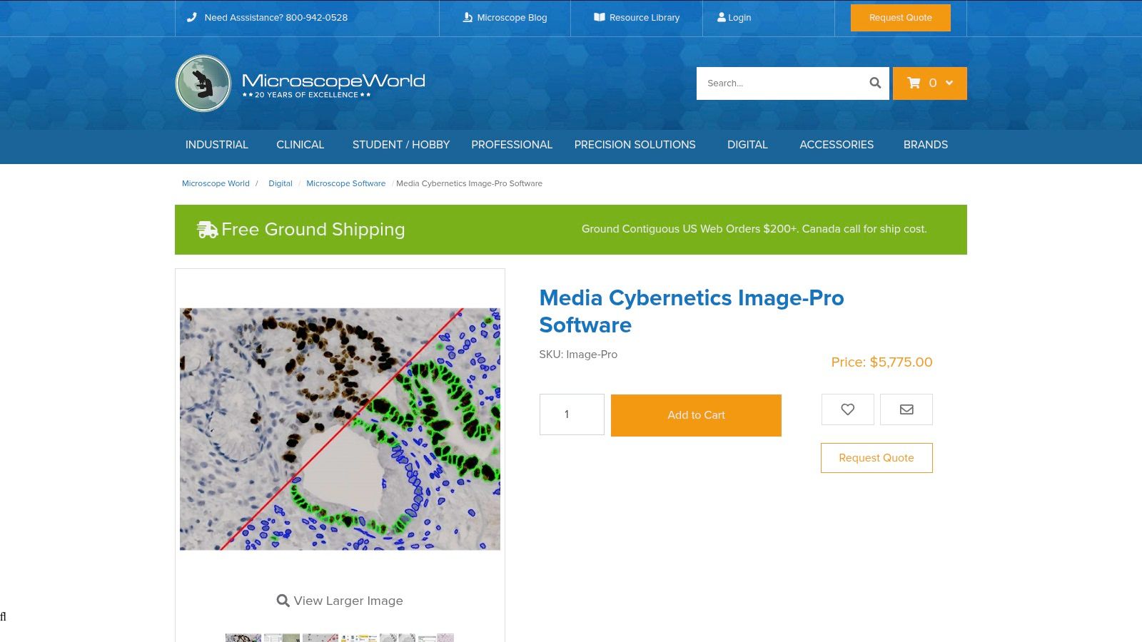
A standout feature is its Smart Segmentation tool, which uses machine learning to identify and delineate complex or faint objects that traditional thresholding methods might miss. This is particularly useful in medical imaging for analyzing cellular structures or tissue samples with subtle variations. The software also includes a Data Collector for aggregating measurements from multiple images into a single dataset, simplifying comparative analysis. However, its comprehensive nature comes with a high price tag, which can be a barrier for smaller labs or startups. Additionally, the licensing relies on a physical USB dongle, an access requirement that some may find inconvenient in modern, multi-device workflows.
Website: https://www.microscopeworld.com/p-4321-media-cybernetics-image-pro-software.aspx
3. Meesoft – Image Analyzer
Meesoft's Image Analyzer stands out as a powerful and entirely free image analysis software for Windows users. It delivers a surprising range of advanced editing and analysis tools, making it an excellent choice for students, hobbyists, or professionals who need capable software without a budget. The platform is designed to be lightweight and efficient, providing a responsive experience even on older hardware. It bridges the gap between basic image viewers and expensive, professional-grade analysis suites.
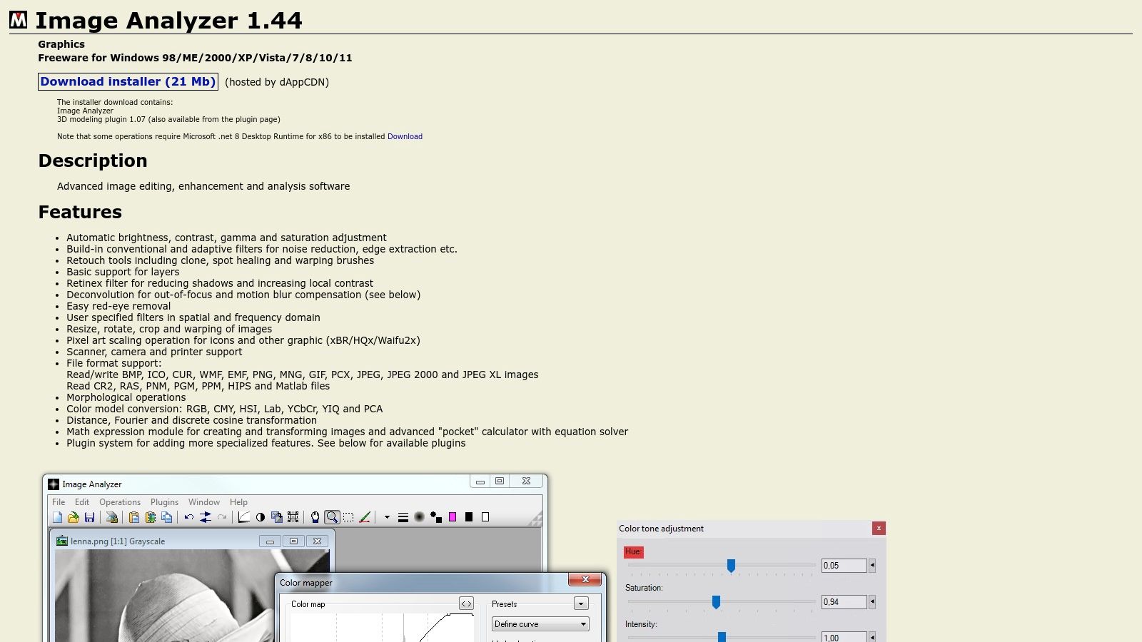
Key features include automatic adjustments for brightness, contrast, and saturation, which simplify initial image processing. It also incorporates a useful set of built-in filters for tasks like noise reduction and edge extraction, alongside practical retouching tools such as clone and spot healing brushes. While it offers extensive functionality for freeware, its primary limitation is the lack of official support or a regular update schedule, which might be a concern for professional environments requiring consistent maintenance. Furthermore, it cannot match the specialized, high-level analytical power found in premium commercial software.
Website: https://www.meesoft.com/Analyzer/
4. 3D Slicer
3D Slicer is a powerful, free, and open-source platform for medical image analysis software and scientific visualization. It stands out due to its extensive support for various imaging modalities, including DICOM, and its robust toolset for image registration, segmentation, and 3D rendering. The platform is highly favored in academic and research settings for its flexibility and strong community backing, providing a cost-effective yet comprehensive solution for complex medical imaging tasks.
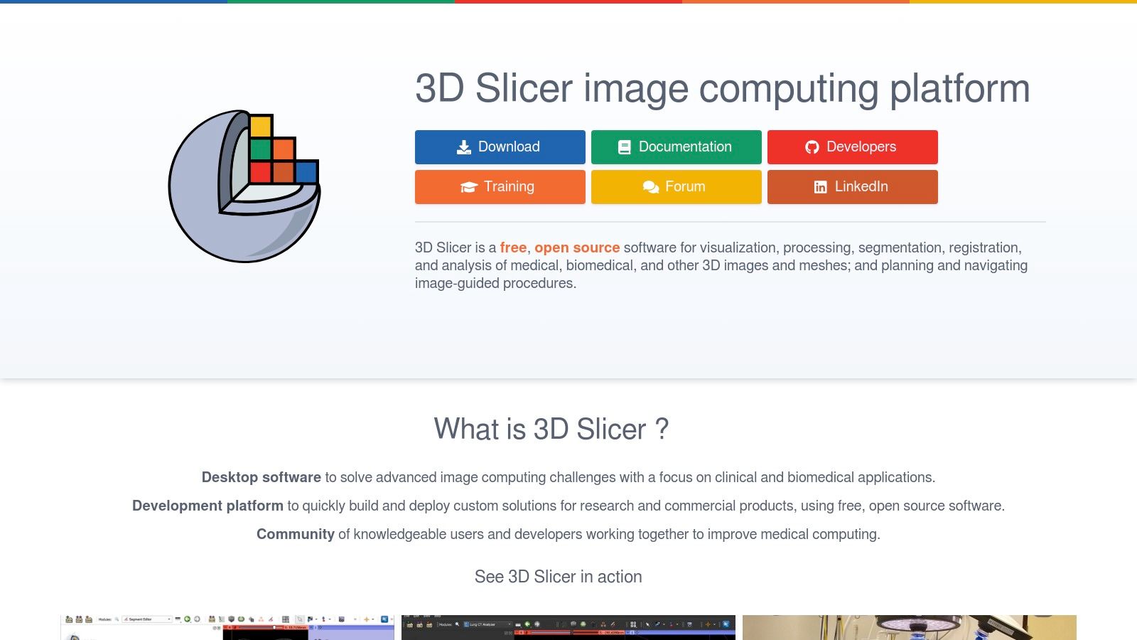
A key advantage of 3D Slicer is its extensive plugin architecture, which allows users to customize and extend its core functionalities for specific research needs, such as analyzing diffusion tensor imaging data. The platform offers excellent interactive visualization for both volumetric images and polygonal meshes. However, its comprehensive capabilities contribute to a steep learning curve, which can be challenging for beginners. Furthermore, being a high-performance tool, it is quite resource-intensive and may require robust hardware to run smoothly, especially when handling large, high-resolution datasets. Despite this, its open-source nature and active community support make it an invaluable resource.
Website: https://www.slicer.org/
5. FastStone Image Viewer
FastStone Image Viewer is a lightweight yet surprisingly capable freeware tool primarily designed for Windows users. While not a dedicated scientific image analysis software platform, its speed, extensive format support, and basic editing capabilities make it an excellent choice for initial image review, organization, and simple modifications in research workflows. It excels at quickly browsing through large datasets of images, including many common raw file formats from various cameras and imaging systems. Its efficiency is a key differentiator, offering a no-frills, high-speed alternative to more resource-intensive programs for preliminary assessment.
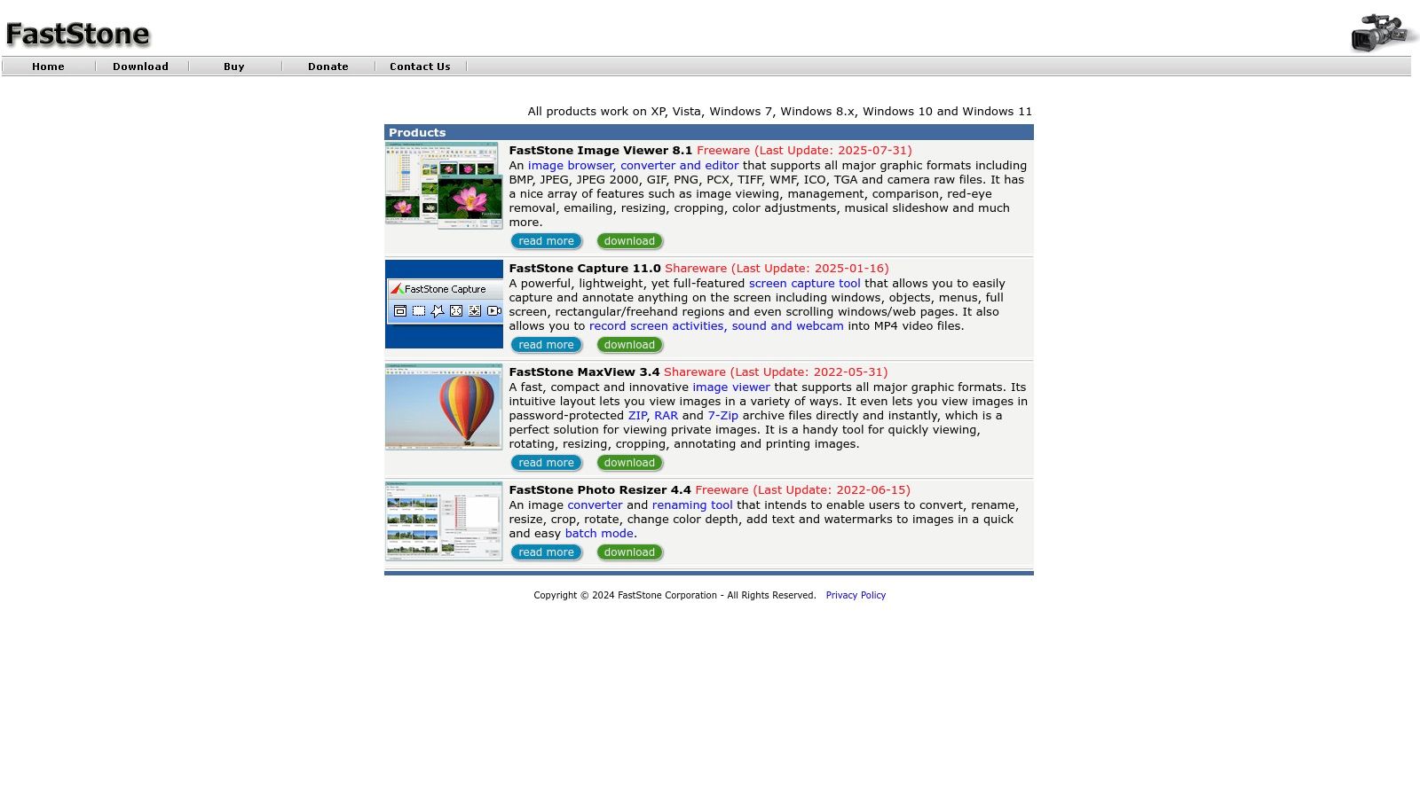
Key features include a high-quality thumbnail viewer with Lanczos resampling, which provides clear previews, and a suite of basic editing tools like cropping, color correction, and red-eye removal. Its batch processing function is particularly useful for researchers needing to rename or convert large batches of files simultaneously. The main advantage is its cost; it's completely free for personal and educational use, making it highly accessible. However, it lacks advanced analytical features and is limited to the Windows operating system, which is a significant drawback for labs using macOS or Linux. It’s best used as a supplementary tool for quick viewing and sorting rather than deep analysis.
Website: http://www.faststone.org
6. Sunny Instruments – MvImage3.0
Sunny Instruments' MvImage3.0 is a specialized image analysis software platform built specifically for integration with motorized microscopes. It excels in life sciences research by automating complex imaging processes and providing a robust suite of tools for in-depth analysis. The software is designed to handle multi-dimensional data, making it ideal for researchers conducting qualitative, quantitative, timing, and positioning analyses on biological samples. Its direct control over microscope hardware streamlines the entire workflow from acquisition to analysis.
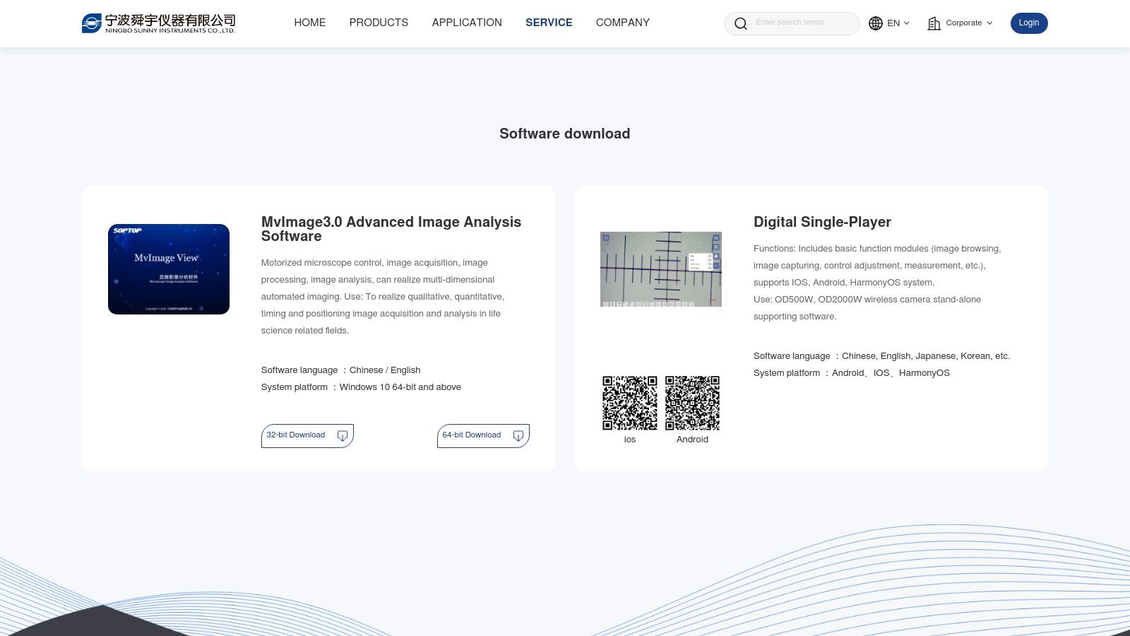
A key strength of MvImage3.0 is its seamless integration, which allows for fully automated, multi-dimensional imaging sequences. This is particularly valuable for time-lapse studies or capturing detailed Z-stacks of cellular structures. While the software offers a comprehensive and user-friendly interface with multilingual support, its primary limitation is its hardware dependency. It is tailored for Sunny Instruments' own equipment and is restricted to Windows 10 64-bit operating systems or newer. This makes it a powerful but niche solution rather than a universally applicable tool.
Website: https://www.sunny-instrument.com/en/download
7. ImageJ
ImageJ is a public domain, Java-based image analysis software developed at the National Institutes of Health (NIH). Its enduring popularity in the scientific community stems from being completely free, open-source, and highly extensible. It provides a core set of tools for processing, editing, and analyzing 2D and 3D images and is celebrated for its cross-platform compatibility, running on Windows, macOS, and Linux. This accessibility makes it a foundational tool in academic research, especially in fields like biology and medicine where funding can be limited.
The platform's greatest strength is its massive ecosystem of user-created plugins, which extend its functionality far beyond the basic package. Researchers can find or create plugins for nearly any specialized task, from particle tracking to complex cell segmentation. However, this flexibility comes with a trade-off. The user interface can feel dated and less intuitive compared to modern commercial software, presenting a learning curve for new users. Furthermore, its performance relies on the Java Runtime Environment, which must be installed and can sometimes introduce compatibility or performance issues, particularly with very large datasets.
Website: https://imagej.net/
8. Digimizer
Digimizer positions itself as a remarkably accessible and user-friendly image analysis software, making it an excellent entry point for researchers or professionals who need precise measurements without a steep learning curve. Its core strength lies in its simplicity, offering both manual measurement tools and automatic object detection in a clean, uncluttered interface. This makes it particularly well-suited for straightforward tasks in fields like medical imaging and material science, where quick, accurate data extraction is paramount.
The platform supports a wide range of image formats and provides tools for basic statistical analysis, allowing users to export their findings easily. Its automatic object detection is customizable, enabling users to define and find objects based on their color or intensity. The software's intuitive design, backed by comprehensive documentation, ensures even beginners can become productive quickly. However, its focus on simplicity means it lacks some of the advanced machine learning and complex workflow automation found in higher-end competitors. A significant limitation is its Windows-only compatibility, which excludes Mac and Linux users from its ecosystem.
Website: https://www.digimizer.com/download/
9. Icy – Open Source Image Processing Software
Icy is a free, open-source bioimage informatics platform that serves as a powerful bridge between user-friendly interfaces and advanced computational capabilities. As a community-driven image analysis software, it excels in versatility, offering tools for everyone from novices to seasoned programmers. It is particularly well-suited for biological and cellular imaging, providing an extensive environment for visualizing, annotating, and quantifying complex biological data without any financial investment.
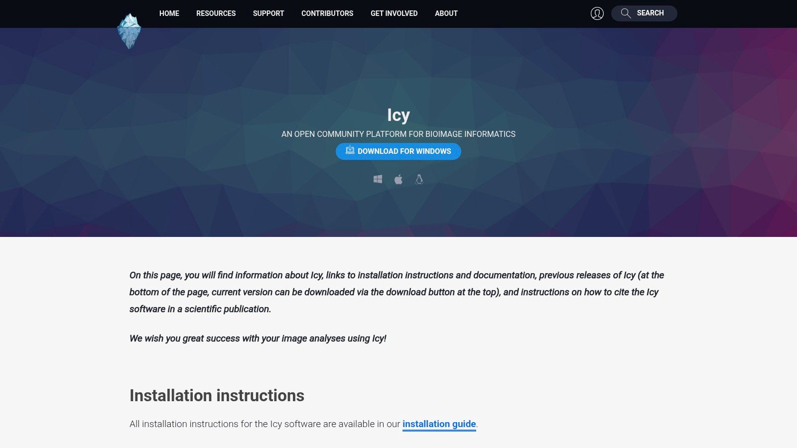
One of Icy's most compelling features is its graphical programming interface, which allows users to build complex analysis workflows by connecting functional blocks, making automation accessible without deep coding knowledge. For developers, it supports scripting in JavaScript, Python, and other languages, enabling unlimited customization. However, the sheer breadth of its features and plugins means there can be a steep learning curve for mastering advanced analyses. Its performance can also be resource-intensive, especially when handling large 3D or time-lapse datasets, requiring a capable workstation for smooth operation.
Website: https://icy.bioimageanalysis.org/download/
10. Cytiva – ImageQuant TL Analysis Software
Cytiva's ImageQuant TL is a specialized image analysis software developed specifically for researchers using Cytiva's Amersham and Typhoon imaging systems. Its primary strength is the seamless integration with this hardware, creating a highly efficient and intuitive workflow for life science applications like Western blotting, gel, and membrane analysis. The platform is designed to simplify complex quantitative tasks, offering a streamlined experience from image acquisition to final data reporting.
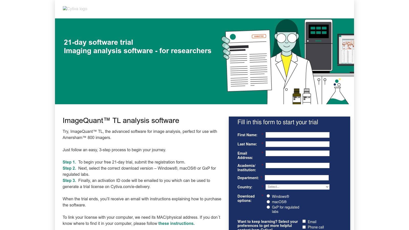
The software excels with its user-friendly interface and dedicated tools for common laboratory analyses, such as automatic lane and band detection, background subtraction, and colony counting. This focused approach ensures that users can achieve accurate and reproducible results without a steep learning curve. While it is perfectly optimized for Cytiva imagers, this specialization means it may lack the broad versatility needed for images from non-Cytiva systems. Pricing is not publicly listed and requires a direct inquiry, but a free 21-day trial is available for evaluation, allowing labs to test its compatibility with their specific workflows before committing.
Website: https://info.cytivalifesciences.com/image-analysis-software.html
11. Optotherm – Image Analysis Software
Optotherm provides highly specialized image analysis software tailored specifically for thermal imaging applications. This focus makes it an indispensable tool in fields like electronics testing, industrial process monitoring, and even certain medical diagnostics where temperature variations are critical indicators. The software is designed to work seamlessly with Optotherm's thermal imaging cameras, offering a cohesive ecosystem for capturing, analyzing, and reporting thermal data in real time.
The platform excels at providing advanced analytical tools like temperature point analysis, region-of-interest statistics, and isotherm visualization, all accessible through a user-friendly interface. Its strength lies in real-time monitoring, enabling engineers and researchers to detect thermal anomalies as they happen. However, its biggest limitation is its hardware dependency, as the software is optimized exclusively for Optotherm's own thermal imaging systems, which restricts its use with third-party cameras. Furthermore, pricing is not publicly listed and requires a direct inquiry, which can slow down the procurement process for interested organizations.
Website: https://www.optotherm.com/shop/category/image-analysis-software-6
12. Bionity – Cytiva Image Analysis Software
Bionity serves as a specialized marketplace and information hub for life science products, prominently featuring Cytiva's powerful image analysis software. Instead of being a software developer, Bionity provides a streamlined portal for researchers and lab managers to discover, compare, and request quotes for Cytiva's imaging solutions. The platform is designed for efficiency, centralizing detailed product specifications, application notes, and related equipment, making it a valuable resource for those specifically seeking tools for cellular and biomolecular analysis.
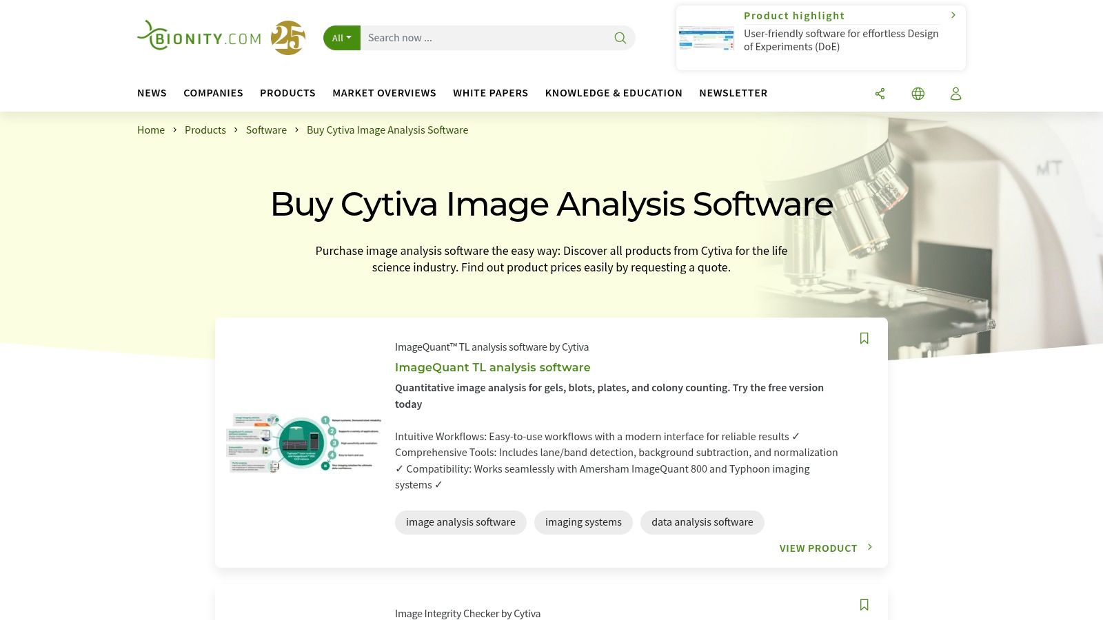
The primary advantage of using Bionity is its focused user experience. The interface simplifies the often-complex procurement process by organizing Cytiva’s software into clear categories and enabling easy quote requests. This direct-to-vendor inquiry model ensures users get accurate, up-to-date pricing and technical support. However, its major limitation is its scope; the platform is exclusively a gateway to Cytiva products, so it is not suitable for users looking to compare software across different manufacturers. The requirement to request a quote rather than seeing direct pricing also adds an extra step to the purchasing workflow.
Website: https://www.bionity.com/en/products/cytiva/image-analysis-software/order_nt/
Image Analysis Software Comparison Overview
| Product | Core Features / Functionality | User Experience / Quality ★ | Value Proposition 💰 | Target Audience 👥 | Unique Selling Points ✨ |
|---|---|---|---|---|---|
| 🏆 Pycad Convert | Multi-format conversion, 2D segmentation, 3D reconstruction | ★★★★☆ Intuitive, AI-integrated | 💰 Scalable, efficient workflow | 👥 Radiologists, researchers, data scientists | ✨ AI-powered ecosystem integration |
| Media Cybernetics – Image-Pro | Advanced segmentation, data collection, machine learning | ★★★★☆ Versatile & customizable | 💰 High cost, license dongle required | 👥 Life sciences, manufacturing | ✨ Smart segmentation, live image capture |
| Meesoft – Image Analyzer | Auto adjustments, filters, retouch, multi-format | ★★★☆☆ Lightweight, beginner-friendly | 💰 Freeware | 👥 General users, beginners | ✨ Free, with editing and analysis tools |
| 3D Slicer | DICOM support, segmentation, 3D visualization | ★★★★☆ Open-source, active community | 💰 Free | 👥 Medical researchers, clinicians | ✨ Extensive plugins, cross-platform |
| FastStone Image Viewer | Image viewing, basic editing, batch operations | ★★★☆☆ Fast, simple UI | 💰 Free for personal/education use | 👥 General users | ✨ Lightweight viewer with raw support |
| Sunny Instruments – MvImage3.0 | Motorized microscope control, multi-dimensional imaging | ★★★★☆ User-friendly, life science focused | 💰 Hardware-specific, limited OS | 👥 Life sciences researchers | ✨ Automated imaging, multi-language |
| ImageJ | Plugin extensibility, multi-format support | ★★★★☆ Powerful, open-source | 💰 Free | 👥 Scientific image analysts | ✨ Highly extensible, large plugin base |
| Digimizer | Manual & automatic measurements, stats, export | ★★★☆☆ Intuitive, beginner-friendly | 💰 Moderate pricing possible | 👥 Medical & material science users | ✨ Precise measurements, easy to use |
| Icy | Plugins, scripting, 3D visualization, workflow automation | ★★★★☆ Flexible, cross-platform | 💰 Free | 👥 Bioimage informatics experts | ✨ Workflow automation, graphical programming |
| Cytiva – ImageQuant TL | Lane/band detection, background subtraction | ★★★★☆ Intuitive workflows | 💰 Pricing on request | 👥 Life science imaging users | ✨ Tailored for Cytiva imaging systems |
| Optotherm – Image Analysis | Thermal imaging analysis, real-time monitoring | ★★★★☆ User-friendly, specialized | 💰 Pricing on request | 👥 Thermal imaging professionals | ✨ Thermal imaging focus, real-time tools |
| Bionity – Cytiva Software | Product discovery, quote requests | ★★★☆☆ Easy to navigate | 💰 Pricing on quote | 👥 Life science industry buyers | ✨ Centralized Cytiva product platform |
Final Thoughts
Navigating the landscape of image analysis software reveals a diverse ecosystem of tools, each with its own strengths, complexities, and ideal use cases. From open-source academic powerhouses like ImageJ and 3D Slicer to specialized commercial platforms such as Image-Pro by Media Cybernetics, the right solution depends entirely on your specific project requirements, technical expertise, and budget. Our exploration has shown that there is no single "best" platform; instead, there is a "best fit" for your unique challenges.
A primary takeaway is the critical trade-off between accessibility and power. Open-source tools like ImageJ offer unparalleled flexibility and a massive community for support, but they often demand a steeper learning curve and significant user investment in customization and plugin management. Conversely, commercial software provides polished user interfaces, dedicated customer support, and validated workflows, which are crucial for clinical and regulated environments, but these benefits come at a direct financial cost.
Key Considerations for Your Selection
When deciding on the right image analysis software, it's essential to move beyond feature lists and consider the practical realities of implementation. Your decision-making process should be guided by a few core questions:
- What is your primary application? A tool designed for cellular biology, like Icy, will have different core functionalities than one optimized for radiological DICOM series analysis, such as 3D Slicer. Define your most frequent and critical tasks first.
- What is the skill level of your end-users? Will your team be composed of computer scientists comfortable with scripting, or clinicians who require an intuitive, graphical user interface? The answer will immediately narrow your options.
- What are your integration and interoperability needs? Consider how the software will fit into your existing data pipeline. Does it need to handle specific file formats like DICOM, integrate with a PACS, or export data in a format compatible with other analytical tools? Tools like PYCAD Convert excel in this specific niche of format handling and preparation.
- Do you require regulatory compliance? If your work falls under regulations like HIPAA or requires FDA clearance, you must prioritize software that offers the necessary documentation, validation, and security features.
Implementation and Beyond
Successfully implementing any image analysis software is a project in itself. The process involves not just installation but also data migration, team training, and workflow validation. It’s a microcosm of the same strategic thinking that goes into building these complex applications from the ground up. As you navigate the world of advanced image analysis, it's insightful to consider the foundational processes involved in creating such sophisticated tools. For a deeper dive into modern software development planning, exploring how these powerful solutions are conceptualized and built can provide valuable context.
Ultimately, the goal of any image analysis software is to transform visual data into quantitative, actionable insights. The right tool acts as a powerful lens, revealing patterns and details that are invisible to the naked eye. By carefully evaluating your needs against the capabilities of the solutions we've discussed, you can empower your team to unlock new discoveries, streamline diagnostic processes, and push the boundaries of medical and scientific innovation.
Ready to streamline your medical imaging data workflow? The team at PYCAD specializes in DICOM and NIfTI file conversion and de-identification, providing the critical first step for any advanced image analysis project. Visit PYCAD to see how our solutions can prepare your data for the powerful analytical tools discussed in this guide.