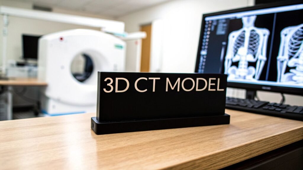
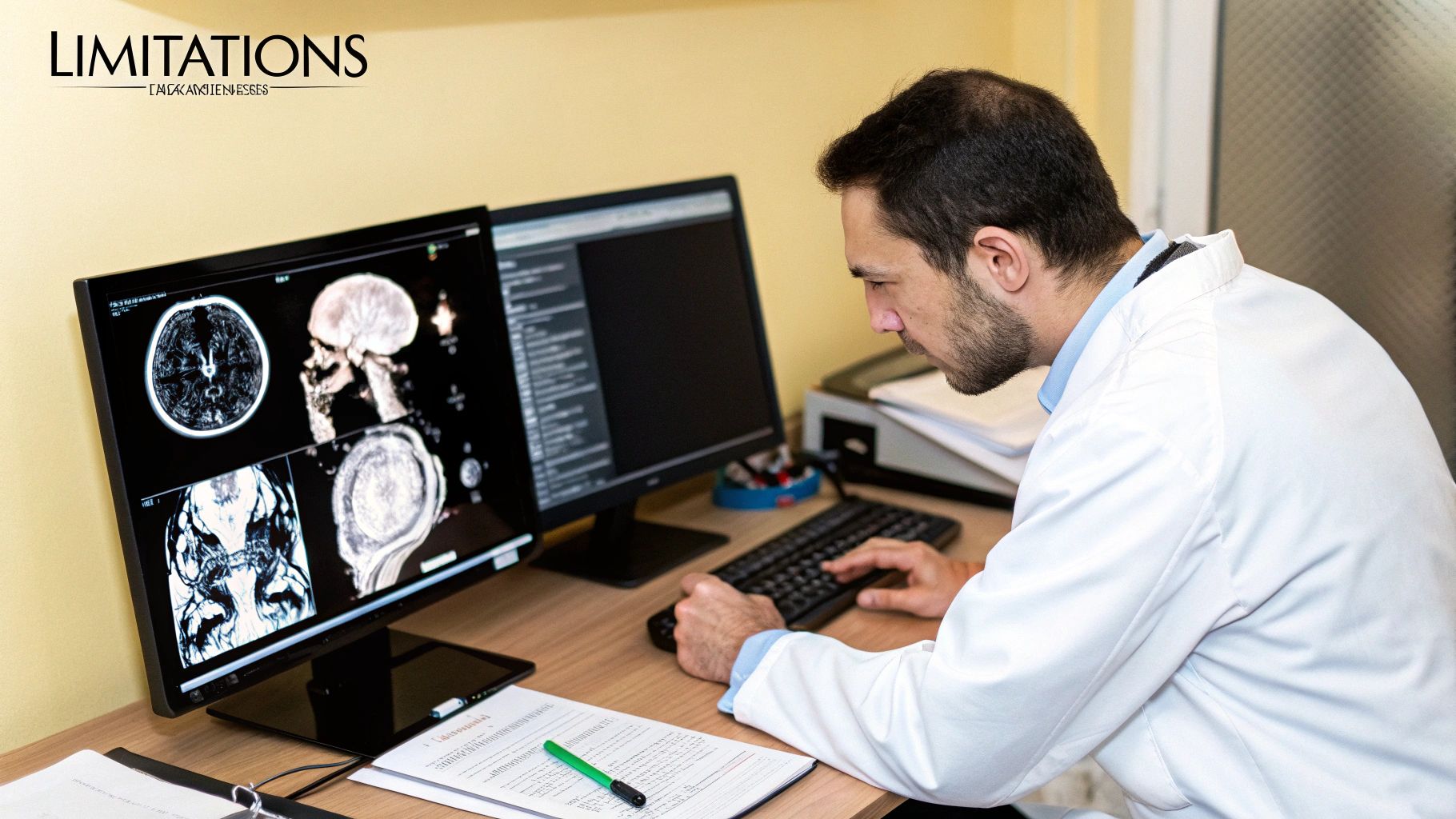
A standard CT scan gives doctors flat, cross-sectional images of the body. Think of it like a loaf of sliced bread—you can see each individual slice clearly. But CT with 3D reconstruction takes this a giant leap forward. It uses powerful software to digitally "restack" those individual slices, creating a detailed, interactive 3D model. This process transforms that flat 2D data into a complete, dimensional view of a patient’s anatomy.
From Flat Slices to Full Dimensions
Imagine trying to understand a complex engine by only looking at a series of separate, paper-thin diagrams. While each diagram offers clues, you’d miss the engine's true shape, depth, and how all its intricate parts fit together. This is the classic limitation of traditional 2D medical imaging.
A Leap in Diagnostic Clarity
3D reconstruction completely changes the game. It’s a bit like assembling a complex LEGO model from hundreds of thin, individual bricks. First, the CT scanner captures hundreds, sometimes thousands, of these cross-sectional images (the bricks). Then, sophisticated software intelligently assembles them, building a complete, rotatable 3D model (the finished LEGO creation).
This gives doctors a holistic and intuitive view of internal structures, from complex bone fractures to the tangled web of blood vessels. It allows them to see and interact with anatomy in a way that flat images simply can't match, providing a much more realistic and comprehensive perspective.
The move from 2D slices to 3D models represents a monumental leap in diagnostic medicine, offering unprecedented clarity that benefits both clinicians performing complex procedures and patients seeking to understand their own health.
The Impact on Medical Practice
This shift in technology has had a profound effect on medical diagnostics and treatment. Once CT technology became widely available, the adoption of 3D reconstruction capabilities grew rapidly. Today, it's estimated that over 80% of advanced CT scanners worldwide come equipped with some form of 3D reconstruction software, which speaks volumes about its clinical value. If you're interested in the timeline, you can learn more about the history of CT scanning and its evolution.
The advantages are clear and have a direct impact on patient care:
- Improved Spatial Understanding: Surgeons can visualize the exact relationship between a tumor and surrounding healthy tissue or blood vessels before ever making an incision.
- Enhanced Diagnostic Accuracy: Radiologists can spot subtle abnormalities, like hairline fractures or small vascular issues, that might be invisible or ambiguous on flat 2D images.
- Better Patient Communication: Showing a patient a 3D model of their own anatomy is infinitely more effective and easier to understand than pointing to grayscale slices on a screen.
To put it simply, let's compare the two approaches side-by-side.
2D Imaging vs 3D Reconstruction at a Glance
This table offers a quick comparison, highlighting how 3D reconstruction builds upon the foundation of traditional 2D imaging to offer deeper insights.
| Feature | Traditional 2D Imaging | CT with 3D Reconstruction |
|---|---|---|
| Visualization | Flat, cross-sectional "slices" | Interactive, rotatable 3D models |
| Depth Perception | Limited; depth is inferred | Excellent; true spatial relationships |
| Diagnostic Use | Good for identifying issues on a specific plane | Superior for complex anatomical assessment |
| Surgical Planning | Provides basic anatomical reference | Allows for virtual "fly-throughs" and precise pre-operative planning |
| Patient Education | Can be difficult for patients to interpret | Intuitive and easy for patients to understand |
While 2D imaging remains a cornerstone of diagnostics, the addition of 3D reconstruction gives medical professionals a much more powerful and complete toolkit for diagnosis, planning, and patient communication.
How a 3D CT Scan Is Actually Created
So, how do we get from a patient lying on a scanner table to a fully interactive 3D model on a doctor's screen? It’s a sophisticated process, but it’s not magic. Think of it less like a single snapshot and more like assembling a detailed sculpture piece by piece.
The entire workflow, which this graphic neatly summarizes, breaks down into three core phases. Each step builds on the last, turning raw scan data into a powerful diagnostic tool.
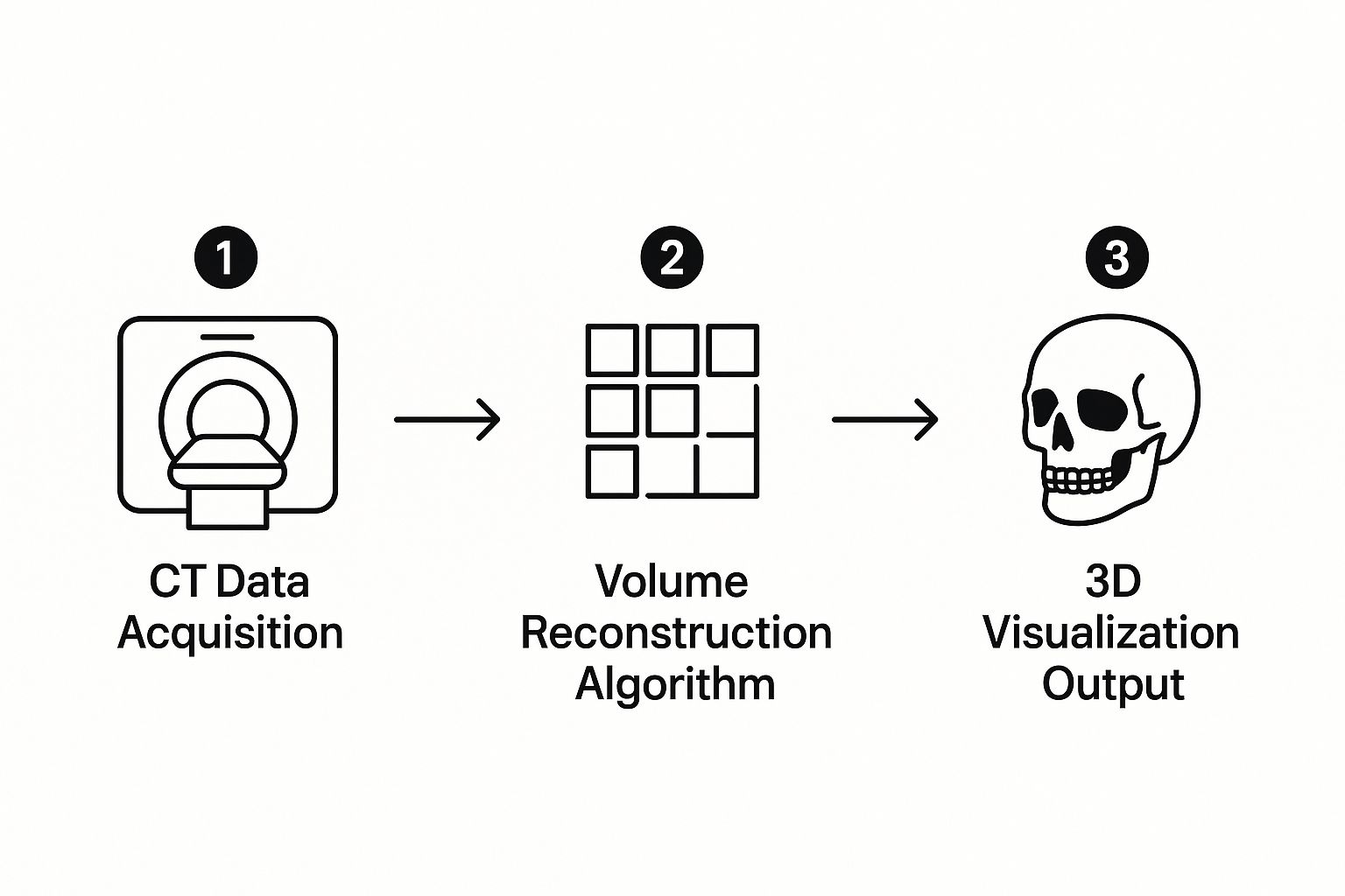
As you can see, it all starts with acquiring the data, followed by some heavy-duty computer processing, and finally, rendering the image for human eyes. Let's walk through it.
Stage 1: Data Acquisition
Everything begins with the Data Acquisition phase. This is the part everyone is familiar with—the actual scan. The patient lies still as the CT scanner’s gantry, the large donut-shaped ring, rotates around them.
As it spins, the machine captures hundreds, sometimes thousands, of individual X-ray images from every possible angle. These aren't 3D images yet. They are incredibly thin, 2D cross-sections of the body, like looking at individual slices of a loaf of bread before it's been put together. Each slice provides a wealth of information on its own distinct plane.
Stage 2: Data Processing and Volume Construction
Once the scan is complete, the raw data moves into the Data Processing stage. Here, the real heavy lifting is done by powerful computer software. The main goal is volume reconstruction, which is exactly what it sounds like: building a complete 3D volume from all those separate 2D slices.
The software acts like a digital craftsman, meticulously stacking and aligning each slice in the correct sequence. To do this, it relies on complex algorithms to:
- Align the Slices: Ensure every image is perfectly registered with the ones before and after it.
- Interpolate Data: Intelligently fill in any microscopic gaps between the slices, creating a smooth, continuous block of anatomical data.
- Create Voxels: Convert the 2D image pixels into voxels, or "volumetric pixels." These are tiny 3D cubes of data, and they are the fundamental building blocks of the final 3D model.
This resulting stack of voxels is the volumetric dataset. It’s no longer just a collection of flat images but a comprehensive, three-dimensional data block representing the scanned part of the patient's anatomy.
Stage 3: 3D Image Rendering
The final step is Image Rendering. This is where the magic happens for the viewer. Specialized software takes that raw volumetric dataset and transforms it into the visible, interactive 3D model a clinician can actually use.
Rendering techniques allow the software to assign colors and different levels of transparency to tissues based on their density. This is how bone can be made to look solid white, while soft tissues and blood vessels can be made semi-transparent, allowing a doctor to see through them to what lies beneath.
This is where the real power of CT with 3D reconstruction shines. A surgeon can virtually spin the model around, slice through it at any angle, and even "fly through" blood vessels to plan a complex procedure. It provides a perspective on the patient's anatomy that a stack of 2D images simply can't match.
The Evolution of Medical Imaging Technology
The story of computed tomography is one of incredible progress, a journey from slow, almost primitive machines to the high-definition, nearly instantaneous imaging we have today. The sophisticated CT with 3D reconstruction you see in modern hospitals didn't just appear out of nowhere; it’s the result of decades of brilliant, often painstaking, innovation focused on making scans faster, clearer, and safer for patients.
Let's rewind to the early 1970s. The very idea of looking inside the human body without a scalpel felt like science fiction. The first-ever CT scan of a patient, which took place on October 1, 1971, was a monumental achievement. But by our standards, the technology was incredibly clunky.
Imagine waiting over 20 minutes for a computer to process a single, grainy image with a resolution of just 80×80 pixels. Despite these early hurdles, the potential was obvious. By 1980, the number of CT exams was already climbing to around 3 million a year. If you're interested in the nuts and bolts of this early history, you can dive deeper with this detailed medical journal review.
Key Technological Breakthroughs
Over the next few decades, a series of breakthroughs pushed CT technology into the modern era. Each innovation tackled a major limitation of the original systems, gradually building the powerful diagnostic tools we rely on now. Getting from those first blurry pictures to today's interactive 3D models required some serious engineering and computational muscle.
These were the game-changing improvements:
- Helical (Spiral) Scanning: This arrived in the late 1980s and was a total game-changer. It allowed the scanner to capture data in a continuous spiral motion as the patient moved through the machine. This single development cut scan times from minutes down to mere seconds, making it possible to image an entire organ in the time it takes to hold your breath.
- Multidetector CT (MDCT): The next big leap came with scanners that had multiple rows of detectors. Think of it like going from a single-lane road to a multi-lane highway. This allowed for capturing many slices at once, massively increasing the speed and volume of data collection and enabling much thinner slices for incredible detail.
- Improved Computing Power: We can't forget Moore's Law. As computers grew exponentially faster, the time it took to perform complex image reconstruction dropped dramatically. A process that once took nearly half an hour can now happen in real-time.
These advancements weren't just about going faster; they were about getting better pictures. Quicker scans meant fewer blurs from patient movement like breathing, and thinner slices provided the high-quality raw data needed to build truly accurate 3D models.
Impact on Patient Care and Safety
This rapid evolution also made a huge difference in the patient's experience and overall safety. Shorter scan times mean less time a patient has to lie perfectly still—a massive benefit for children, the elderly, or trauma victims who can't hold a position for long.
On top of that, new detector materials and smarter reconstruction algorithms have led to major reductions in the required radiation dose. Today's systems can create stunningly clear images using just a fraction of the radiation needed by older machines, which is always a top priority in medicine. The development of CT with 3D reconstruction was really the culmination of all these efforts, turning a novel diagnostic device into a tool that modern medicine simply can't do without.
How 3D Reconstruction Is Transforming Patient Care
https://www.youtube.com/embed/JeuRd9OquVE
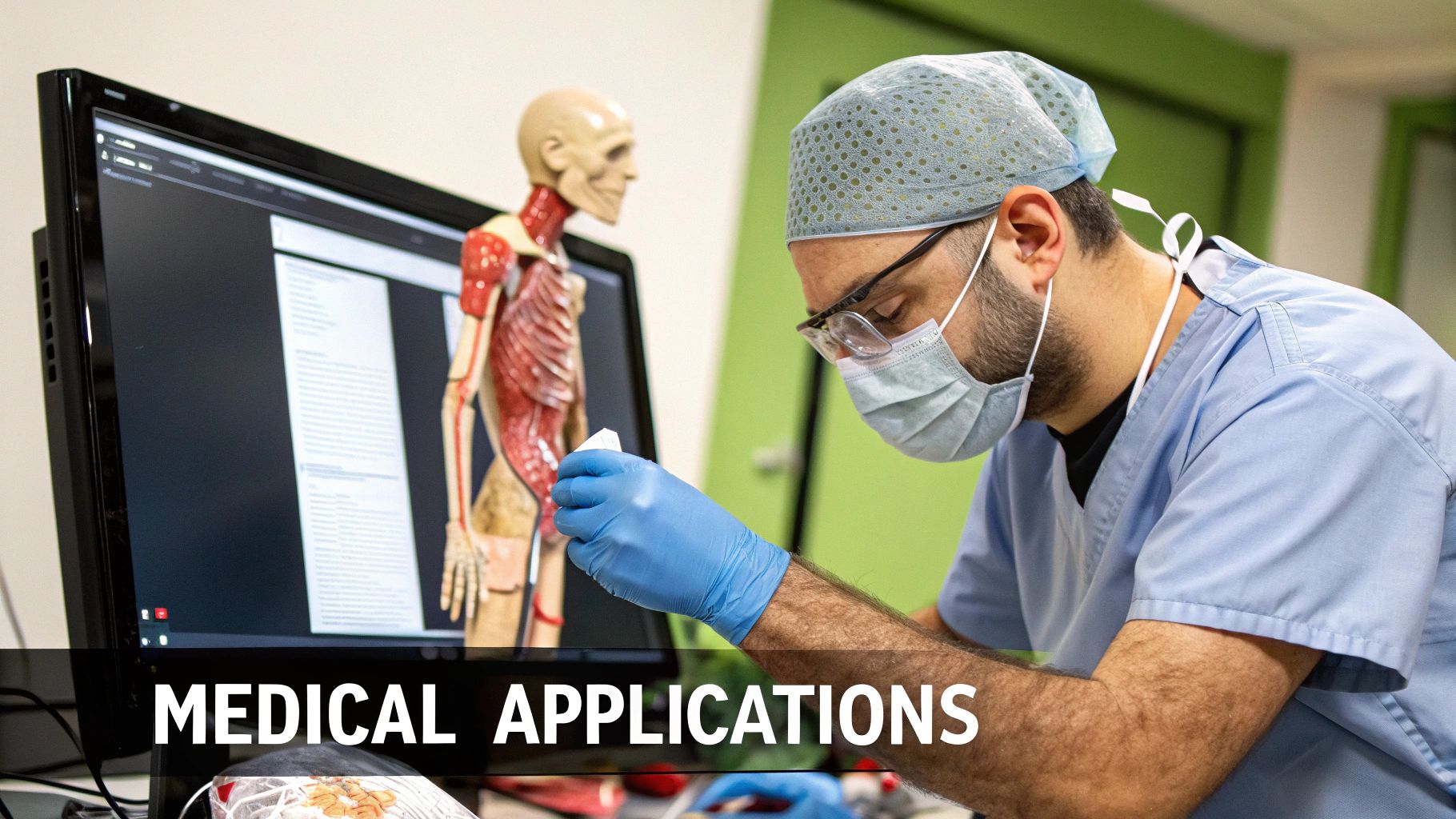
While the technology behind 3D CT scans is fascinating, what really matters is the direct impact it has on people's lives. These dynamic 3D models aren't just a neat visual trick; they have become essential tools in operating rooms and diagnostic clinics, completely changing how doctors approach treatment. It's a leap from staring at flat, grayscale slices to holding a detailed, rotatable map of a patient's internal anatomy.
This incredible level of clarity directly translates into safer, more precise medical procedures. For patients, that means shorter hospital stays, fewer complications, and better results overall.
Precision in Surgical Planning
One of the most powerful applications is in planning surgeries before they ever begin. Surgeons can now take a virtual "tour" of a patient's anatomy, exploring every angle and crevice without making a single cut. Think about an oncologist preparing to remove a tricky liver tumor. A 3D model reveals the tumor's exact size and shape, but more critically, it shows precisely how it's intertwined with major blood vessels and surrounding healthy tissue.
This digital roadmap allows the surgeon to plot the safest route for removal, minimizing damage to healthy organs and reducing risks. A study on liver cancer surgery, for instance, showed how a 3D model accurately calculated the remaining liver volume—a crucial piece of information that determined if the surgery was even safe to perform.
Or consider an orthopedic surgeon dealing with a badly shattered bone. Using a 3D reconstruction, they can meticulously reassemble the fragments on a screen, almost like a high-stakes jigsaw puzzle. This lets them select the perfect implants and map out their entire strategy long before the patient is even in the operating room.
By providing a comprehensive, interactive view of the surgical field, 3D models empower surgeons to anticipate challenges, refine their strategy, and operate with greater confidence and accuracy.
Guiding Complex Interventions
The benefits of 3D reconstruction ripple out across many different medical fields, guiding treatments that are minimally invasive but extraordinarily complex. For many specialists, these detailed models are now the standard of care.
The table below breaks down just a few key examples of how different specialties use this technology.
Key 3D CT Applications by Medical Specialty
| Medical Specialty | Primary Use Case | Key Benefit |
|---|---|---|
| Orthopedics | Planning complex fracture repair and joint replacement surgery. | Enables precise implant selection and placement, improving long-term outcomes. |
| Oncology | Mapping tumors and their relationship to vital structures. | Allows for complete tumor removal while preserving maximum healthy tissue. |
| Cardiology | Assessing complex coronary artery disease and heart structures. | Guides minimally invasive procedures like stent placement and valve repair. |
| Vascular Surgery | Navigating intricate blood vessel networks to treat aneurysms. | Acts as a GPS for catheters and stents, increasing procedural safety. |
| Craniofacial Surgery | Reconstructing facial bones after severe trauma or for birth defects. | Ensures both functional and aesthetic restoration with incredible accuracy. |
As you can see, the ability to see and interact with patient anatomy in three dimensions helps inform better decisions and leads to safer procedures across the board.
The incredible advancements in 3D reconstruction and patient care are largely powered by highly specialized digital solutions. For a deeper look into how these powerful tools are created, you might be interested in learning about customized healthcare software development. This ability to understand complex anatomy in three dimensions ultimately leads to a higher standard of care for everyone.
The Role of AI in Sharpening 3D Medical Images
While CT with 3D reconstruction already gives us an incredible view inside the human body, the next step is to make the process smarter, not just clearer. This is where artificial intelligence (AI) comes in, acting less like a replacement and more like an expert co-pilot for medical professionals. Its real strength lies in handling repetitive, time-consuming work and flagging tiny details a human eye might miss after hours of staring at screens.
The goal isn't to take the radiologist out of the picture. It's to give them powerful tools that help them work faster and with more confidence. This teamwork is what's really pushing medical diagnostics forward. If you're new to the topic, it helps to start by getting to grips with AI basics.
Automating the Segmentation Process
One of AI's biggest immediate impacts is on segmentation. This is the painstaking task of tracing the outlines of specific organs, blood vessels, or tumors within the 3D model. Done by hand, it can eat up a huge chunk of a specialist's day.
AI algorithms change the game entirely. Trained on vast libraries of medical scans, they learn to recognize and isolate these structures automatically, often in a matter of seconds. This frees up clinicians from tedious digital drawing, letting them jump straight into what they do best: diagnosing conditions and planning treatments.
Think of it this way: AI gives medical professionals a massive head start. It hands them a clean, pre-segmented 3D model, so they can immediately begin the critical work of analysis and interpretation.
Enhancing Image Quality and Reducing Dose
AI is also making CT scans safer for patients. Algorithms are exceptionally good at "denoising," a process where they intelligently clean up the visual static from an image.
This means we can now create sharp, diagnostically useful 3D models from lower-dose CT scans. The result is less radiation exposure for the patient without compromising the quality of the image the doctor needs to see.
The drive for speed and detail has always pushed CT technology. Some modern scanners can complete a full rotation in a mind-boggling 0.24 seconds. AI builds on this, enabling automated quantitative analysis that adds another layer of accuracy to the entire workflow.
Predicting Outcomes and Guiding Treatment
Looking beyond just creating images, AI is starting to play a role in prognosis. By analyzing subtle patterns within a 3D reconstruction—like the shape, size, and texture of a tumor—AI models can help predict how a disease might behave over time.
This is a huge step toward more personalized medicine, where a treatment plan can be fine-tuned based on what the AI predicts is most likely to happen. It’s this predictive power that positions AI not just as a tool for today, but as a critical partner in the future of healthcare.
Common Questions About 3D CT Scans
If your doctor has mentioned a CT with 3D reconstruction, you probably have a few questions. It’s completely normal. Hearing about advanced imaging can be a bit intimidating, but getting clear answers can give you confidence and a better understanding of your own healthcare.
Let's tackle the most common concern right away: radiation safety. It's a valid question. Modern CT scanners, however, are built with patient safety as the top priority. Radiologists and technicians operate on a principle called ALARA—As Low As Reasonably Achievable. This means they use the absolute minimum radiation dose necessary to get the high-quality images your doctor needs.
While a CT scan does involve more radiation than a simple X-ray, the detailed information it provides is often essential for diagnosing and treating complex conditions. The diagnostic benefits almost always far outweigh the minimal risks.
What Is the Real Difference?
So, what really separates a regular CT scan from one that includes 3D views? Think of it this way: a standard CT gives your doctor a stack of flat, two-dimensional images, like individual slices of a loaf of bread. A CT with 3D reconstruction takes all those individual slices and uses powerful software to digitally reassemble the entire loaf.
This creates an interactive, three-dimensional model that doctors can turn and examine from any angle. It’s a game-changer for situations where understanding spatial relationships is everything.
For instance, 3D views are invaluable for:
- Surgical Planning: A surgeon can see the exact relationship between a tumor and the delicate blood vessels surrounding it.
- Complex Fractures: An orthopedic specialist gets a clear blueprint for how to precisely reassemble shattered bone fragments.
- Vascular Issues: Doctors can map the exact twists and turns of blood vessels to plan a safe treatment for an aneurysm.
In essence, 3D reconstruction turns a flat paper map into a detailed, interactive globe. It doesn’t just show where things are located; it reveals how they are connected and oriented in three-dimensional space. That depth of understanding is often the key to a safer procedure and a better outcome.
At PYCAD, we're focused on bringing AI into medical imaging to make these powerful diagnostic tools even more precise and efficient. Our solutions are built to support the future of diagnostics. To see how we can enhance your medical imaging capabilities, visit us at https://pycad.co.