You've probably seen a standard 3D CT scan—it gives doctors a detailed, static model of your insides. It’s an amazing piece of technology, but it’s still just a snapshot. A 4D CT scan adds the fourth dimension, time, into the mix.
Think of it this way: a 3D scan is like a high-resolution photograph of a moving car. You see every detail of the car, but you have no idea how fast it's going or in what direction. A 4D CT scan is like a movie of that car, showing its complete journey. It lets doctors see your organs moving in real-time, like watching your lungs breathe or your heart beat.
This isn't just a cool tech upgrade; it's a fundamental shift in how we see the human body.
What Is a 4D CT Scan and Why Does It Matter
Let's stick with the athlete analogy. A single photo gives you a crystal-clear image of a tennis player mid-swing. But to truly understand their form, power, and technique, you need to watch a slow-motion video. You see the wind-up, the point of impact, and the follow-through. That’s the leap from 3D to 4D imaging.
A 3D CT scan provides an incredible, but frozen, anatomical model. The 4D CT scanner captures a rapid succession of these 3D images and stitches them together. The result is a video loop that brings anatomy to life, revealing the rhythm and function of our internal systems.
From Static Pictures to Living Anatomy
Seeing things in motion is a game-changer for medicine. Our most critical organs are constantly on the move—the heart, the lungs, the digestive tract. Capturing this movement provides insights that a static image simply can't.
- Pinpointing a Diagnosis: It’s not just about what an organ looks like, but how well it’s working. Cardiologists can watch the heart muscle contract, spotting areas that are weak or damaged from a heart attack.
- Targeting Treatments with Precision: When treating a lung tumor with radiation, that tumor is a moving target. It shifts with every single breath. A 4D scan maps that entire movement pattern, allowing oncologists to aim the radiation beam precisely at the tumor while avoiding healthy surrounding tissue.
- Guiding Complex Procedures: For surgeons performing delicate, minimally invasive procedures, 4D imaging acts like a real-time GPS, helping them navigate around moving organs with far greater accuracy.
A study comparing different imaging techniques revealed that 4D CT can track a tumor's motion with an accuracy of within 1 to 2 millimeters. That level of precision makes it an indispensable tool for modern radiation therapy.
How Time Becomes the Fourth Dimension
So, how does it all work? The secret lies in syncing the scan with the body's own rhythms, usually the breathing cycle. As the patient breathes normally, the CT scanner captures image after image at high speed. At the same time, a separate device tracks the patient's breathing pattern.
Powerful software then gets to work, sorting all those images into digital "bins." Each bin represents a specific moment in the respiratory cycle—say, 10% inhaled, 50% inhaled, or full exhalation. When these sorted 3D images are played back in order, they create that smooth, looping video of the organ moving through a full breath.
And that’s how time gets woven into the imaging process. It’s this jump from a static view to a dynamic one that opens up a whole new world of medical understanding.
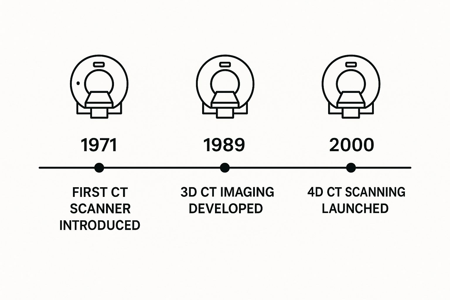
How CT Scans Evolved From Still Images To Dynamic Video
The journey from grainy stills to live-motion imaging is surprisingly recent. Back then, the idea was simple: look inside the body without making an incision.
Picture each CT slice as a page in a flipbook. When flipped at speed, those static frames turn into fluid movement—just like early CT machines dreamed of doing.
In the 1970s, raw CT data felt like a slow-motion novel. The first human scan in October 1971 took almost nine days to collect, plus hours of computation, and yielded a single low-resolution image. Learn more about those pioneering steps history of computed tomography at NCBI.
The Race For Speed And Clarity
As clinicians saw the promise, engineers raced to speed up scans. Reducing rotation times on the gantry, the ring that holds X-ray sources and detectors, was the first milestone.
- Sub-Second Gantry Rotation: Cut scan times from minutes to seconds, slashing motion blur.
- Multidetector CT (MDCT): Expanded from a single detector to 4, 16, 64 and more—vastly increasing data capture.
- Compute Power Advances: Enabled rapid reconstruction of high-resolution 3D models in near real time.
These breakthroughs set the stage for the final leap: weaving time into the 3D story.
Adding The Fourth Dimension Time
Once 3D images arrived in seconds, the question became “What if we could watch them move?” By syncing scans with a patient’s breathing or heartbeat, CT moved from static snapshots to something far more dynamic.
By capturing multiple 3D frames across a physiological cycle and stitching them into a single sequence, CT scanning gained the time dimension.
Key innovations that turned this vision into reality:
- Rapid Gantry Rotations: Now sub-second, allowing precise tracking of moving anatomy.
- Parallel Detector Arrays: Capture vast volumes on each pass, giving temporal depth.
- High-Performance Reconstruction: Sorts images by physiological phase and renders smooth video loops.
Bringing these elements together transformed CT from a static snapshot into a true four-dimensional window on anatomy and function.
What to Expect During Your 4D CT Scan
Hearing you need any kind of specialized medical scan can be a little intimidating. But when you know what to expect, that uncertainty tends to fade. A 4D CT scan is actually a very straightforward procedure, and understanding how it works can make the whole experience feel much more comfortable.
The entire process is built to do one thing: capture a crystal-clear moving picture of what's happening inside your body. Let's walk through the simple, well-planned steps from start to finish.
Preparing for Your Scan
Your prep work actually starts before you even leave the house. The medical team will give you a list of specific instructions that are crucial for getting the best possible images from your scan.
These instructions often include things like:
- Fasting: You'll likely be asked not to eat or drink for several hours before your appointment. This helps clear out your digestive system, which can prevent shadows and improve the quality of the scan.
- Contrast Agents: For some scans, you might be given a special dye called a contrast agent. This harmless substance helps highlight certain tissues or blood vessels, making them pop on the final image. You might drink it or have it administered through an IV.
- Medication Review: Make sure your care team knows about any medications you’re taking. They’ll tell you if you need to pause anything for the day.
Following these guidelines is the best way to help the radiology team get the accurate results they need.
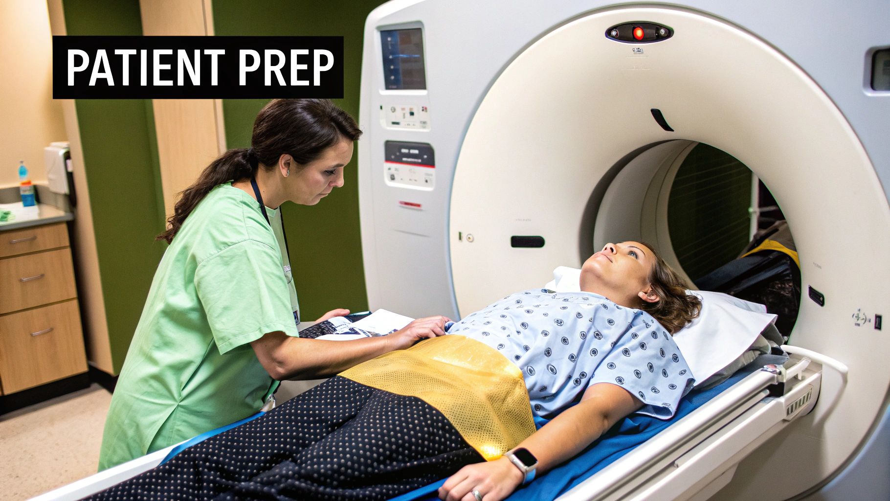
Inside the Scanning Room
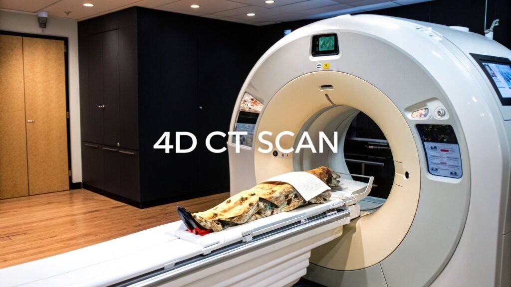
When it's time, a technologist will bring you into the scanning room. You'll see the CT scanner—it looks like a large, modern donut with a comfortable, motorized table that slides right through the middle. You’ll just need to lie down on the table, and the technologist will make sure you're positioned perfectly.
The magic of a 4D CT scan is its ability to capture motion, which almost always means tracking your breathing. To do this, the scanner has to perfectly time its pictures with your body's natural rhythm.
This timing is often handled by a small, simple device. For example, a system like Varian's RPM (Real-Time Position Management) uses a camera to watch a tiny reflective box placed on your chest or abdomen. As you breathe, the box rises and falls, telling the scanner exactly where you are in your breathing cycle.
Some newer systems can even track your internal anatomy, like your diaphragm, without needing any external markers at all. This "deviceless" method makes the setup even simpler. Once you're set, all you have to do is lie still and breathe normally. You'll hear the machine whir as it works, capturing hundreds of images from different angles.
From Raw Data to a Dynamic Video
The final step is where the real magic happens, and it all takes place on a computer after your scan is done. The system takes all the individual 3D image "slices" it just collected.
Powerful software then gets to work, intelligently sorting every single image based on the breathing data it tracked. It organizes them into a perfect time-lapse sequence. Think of it like taking hundreds of still photos of a flower blooming and then arranging them in the exact order they were taken.
When played back, these images create a seamless, looping video—the 4D rendering of your organ in motion. This allows your doctor to see precisely how a tumor shifts when you breathe or how your heart is pumping, leading to a much more accurate diagnosis and a truly personalized treatment plan.
Here’s how 4D CT scanning is making a real difference in patient care, moving from a fascinating concept to a clinical workhorse.
The true test of any medical technology isn't how clever it is, but how it impacts real patients. Adding the dimension of time to a CT scan is a brilliant idea, but it's in the day-to-day work of hospitals and cancer centers where 4D CT truly proves its worth. It gives doctors a dynamic roadmap of the human body, one that finally accounts for the one thing that's always constant: motion.
This ability to see anatomy move over time has opened the door to a new level of precision in a few key areas. It's fundamentally changing how we diagnose and treat some of our most complex diseases.
Pinpoint Accuracy in Cancer Treatment
Nowhere is the impact of 4D CT felt more than in radiation oncology. For years, treating tumors in organs that move—think lungs, liver, or anything affected by breathing—was a huge challenge. A tumor isn't a static bullseye; it shifts with every single breath.
A standard 3D CT scan gives you just one snapshot in time. To compensate for the tumor's movement, oncologists had to irradiate a larger area around it, creating a "margin" just to be safe. This was the only way to ensure the moving target got the full dose, but it also meant healthy tissue was inevitably caught in the crossfire, increasing the risk of side effects.
A 4D CT scan completely flips that script.
By capturing the tumor's movement through an entire breathing cycle, the scan creates a short video loop. It shows the oncologist the exact path the tumor travels.
With this information, they can design radiation treatments that actually follow the tumor. This technique, called respiratory gating, syncs the radiation beam to the patient's breathing, turning it on only when the tumor is perfectly aligned and switching it off as it moves away. This spares a tremendous amount of surrounding healthy tissue.
The clinical need for this was obvious once the technology became available. As 4D CT scanning was adopted in the late 1990s and early 2000s, it took off in oncology because it directly solved this problem. Studies showed that breathing can move a tumor by several centimeters—an enormous distance in medical terms—which explained why those old, larger treatment margins were so necessary. You can learn more about the clinical evolution of 4D imaging from research published at NCBI.
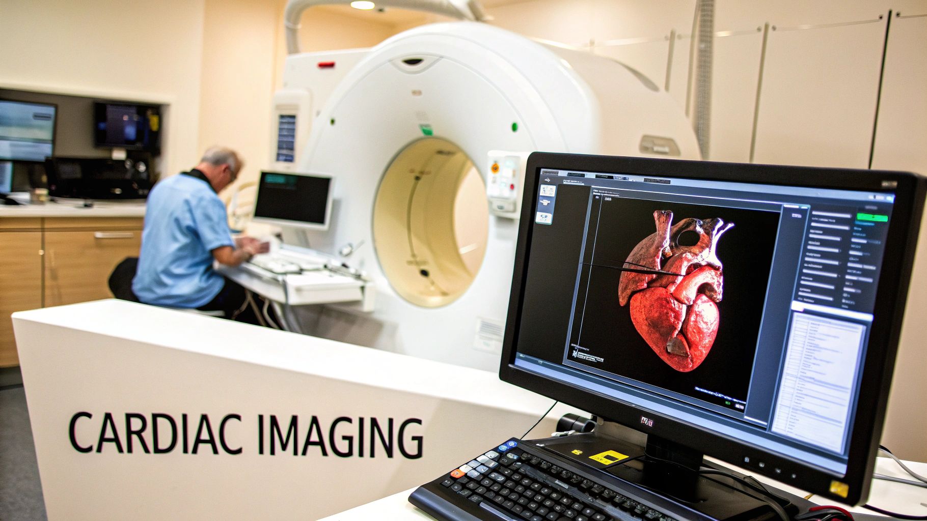
A Clearer View of the Heart
Beyond cancer treatment, 4D CT offers incredible insights for cardiology. The heart is the ultimate moving target, in constant motion as it pumps blood. A static image can show you the heart's anatomy, but it can't tell you how well that anatomy is working.
With 4D CT, cardiologists can actually see the heart in action. This allows them to:
- Assess Wall Motion: Watch the heart muscle contract and relax in real-time to spot areas weakened by a past heart attack or disease.
- Evaluate Valve Function: See if the heart valves are opening and closing correctly or if there are signs of leakage or stenosis (narrowing).
- Measure Cardiac Output: Calculate precisely how much blood the heart pumps with each beat—a vital indicator of overall heart health.
This dynamic view gives a far more complete diagnostic picture than a 3D model ever could. It leads to better, more confident decisions about everything from medication to major surgical procedures.
Guiding Complex Medical Procedures
The technology also acts as a real-time GPS for surgeons and interventional radiologists during delicate procedures. When you're trying to guide a needle or catheter to a tiny target inside the body, you have to account for organ movement.
Think about performing a biopsy on a small lesion in the liver. Every time the patient breathes, your target moves. A 4D CT scan gives the doctor a live map of that movement, letting them anticipate where the organ will be. This helps them guide their instruments with far greater confidence and safety, which means shorter procedures and fewer risks for the patient.
To put it all in perspective, here are some of the most common ways this technology is being put to work.
Primary Applications of 4D CT Scanning
This table highlights the key medical fields where 4D CT scanning provides specific diagnostic or therapeutic advantages.
| Medical Field | Specific Use Case | Benefit of 4D Imaging |
|---|---|---|
| Radiation Oncology | Treating tumors in the lung, liver, and abdomen | Allows for "respiratory gating" to target the tumor precisely as it moves, sparing healthy tissue from radiation. |
| Cardiology | Assessing heart muscle and valve function | Provides a dynamic view of the beating heart to diagnose conditions like wall motion abnormalities or valve leakage. |
| Interventional Radiology | Guiding biopsies and ablations in moving organs | Acts as a real-time navigation tool, increasing the accuracy and safety of minimally invasive procedures. |
| Pulmonology | Evaluating diaphragm and lung function | Shows the full range of lung motion, helping diagnose issues related to breathing mechanics that a static scan would miss. |
Ultimately, the ability to see and predict movement is a fundamental advantage that is reshaping modern medicine.
The Pros and Cons of 4D CT Technology
Like any powerful medical tool, 4D CT scanning has its own distinct advantages and a few practical hurdles. To really grasp its place in modern medicine, you have to look at both sides of the coin. On one hand, its ability to show us the body in motion has been a game-changer for precision treatments. On the other, that incredible capability comes with some technical and clinical trade-offs.
Looking at the full picture helps us see where this technology truly excels and where there's still work to be done. The biggest wins are in improved accuracy and a better understanding of how organs actually function. The main challenges? They usually come down to radiation exposure and the sheer complexity of the whole process.
The Power of Seeing Things in Motion
The single greatest advantage of a 4D CT scan is right there in the name: it adds the dimension of time. It lets clinicians see anatomy as it moves, giving them a level of insight that a static 3D image could never provide. This leads directly to safer and more effective treatments.
- Pinpoint Tumor Targeting: Think about tumors in the lungs or abdomen—they move every time a person breathes. A 4D scan actually maps out this entire range of motion. For radiation oncologists, this means they can aim their treatments with incredible precision, hitting the tumor while protecting the healthy tissue around it.
- A Live Look at Organ Function: Instead of just a snapshot, doctors can watch the heart beat or see the lungs expand and contract in real-time. This kind of functional data is priceless for diagnosing conditions that are less about an organ's structure and more about its performance.
- Safer Guided Procedures: When a surgeon is guiding a biopsy needle or a catheter, seeing organ movement in real-time is a massive advantage. It makes minimally invasive procedures far more accurate and significantly lowers the risk to the patient.
By capturing an organ's full range of motion, a 4D CT scan provides a dynamic roadmap for treatment. This is a fundamental improvement over static imaging, which only offers a single, frozen moment in time.
Facing the Practical Challenges
For all its strengths, 4D CT isn't a perfect solution for every situation. There are real limitations that both doctors and patients need to consider when choosing an imaging strategy. The main concerns typically revolve around radiation dose, image quality, and cost.
It's worth remembering that the very foundation of 4D CT was built on hardware evolution. The fourth generation of CT scanners, which introduced a stationary ring of detectors and a rotating X-ray tube, was a major leap. This design cut down on image distortions and slashed scan times from minutes to seconds, finally making it possible to capture moving organs. This innovation, detailed in the history of CT innovations at Siemens Healthineers, paved the way for the 4D CT we use today.
But this ability to gather so much data so quickly is also linked to some of the technology's current limitations.
Analyzing the Trade-Offs
Deciding to use a 4D CT scan always involves a careful balancing act. The medical community understands these trade-offs well and manages them meticulously to protect patients while getting the best possible diagnostic information.
- Increased Radiation Dose: This one is unavoidable. Capturing a series of 3D images over time simply requires more radiation than a single, static scan. Modern scanners have all sorts of dose-reduction features, but the total exposure is still higher, a fact that clinicians weigh carefully.
- Potential for Image Artifacts: The final 4D "video" is reconstructed based on the patient's breathing cycle during the scan. If a patient breathes irregularly, it can throw off the reconstruction and create distortions, or "artifacts," in the final image that might compromise its quality.
- Cost and Complexity: Let's be frank: 4D CT scanners are sophisticated, expensive machines. They demand specialized software and highly trained technologists to run them, which drives up the cost and often means they're only available at larger hospitals or specialized centers.
In the end, the goal is always to use the incredible diagnostic power of the 4D CT scan in a way that minimizes its challenges, ensuring it’s reserved for situations where the benefits truly outweigh any risks.
The Future Of Imaging AI And Smarter Scans
The evolution of the 4D CT scan feels more like a marathon than a sprint. Right now, artificial intelligence is stepping in to boost image clarity, refine motion tracking, and lower patient exposure.
Take, for example, a software model that processes gigabytes of scan data in seconds. It can outline a tumor’s full motion trail—saving clinicians hours and cutting out variability at the planning stage.
AI Driven Enhancements
When you look at where AI is headed in this workflow, three areas stand out:
- Automated Data Analysis: Advanced algorithms review dozens of breathing cycles, marking organ movement with superhuman precision and easing the burden on radiologists.
- Predictive Motion Modeling: Instead of reacting to every inhale and exhale, the next wave of AI will forecast a patient’s breathing pattern in real time, keeping treatment beams locked on target.
- Intelligent Dose Reduction: New AI methods reconstruct clear images from lower-dose scans, trimming radiation while preserving every diagnostic detail.
By intelligently reconstructing images from less data, AI could make the benefits of a 4D CT scan accessible with a radiation profile closer to that of a 3D scan. This would represent a monumental step forward in medical imaging safety.
Building these systems depends on solid backend frameworks. Recent progress in development platforms—from open-source libraries to no-code backend AI tools—is smoothing out the road from prototype to patient-ready software.
Beyond AI Other Technological Horizons
Hardware improvements are moving in step with smarter software. Next-generation detectors promise to push frame rates higher, giving motion videos an almost cinematic fluidity.
We’re also on the brink of combining modalities. Imagine a single device that pairs a 4D CT scan with PET imaging, mapping both structure and metabolic activity in one seamless view.
The result? A multidimensional perspective that could unlock highly targeted therapies and paint a clearer picture of disease than ever before.
Answering Your Questions About 4D CT Scans
Even with a good grasp of the technology, it's completely normal to have some questions pop up before you go in for a 4D CT scan. Let's walk through a few of the most common ones to help you feel more prepared for the experience.
Is a 4D CT Scan Safe?
This is often the first question on everyone's mind: what about the radiation? It's true that a 4D CT scan involves X-rays, and because it's essentially a series of scans strung together, the total radiation exposure is higher than a single 3D CT.
However, every radiologist and technician lives by a core principle called ALARA, which means "As Low As Reasonably Achievable." They are experts at dialing in the absolute minimum radiation dose needed to get a clear, diagnostically useful image. Your medical team always carefully considers the tiny risk from radiation against the massive benefit of getting a precise, dynamic picture of the treatment area. That moving image is often essential for planning the most effective therapy.
How Is This Different From a 4D Ultrasound?
It's easy to get these two mixed up since they both produce moving "4D" images. The real difference is how they create those images and what they're best at seeing.
- 4D CT Scan: Think of this as the expert for seeing dense structures. It uses X-rays to build incredibly detailed images of bones, organs, and especially tumors in motion. This makes it the gold standard for things like planning radiation therapy.
- 4D Ultrasound: This technology uses sound waves—no radiation at all. You probably know it from the amazing moving images of babies during pregnancy. It's fantastic for looking at soft tissues and tracking blood flow.
Basically, your doctor picks the right tool for the job. If they need to precisely map a tumor's movement for treatment, the 4D CT scan is the way to go. If they need to see soft tissues without radiation, ultrasound is the winner.
How Long Will the Appointment Take?
While the actual scan time can be surprisingly quick—often just a few minutes—you should probably block out about an hour or so for the whole visit.
This extra time covers all the important steps: getting checked in, changing into a hospital gown, maybe having an IV line placed if you need a contrast dye, and getting positioned perfectly on the scanner bed. The team takes their time to make sure you're comfortable and everything is set up just right before they even start the scan.
Will I Have to Hold My Breath?
For the vast majority of 4D CT scans, the answer is no! In fact, the whole point of the technology is to see how your body moves while you breathe naturally.
The most important instruction you'll get is to just lie still and breathe normally and steadily. This lets the scanner create a perfect map of how your organs and the target area shift during a regular breathing cycle, which is exactly the information your doctors need.
At PYCAD, we're passionate about advancing what's possible in medical imaging with the help of AI. Our tools are designed to sharpen diagnostic accuracy and simplify workflows, giving healthcare providers the powerful insights they need. Discover how we're contributing to the future of medical technology by visiting us at https://pycad.co.
Keep Reading
Related Articles
Explore the full Quantitative Imaging HubAll services, tools, and guides on this topic — in one place.
Visit Hub →