
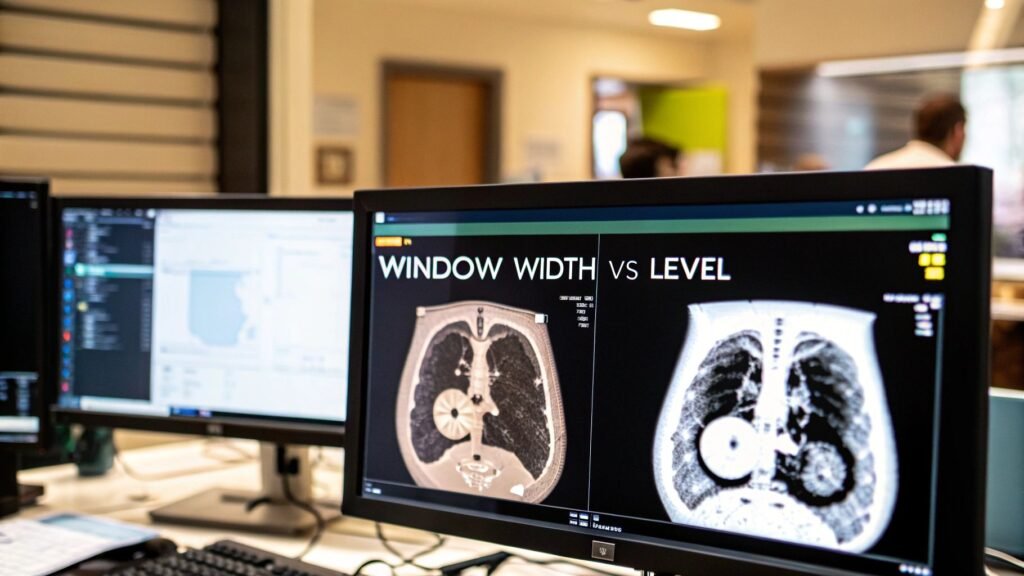
Window width vs window level is the clinical pair on a CT: width is contrast (how many HU you spread across the greys), level is brightness (which HU sits at the midpoint). Same two sliders 330 calls center and width. This page compares them. It does not apply them in Python.
If you meant apply WC/WW to a DICOM series in Python → CT scan window settings. If you meant what HU is → what Hounsfield units are. MRI intensity is a different physics; CT presets do not transfer.
The two sliders
| Parameter | Job | What you see | When you reach for it |
|---|---|---|---|
| Window width | Range of HU on the screen | Contrast | Pull apart tissues that sit close on the scale (grey vs white matter) |
| Window level | Center of that range | Brightness | Park the midpoint on the tissue you are reading (bone, lung, brain) |
Width without the right level is high contrast on the wrong organ. Level without a sane width is a washed-out or clipped picture of the right organ. You set both for one question, not “to make the image look good.”
Why a monitor needs a window
Every CT pixel is an HU value. Water is 0, air is −1000, fat sits around −100, cortical bone around +1000. That scale is Hounsfield units, not this page. A typical display shows about 256 greys. Map two thousand densities onto 256 shades and liver vs a small lesion in it become the same grey.
Windowing is the lens: pick a band of HU, stretch it across the greys, clip everything outside to black or white.
- Level = the HU at the middle of the band. A bleed you expect near +60 HU wants the level near that number.
- Width = how wide the band is. Narrow = high contrast on similar densities. Wide = a survey of air, soft tissue, and bone at once.
Pitfalls
- Width too wide. A “general overview” crams too many HU into 256 greys. Similar tissues merge. Soft-tissue questions die here.
- Level on the wrong tissue. Hunting a lung nodule near −600 HU with a bone level near +400 HU makes the nodule black. The level has to sit on the target.
Ask what anatomy you need to see, then set both. Named presets (the three below, plus liver/soft-tissue pairs) live as a Python apply-page at CT scan window settings.
Clinical windows
| Goal | Width | Level | Example |
|---|---|---|---|
| Subtle soft-tissue difference | Narrow (~80 HU) | On the tissue (~+40 HU brain) | Grey vs white matter; early infarct |
| Survey of mixed densities | Wide (~2000 HU) | Mid-range (~+200 HU) | Chest: air, soft tissue, bone in one look |
| Isolate dense structure | Moderate–wide (~1500–2000 HU) | High (~+300 to +400 HU) | Hairline fracture |
| Lung parenchyma | Wide (~1500 HU) | Low (~−600 HU) | Nodule, PE filling defect, emphysema |
Lung window — PE
Width about 1500 HU, level about −600 HU. The low level parks the greys on air-filled lung. The wide width still holds vessels and a denser clot as a filling defect. A soft-tissue window hides that anatomy.
Brain window — stroke
Width about 80 HU, level about +40 HU. The narrow width exaggerates the small density gap between grey and white matter, which is where early swelling shows. A wide window turns that into one grey.
Bone window — hairline fracture
Width about 2000 HU, level about +400 HU. High level on cortex; wide width so marrow vs cortex still has steps. The same crack is invisible on a soft-tissue window.
Short answers
Width too narrow? Contrast goes extreme. Similar tissues separate, everything outside the band clips to black or white, and you lose context. Use it on purpose (brain), not as a default.
CT presets on MRI? No. CT windowing is tied to HU. MRI intensity is T1/T2 and sequence-dependent. There is no portable “brain window” across modalities.
How to pick the level? Put it on the HU of the tissue you are reading: brain near +40, bone +300 to +500, lung near −600. Width then decides how much neighborhood you keep.
PYCAD builds viewers and pipelines where these two sliders have to stay a clinical control, not a decoration. Case studies.