Why Cardiac Segments Matter in Modern Cardiology

Cardiac segments provide a vital framework for understanding and treating heart conditions. This anatomical framework is essential in modern cardiology, having evolved from a basic concept to a sophisticated system used daily by healthcare professionals. It forms the basis for accurate diagnoses and effective treatment plans.
The standardized language of cardiac segments allows for clear communication between healthcare providers in various settings. This shared understanding connects imaging specialists, interventional cardiologists, and other members of the cardiac care team. For instance, describing abnormalities within specific segments ensures everyone understands the issue, regardless of their specialization. This precise communication is critical for coordinated and efficient patient care.
Furthermore, this standardized approach is directly related to improved patient outcomes. By identifying the precise location of damage or dysfunction, healthcare providers can make better-informed treatment decisions. This targeted approach results in more effective interventions and improved long-term prognoses.
The development of cardiac segments has been closely linked to advancements in imaging technologies. By the late 20th century, echocardiography and SPECT nuclear cardiology had paved the way for refined segmentation models of the left ventricle. The 17-segment model became the standard, dividing the heart into basal, mid-cavity, and apical sections, with a separate segment for the apical cap. This model's simplicity and reliability in diagnosing regional dysfunction solidified its place in clinical practice. Learn more about the evolution of cardiac segmentation here. This standardized approach is vital for accurate diagnoses and treatment planning across a wide spectrum of cardiovascular problems.
Importance of Standardized Segmentation
The standardization provided by cardiac segmentation is critical for several key reasons:
- Accurate Diagnosis: Identifying the affected segments allows for precise diagnosis of the problem.
- Targeted Treatment: This precision informs treatment choices, including medication, interventional procedures, or surgery.
- Consistent Communication: All members of the cardiac care team use the same terminology, reducing errors and improving collaboration.
- Research and Development: Cardiac segments provide a consistent framework for cardiovascular research, enabling comparisons of data across different studies.
This framework allows clinicians to pinpoint the specific area of concern within the heart, similar to using coordinates on a map. This level of precision is fundamental to effective diagnosis and treatment in modern cardiology.
Mastering the 17-Segment Model of Cardiac Analysis
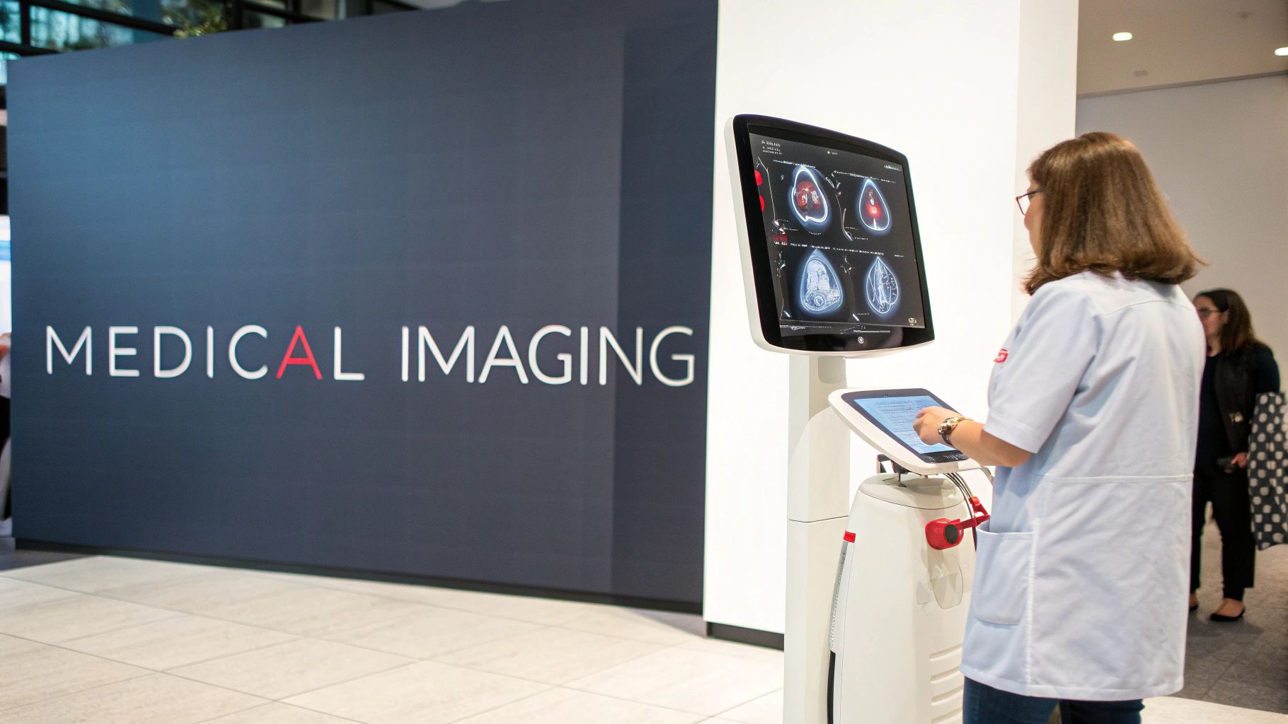
This section explores the 17-segment model, a widely used standard in cardiology. This model provides a structured framework for evaluating the heart, essential for accurate diagnoses and effective treatment plans. Understanding each segment's location is crucial for medical professionals.
This precision allows them to identify specific areas of concern, leading to more targeted and effective interventions.
Understanding the Segment Locations
The 17-segment model divides the left ventricle into three distinct levels: basal, mid-cavity, and apical. Imagine slicing a cylinder into three equal parts. Each of these levels is then further subdivided into segments, numbered clockwise as viewed from the apex of the heart. The basal and mid-cavity levels each contain six segments, while the apical level has five. The apical cap, representing the very tip of the left ventricle, completes the model.
This methodical approach ensures clear communication among healthcare providers. For example, if an echocardiogram reveals an issue in segment 12, every member of the cardiac team immediately understands the exact location being referenced. This standardized system minimizes ambiguity and fosters efficient collaboration.
Linking Segments to Coronary Arteries
Each segment in the 17-segment model corresponds to a specific coronary artery territory. This means each segment receives blood supply from a particular branch of the coronary arteries. Understanding this link is vital for diagnosing and managing conditions like myocardial infarctions (heart attacks).
If a segment shows signs of damage, it can pinpoint the likely blocked coronary artery. This informs treatment strategies, such as where to place a stent to restore blood flow.
To further illustrate the relationship between segments and their corresponding arterial supply, let's take a closer look at the table below.
The 17 Cardiac Segments and Their Locations
This table presents all 17 cardiac segments organized by anatomical level, with their standard nomenclature and corresponding coronary artery supply.
| Segment Number | Segment Name | Anatomical Level | Coronary Artery Territory |
|---|---|---|---|
| 1 | Basal Anterior | Basal | Left Anterior Descending (LAD) |
| 2 | Basal Anteroseptal | Basal | LAD |
| 3 | Basal Inferoseptal | Basal | Right Coronary Artery (RCA) |
| 4 | Basal Inferior | Basal | RCA |
| 5 | Basal Inferolateral | Basal | RCA/Left Circumflex (LCx) |
| 6 | Basal Anterolateral | Basal | LCx |
| 7 | Mid Anterior | Mid-cavity | LAD |
| 8 | Mid Anteroseptal | Mid-cavity | LAD |
| 9 | Mid Inferoseptal | Mid-cavity | RCA |
| 10 | Mid Inferior | Mid-cavity | RCA |
| 11 | Mid Inferolateral | Mid-cavity | RCA/LCx |
| 12 | Mid Anterolateral | Mid-cavity | LCx |
| 13 | Apical Anterior | Apical | LAD |
| 14 | Apical Septal | Apical | LAD |
| 15 | Apical Inferior | Apical | RCA |
| 16 | Apical Lateral | Apical | LCx |
| 17 | Apical Cap | Apical | LAD/RCA/LCx |
As you can see, the table clearly outlines which coronary artery supplies each individual segment. This detailed mapping is fundamental for accurate interpretation of cardiac imaging.
Practical Applications of the 17-Segment Model
The 17-segment model is a powerful tool with practical applications in daily cardiology. It’s essential for interpreting various cardiac imaging modalities, including echocardiography, cardiac MRI, and nuclear imaging studies. By analyzing individual segment function, cardiologists can accurately assess overall heart performance and identify regional abnormalities.
This precise information is critical for making informed treatment decisions. For instance, a patient with decreased function in multiple segments may require a different approach than a patient with a localized area of dysfunction. The 17-segment model allows for a more detailed understanding of cardiac function, going beyond a simple assessment of the heart as a whole. This granularity is vital in modern cardiac care, facilitating precise communication and contributing to improved patient outcomes.
Visualizing Cardiac Segments: Imaging Technologies Compared

This section compares different imaging technologies for visualizing and analyzing cardiac segments. Each modality offers unique strengths and limitations, contributing to a comprehensive understanding of cardiac function. This understanding is essential for accurate diagnoses and effective treatment plans.
Echocardiography: Accessibility and Real-Time Imaging
Echocardiography uses ultrasound to create real-time images of the heart. This widely available and cost-effective modality is particularly useful for assessing wall motion abnormalities and valvular function, making it a first-line tool for evaluating cardiac segments.
However, image quality can be limited, especially for patients with obesity or lung disease. Furthermore, the operator's skill significantly impacts the quality of the echocardiographic images.
This underscores the importance of experienced sonographers and cardiologists for accurate interpretations. Despite these limitations, echocardiography remains a cornerstone of cardiac assessment due to its accessibility and real-time capabilities.
Cardiac MRI: Precision and Tissue Characterization
Cardiac MRI offers superior image quality and tissue characterization compared to echocardiography. It provides detailed anatomical information and can identify subtle abnormalities within cardiac segments.
This precision makes cardiac MRI invaluable for diagnosing conditions like myocardial infarction and cardiomyopathies. Additionally, cardiac MRI can quantify myocardial strain, offering insights into early signs of dysfunction.
However, cardiac MRI is more expensive and less readily available than echocardiography. The procedure can also be challenging for patients with pacemakers or claustrophobia.
Nuclear Imaging Techniques: Functional Assessment
Nuclear imaging techniques, including SPECT and PET scans, provide functional information about cardiac segments. These techniques assess myocardial perfusion (blood flow) and metabolism, revealing areas of ischemia or damage.
This information is crucial for understanding the extent of coronary artery disease and evaluating heart tissue viability. However, nuclear imaging involves exposure to radiation, although the dose is generally considered safe.
These techniques typically have lower spatial resolution than cardiac MRI, making it harder to pinpoint the exact location of abnormalities within smaller segments.
Multi-Modality Imaging: A Comprehensive Approach
Leading cardiac centers increasingly use multi-modality imaging, combining the strengths of different technologies for a more complete assessment of cardiac segments. For example, echocardiography might be used for initial evaluation, followed by cardiac MRI for detailed tissue characterization or nuclear imaging for functional assessment.
This integrated approach provides a more comprehensive understanding of cardiac structure and function. This, in turn, leads to better clinical decision-making and improved patient outcomes. Physicians can capitalize on each modality's individual strengths.
Comparing Imaging Modalities
| Feature | Echocardiography | Cardiac MRI | Nuclear Imaging |
|---|---|---|---|
| Cost | Lower | Higher | Moderate |
| Availability | Widely available | Less available | Moderately available |
| Image Quality | Moderate | Excellent | Moderate |
| Tissue Characterization | Limited | Excellent | Limited |
| Functional Assessment | Moderate | Moderate | Excellent |
| Real-Time Imaging | Yes | No | No |
| Radiation Exposure | None | None | Yes |
By understanding the advantages and limitations of each imaging modality, clinicians can select the most appropriate technique for each patient’s specific needs. This personalized approach optimizes diagnostic accuracy and contributes to better treatment outcomes.
Cardiac Segments in Action: Real Clinical Applications
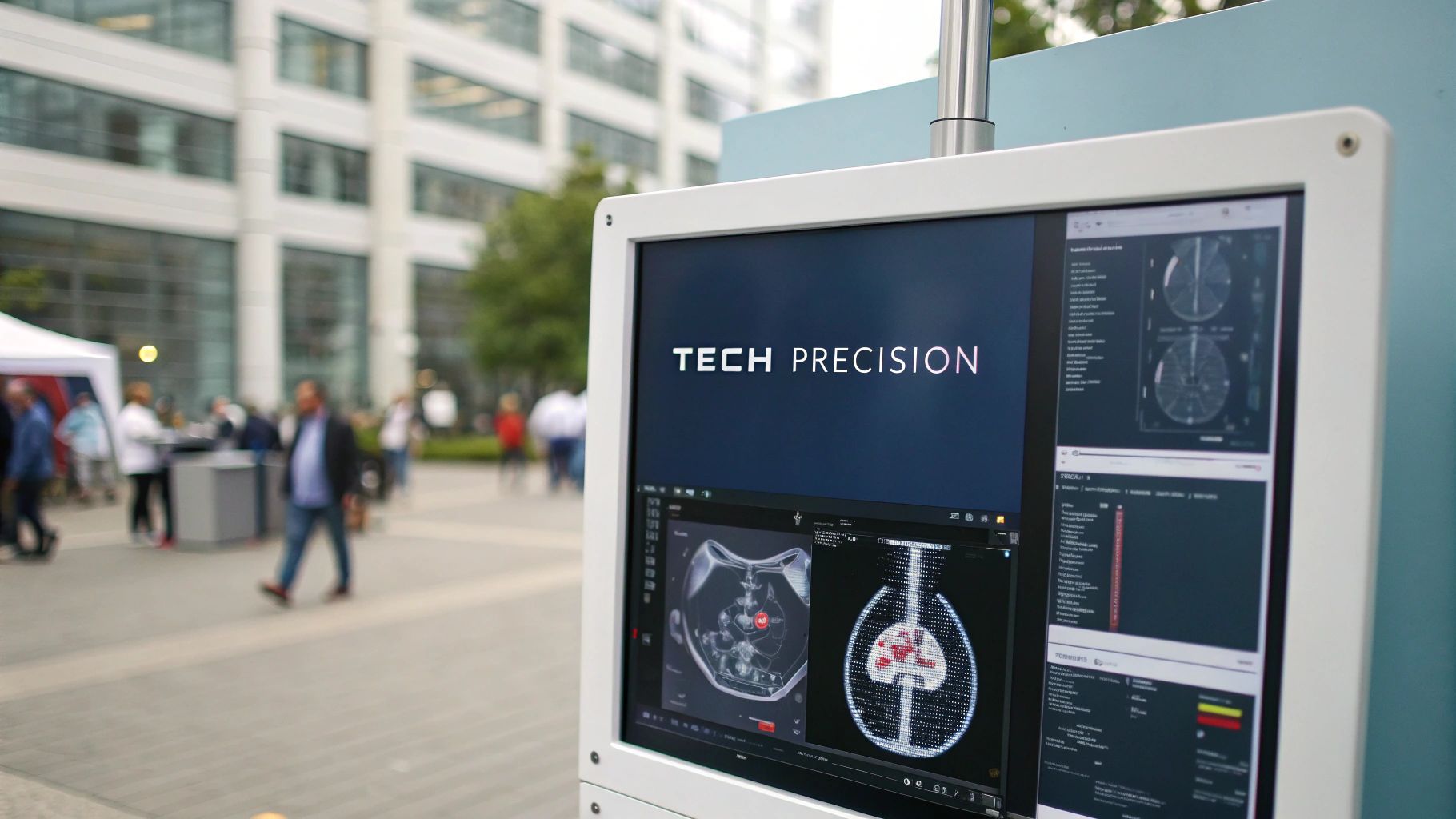
This section explores how analyzing cardiac segments translates into real-world patient care. We'll examine how pinpointing abnormalities within these specific heart segments informs crucial treatment decisions and ultimately, improves patient outcomes across a range of cardiac conditions.
Guiding Treatment Decisions in Myocardial Infarction
In a myocardial infarction (heart attack), quickly identifying the affected cardiac segments is paramount. By pinpointing the segments exhibiting reduced or absent function, cardiologists can identify the blocked coronary artery causing the infarction.
This precise localization is essential for rapid, targeted intervention. It guides decisions during an angioplasty, indicating which artery needs immediate attention. This focused approach leads to more effective treatment and minimizes long-term heart damage.
Managing Complex Cardiomyopathies with Segment Analysis
Cardiomyopathies, diseases affecting the heart muscle, often present complex dysfunction patterns. Cardiac segment analysis helps decipher these complexities by identifying the specific areas of the heart muscle most affected.
This targeted information guides treatment decisions, informing medication choices and other therapies. It can also influence decisions about device implantation, such as pacemakers or implantable cardioverter-defibrillators (ICDs). Segment-based analysis offers a deeper understanding of the disease, allowing for more tailored treatment strategies.
Surgical Planning with Precise Segmental Information
Cardiac segments play a crucial role in surgical planning, especially for complex procedures. Understanding which segments are compromised allows surgeons to preoperatively map the optimal surgical approach and anticipate potential challenges.
For example, in coronary artery bypass surgery, identifying dysfunctional segments helps surgeons decide which arteries require grafting. In valve repair or replacement surgery, segment analysis informs the procedure type and specific techniques used. Advancements in cardiac surgery, particularly in pediatric heart disease, have greatly benefited from cardiac segmentation. Procedures like the arterial switch operation and the Norwood procedure for hypoplastic left heart syndrome rely heavily on the precise anatomical knowledge segmentation provides. Learn more about these procedures here.
Risk Stratification and Long-Term Management
Analyzing cardiac segments helps clinicians stratify patients based on their risk of future cardiac events. By identifying the extent and location of heart damage, doctors can better predict a patient's prognosis and create a personalized long-term management plan.
This individualized approach may involve closer monitoring, specific medication regimens, or lifestyle modifications. Ultimately, it empowers both patient and physician to make informed, proactive decisions, leading to improved long-term outcomes and a better quality of life.
Leading Cardiologists' Frameworks for Action
Leading cardiologists increasingly utilize established frameworks that convert segment-based findings into actionable clinical strategies. These frameworks provide a structured approach to interpreting data from cardiac segment analysis, ensuring consistent, evidence-based decisions.
These frameworks consider not only the location and extent of segmental dysfunction but also factors like patient age, comorbidities, and overall health. This holistic approach enables the development of personalized treatment plans that address individual patient needs and optimize long-term health outcomes.
Breaking Boundaries: Cardiac Segments in Research
Cardiac segments, crucial for clinical practice, are also driving innovative research in cardiovascular medicine. This established framework is accelerating discoveries in disease mechanisms, treatment strategies, and improving diagnostic accuracy. Researchers are actively exploring new technologies and approaches based on cardiac segments, striving to enhance outcomes for patients facing complex heart conditions.
AI-Powered Segment Analysis: Enhancing Diagnostic Precision
A promising research area involves leveraging Artificial Intelligence (AI) to analyze cardiac segments. AI algorithms, such as those found in TensorFlow, can detect subtle anomalies within these segments that might be overlooked by human observation. This increased precision enhances diagnostic accuracy, proving particularly valuable for the early detection of cardiovascular diseases. Moreover, AI can automate the segment analysis process, saving valuable time and lessening the workload for clinicians. This translates to quicker diagnoses and a faster initiation of necessary treatments.
Advanced Strain Imaging: Revealing Early Dysfunction
Researchers are also investigating advanced strain imaging techniques. These techniques provide in-depth information about the deformation of cardiac segments throughout the cardiac cycle. This allows for the detection of dysfunction before the onset of noticeable symptoms, even before abnormalities are detectable through traditional methods. This early detection is paramount for preventing disease progression and enhancing long-term outcomes. Advanced strain imaging holds the potential to identify at-risk patients and initiate preventative measures sooner.
Personalized Treatment Protocols: Targeting Specific Segments
Research centers are diligently developing personalized treatment protocols based on cardiac segment analysis. This targeted approach aims to optimize treatment effectiveness for each patient. By focusing on specific segments exhibiting dysfunction, therapies can be precisely administered where they are most needed. For example, in patients with heart failure, segment-specific therapies might concentrate on regenerating damaged tissue within the affected segments. This personalized approach marks significant progress toward more effective treatments for complex cardiac conditions.
To better illustrate current research applications, the table below provides a comparison of different areas utilizing cardiac segmentation and their potential clinical impact.
Research Applications of Cardiac Segments
| Research Area | Segmentation Application | Technological Approach | Potential Clinical Impact |
|---|---|---|---|
| Disease Mechanisms | Identifying patterns of segment involvement in different diseases | AI-powered image analysis, advanced strain imaging | Improved understanding of disease progression and development of new diagnostic criteria |
| Treatment Development | Evaluating treatment efficacy in specific segments | Image-guided therapies, targeted drug delivery | Development of more effective and personalized treatments |
| Diagnostic Precision | Detecting subtle abnormalities and early signs of dysfunction | AI-powered segment analysis, multi-modality imaging | Earlier and more accurate diagnosis of heart conditions |
This table highlights how cardiac segmentation research translates to tangible patient benefits. The diverse technological approaches, from AI-powered analysis to advanced imaging, contribute to a more comprehensive understanding and management of heart conditions.
Researchers continue to push the boundaries of cardiac segment analysis. This ongoing exploration leads to innovations that improve diagnostic capabilities, personalize treatment strategies, and ultimately, enhance the lives of individuals with cardiac conditions.
The Future of Cardiac Segments: What's Coming Next
Cardiac segment analysis is about to undergo a major transformation. Leading cardiologists and researchers are exploring exciting innovations that promise to reshape how we understand and treat heart conditions. These advancements are pushing the boundaries of cardiac care, offering the potential for earlier diagnoses, more personalized treatments, and improved patient outcomes.
4D Imaging and Real-Time Segment Assessment
One exciting development is the emergence of 4D imaging, which adds the dimension of time to traditional 3D imaging. This allows for real-time assessment of cardiac segment movement and function, providing a dynamic view of the heart in action.
This technology offers a deeper understanding of complex heart mechanics. It can also reveal subtle abnormalities that might be missed with static images. This dynamic visualization can lead to more accurate diagnoses and inform more effective treatment strategies.
Machine Learning for Predictive Analysis
Artificial intelligence (AI) is rapidly transforming many fields, including medicine. In cardiology, machine learning algorithms are being developed to predict segment dysfunction before it becomes clinically apparent.
By analyzing large amounts of data from various sources, including imaging studies and patient records, these algorithms can identify patterns and risk factors associated with future segmental problems. This predictive capability has the potential to revolutionize preventative cardiology, allowing for early interventions. These interventions can delay or even prevent the onset of heart disease. Learn how Artificial Intelligence in contact centers is enhancing performance.
Integrating Segment Analysis with Genetic and Molecular Data
The future of cardiac segment analysis lies in integration. Researchers are working to combine segment analysis with genetic and molecular data.
This approach aims to create a more personalized understanding of heart disease, linking specific segment abnormalities with underlying genetic predispositions or molecular markers. This integrated approach could lead to the development of targeted therapies based on a patient's unique genetic and molecular profile, paving the way for truly personalized medicine.
Computational Modeling and Virtual Cardiac Twins
Computational modeling is another exciting area of development. Researchers are creating virtual cardiac twins, personalized computer models of individual hearts based on patient-specific data.
These virtual hearts allow for simulation of different treatment strategies, such as medication adjustments or surgical interventions, without the need for invasive procedures. This allows clinicians to optimize treatment plans and predict patient responses before implementing them in real life, potentially improving outcomes and reducing risks. One example is the research being conducted by Dr. Alison Pouch at the University of Pennsylvania, focusing on the biomechanical assessment and simulated repair of systemic semilunar valves.
Segment-Based Regenerative Approaches
Finally, segment-based regenerative approaches offer hope for revolutionizing heart failure treatment. Researchers are exploring ways to regenerate damaged heart tissue within specific segments using stem cells or other regenerative therapies.
This targeted approach aims to restore function to the affected segments, potentially reversing the progression of heart failure and improving patients' quality of life. While still in its early stages, this research holds immense promise for the future of heart failure management.
These emerging technologies and research directions are transforming cardiac segment analysis. By embracing these advances, healthcare providers can prepare for the next generation of cardiac care and improve outcomes for patients with a wide range of heart conditions. These innovations represent a significant step forward in our ability to understand, diagnose, and treat heart disease.
Interested in leveraging the power of AI in medical imaging? Learn more about PYCAD's innovative solutions for enhanced diagnostics and operational efficiency at PYCAD.
