
The Game-Changing Evolution of CBCT Dentistry
CBCT imaging has fundamentally changed how dental professionals diagnose and plan treatments. This technology represents a major advancement from basic 2D imaging by providing detailed, three-dimensional views of oral structures. Dental practices increasingly adopt CBCT to improve accuracy and outcomes for procedures like implant placement, jaw surgery, and root canal treatments.
Let's explore how CBCT has transformed dental imaging and care delivery through its key developments:
| Year | Technology Milestone | Impact on Dentistry |
|---|---|---|
| 1996 | First dental CBCT system (NewTom 9000) introduced | Enabled 3D imaging with lower radiation than CT |
| 2001 | Introduction of flat panel detectors | Improved image quality and reduced scan time |
| 2005 | Development of compact CBCT units | Made technology accessible to more dental practices |
| 2010 | Integration with treatment planning software | Enhanced precision in implant planning |
| 2015 | Advanced scatter correction algorithms | Further reduced radiation dose while maintaining quality |
| 2020 | AI-assisted image analysis capabilities | Improved diagnostic accuracy and workflow efficiency |
From 2D to 3D: A New Era in Dental Imaging
Traditional 2D x-rays provided limited information about depth and spatial relationships between structures. This made accurate diagnosis challenging and could impact treatment success. CBCT addressed these limitations by generating detailed 3D images that show intricate anatomy of teeth, bone, and soft tissues with exceptional clarity.
The first commercial CBCT system, the NewTom 9000, was introduced in Europe in 1996 by QR of Verona. This marked a key advance in dental imaging by enabling 3D visualization while using significantly less radiation than medical CT scans.
The Impact of CBCT Across Dental Specialties
CBCT technology has benefited multiple dental specialties:
- Implantology: Precise planning of implant position and size based on 3D bone measurements
- Endodontics: Better visualization of complex root anatomy and hidden canals
- Orthodontics: Detailed assessment of facial structures for treatment planning
- Oral Surgery: Enhanced surgical planning through 3D visualization of anatomy
Advantages of CBCT in Modern Dentistry
Key benefits of CBCT technology include:
- Improved Diagnostic Accuracy: 3D scans enable more accurate diagnosis versus 2D x-rays
- Enhanced Treatment Planning: Better visualization leads to more predictable outcomes
- Increased Patient Satisfaction: Clear 3D images improve patient understanding and acceptance
- Reduced Radiation: CBCT uses less radiation compared to traditional CT scanning
As the technology continues to improve, CBCT will likely bring even more advances in image quality and diagnostic capabilities to dental practices.
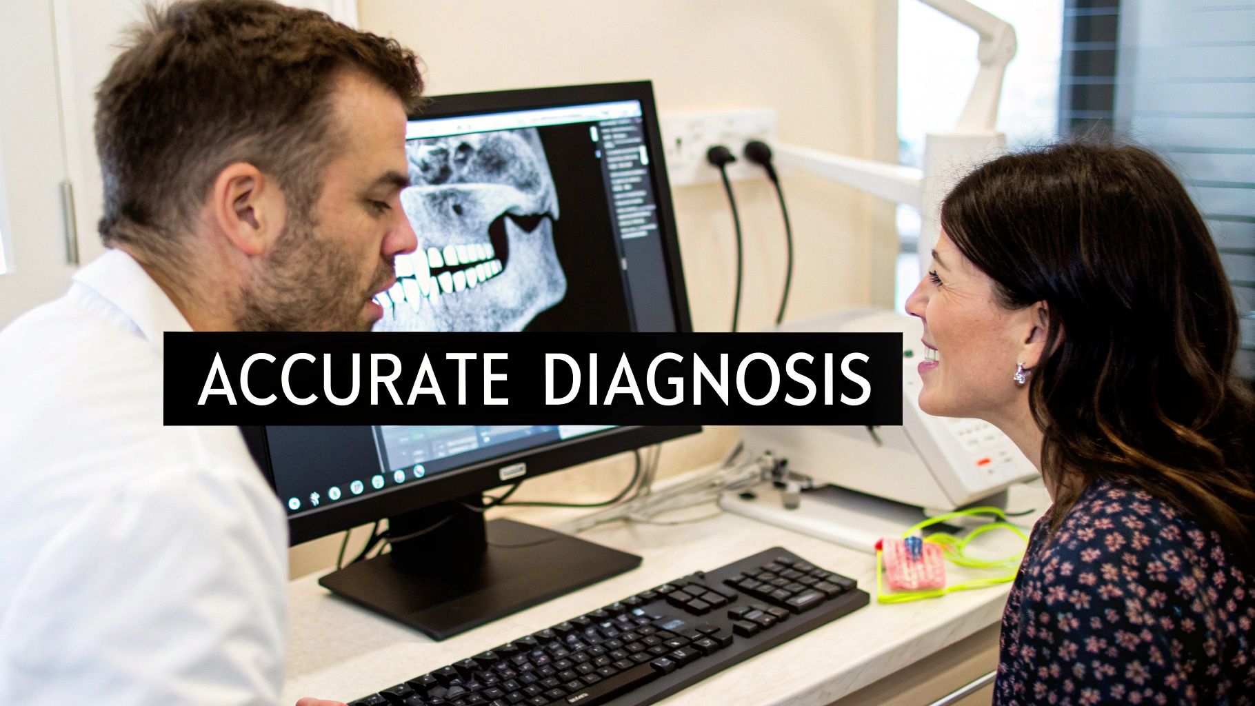
Advancing Clinical Practice with CBCT
CBCT technology has significantly improved dental imaging and patient care across multiple specialties. This advanced imaging system provides detailed insights that help dental professionals make more informed decisions in implantology, endodontics, and orthodontics. In implant procedures, CBCT enables precise evaluation of bone density and volume – key factors for successful implant placement and long-term outcomes.
CBCT in Implantology: Planning for Success
The 3D modeling capabilities of CBCT allow dentists to plan implant placement with exceptional detail. This technology helps assess critical factors like nerve location, bone quality, and optimal implant positioning. By enabling guided surgery techniques, CBCT makes procedures more precise while reducing treatment time. These improvements lead to better results and increased patient satisfaction.
CBCT in Endodontics: Revealing Complex Tooth Anatomy
Root canal procedures require intricate work inside the tooth structure. CBCT provides clear views of complex root canal systems and can detect hidden canals or fractures that standard X-rays might miss. Having this detailed anatomical information helps ensure thorough cleaning of the canal system and reduces chances of reinfection. The end result is higher success rates for endodontic treatments.
CBCT in Orthodontics: Complete Analysis for Custom Treatment
Orthodontic care involves more than teeth alignment – it requires understanding the entire facial structure. CBCT creates comprehensive views of jaw joints, airways, and bone patterns. This helps orthodontists identify skeletal issues and create individualized treatment approaches. The technology also makes it easier to track progress and predict outcomes, giving both doctor and patient more confidence in the treatment plan.
CBCT has become essential in many dental fields, providing vital 3D images for diagnosis and treatment planning. During a single rotation around the patient's head, the system can capture up to 600 distinct images to create detailed volumetric data. Learn more about this technology here. By providing complete diagnostic information, CBCT helps dental professionals make better clinical decisions and deliver improved patient care across multiple specialties.
Mastering Modern CBCT Technology
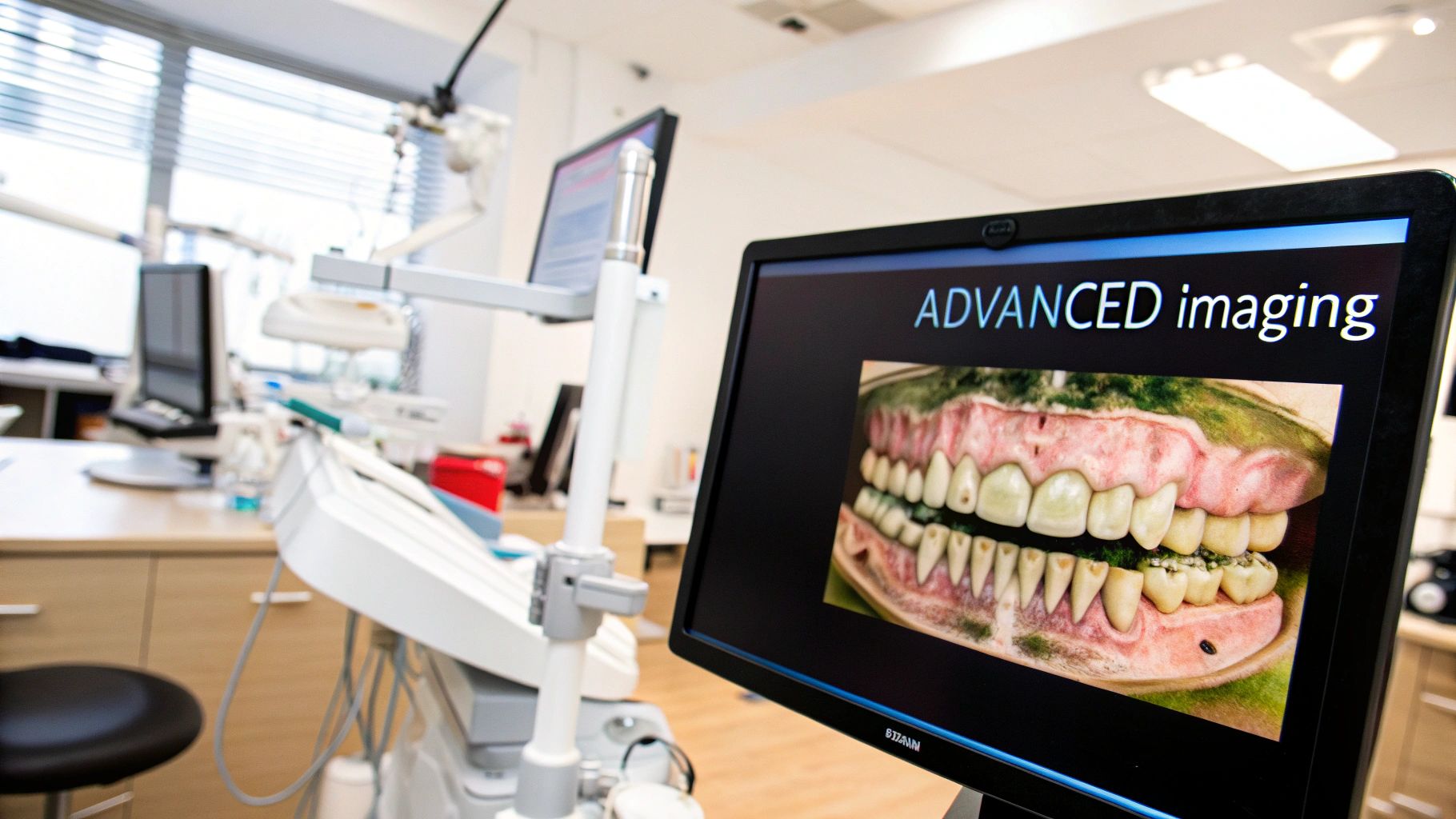
CBCT systems have fundamentally changed dental imaging capabilities. This section covers essential technical aspects of these systems to help dental practices get the most value from this technology. Understanding the core components helps dental professionals achieve better scan quality, optimize protocols, and choose equipment that fits their practice needs. Most importantly, this knowledge directly improves patient care outcomes.
Optimizing Image Quality and Scan Protocols
Getting the best possible image quality depends on several key settings. The field of view (FOV) must match your specific diagnostic needs – use a smaller FOV for single tooth implants and larger FOV for full jaw procedures. This targeted approach maintains diagnostic accuracy while keeping radiation exposure as low as possible.
Image resolution settings also significantly impact quality. While higher resolution provides more detail for procedures like endodontic diagnosis, it increases scan time and file sizes. Finding the right balance is essential. Proper exposure settings are equally important and should be customized based on the patient and type of scan needed.
Selecting and Maintaining CBCT Equipment
When investing in a CBCT system, carefully evaluate the FOV options, resolution capabilities, and software features. If your practice focuses on implants or orthodontics, choose a system with strong capabilities in those areas. Key considerations include 3D modeling, virtual implant planning, and airway analysis tools. The system should also work smoothly with your existing practice software.
Regular maintenance keeps your CBCT system working optimally. Schedule routine calibration and quality checks to ensure consistent image quality. A solid service agreement and reliable technical support help minimize any disruptions when issues arise. Well-maintained equipment is essential for accurate diagnosis and quality patient care.
Advances in CBCT Technology
CBCT imaging keeps improving in both quality and radiation efficiency. Modern systems use flat-panel detectors instead of older image intensifiers, providing better contrast and detail. Learn more about these developments here. Artificial intelligence (AI) integration is also making CBCT analysis more accurate and efficient. These ongoing advances will enable even more precise diagnosis and treatment planning going forward.
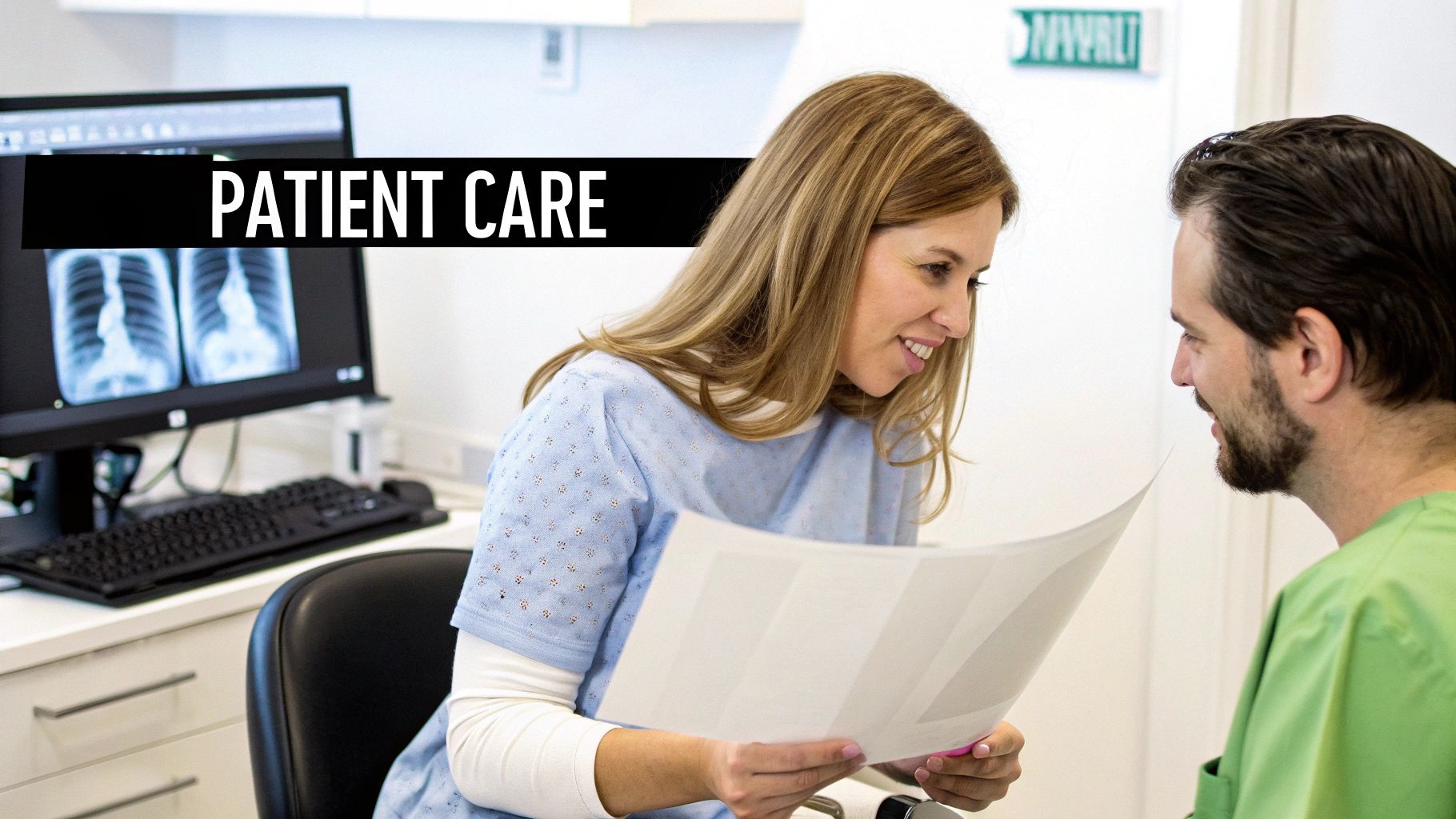
Elevating Patient Experience Through CBCT
CBCT imaging does more than just improve dental diagnoses – it fundamentally changes how patients interact with and understand their dental care. When patients can clearly see and grasp their dental conditions through detailed 3D images, they become more engaged partners in their treatment journey.
Visualizing Treatment: Building Trust and Understanding
The ability to clearly show complex dental issues is one of CBCT's greatest strengths. For example, when patients need a bone graft before an implant, they can see the actual bone deficiency in 3D. This clear visual helps them understand both their condition and why specific treatments are needed.
The dynamic nature of CBCT images allows dentists to zoom and rotate views while explaining findings to patients. This detailed visualization helps reduce anxiety and clears up misconceptions about dental procedures. Patients who fully understand their treatment plan are more likely to follow care instructions and maintain good oral health habits.
Increasing Treatment Acceptance: Showing the Value
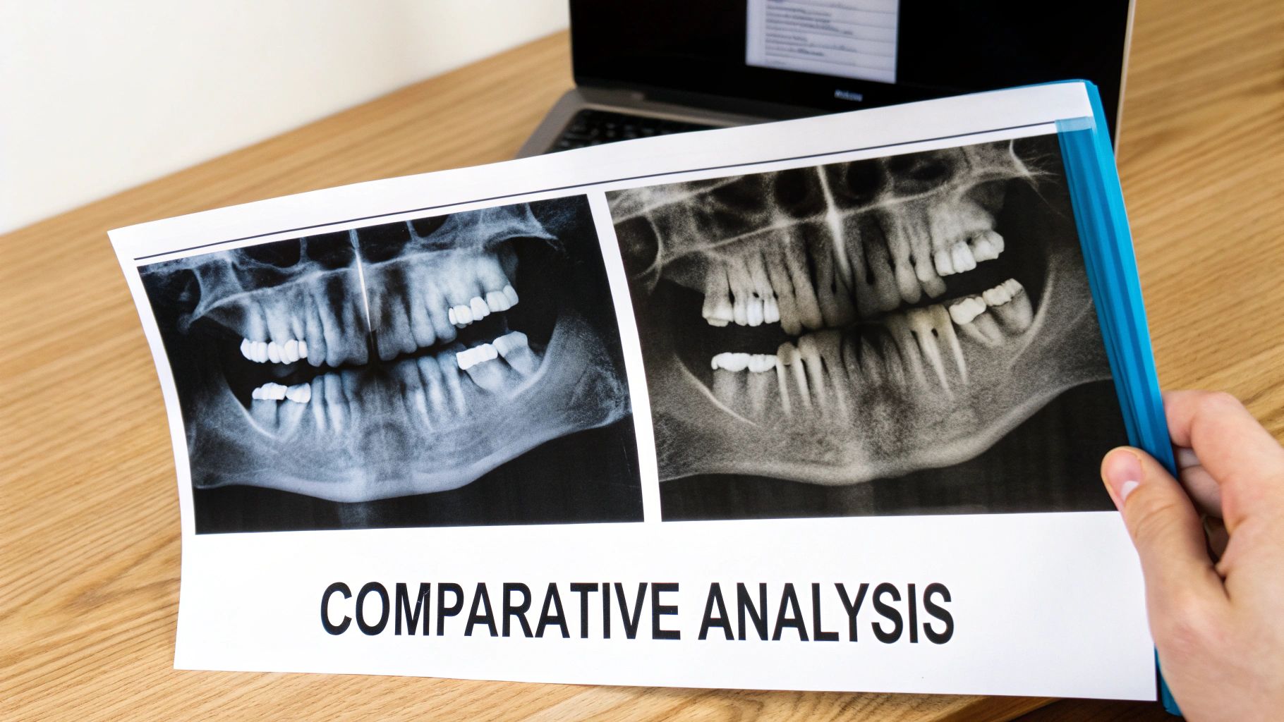
CBCT scans serve as powerful tools for showing patients the real benefits of proposed treatments. When patients can see detailed before-and-after comparisons, whether for teeth straightening or crown restorations, they better understand the value of investing in dental care.
Take orthodontic treatment as an example – patients can view 3D models showing how their teeth will look after alignment. These visual previews of expected results help patients make confident decisions about their dental care. Studies show that visual aids like CBCT increase treatment acceptance rates by up to 30%.
Enhancing Communication and Education: Empowering Patients
CBCT has changed how dental practices operate by providing precise 3D imaging that's essential for complex procedures like implants and root canals. For more details on CBCT's impact on modern dentistry, check out this comprehensive overview from Spear Education.
Patients can easily access and review their CBCT scans at home, helping them make thoughtful decisions about their care. They can share images with family members and take time to process information at their own pace. This level of access and understanding creates true partnerships between dentists and patients, leading to better treatment outcomes and higher satisfaction with dental care.
Building Your Digital CBCT Workflow
Getting the most out of CBCT imaging requires careful planning and setup. A well-designed workflow helps dental practices manage scans efficiently while delivering excellent patient care. Here's how to build an effective system.
Data Management: The Foundation of Your Workflow
Managing CBCT data properly is essential. You need a reliable system to store and access patient scans while maintaining security and privacy standards. Many practices now use cloud storage for easy access and automatic backups. Regardless of your storage choice, having robust backup systems protects your valuable patient data.
Your CBCT system should work smoothly with your practice management software. This allows patient information to flow automatically between systems, reducing manual data entry and potential errors.
Software Selection: Choosing the Right Tools
The software you select has a major impact on your daily workflow. Look for programs that offer key features like 3D reconstruction, implant planning tools, and diagnostic capabilities. The interface should be straightforward enough that your team can learn it quickly.
Make sure any software you choose works well with your existing systems. Good compatibility between your CBCT scanner, imaging software, and practice management system keeps everything running smoothly.
| Software Name | Key Features | Specialty Focus | Integration Capabilities |
|---|---|---|---|
| Example Software A | Implant Planning, 3D Modeling | Implantology, Oral Surgery | Seamless integration with major practice management systems |
| Example Software B | Airway Analysis, TMJ Assessment | Orthodontics, Sleep Apnea | DICOM compatible, open API for custom integrations |
| Example Software C | Endodontic Measurement Tools, Root Canal Visualization | Endodontics | Limited integrations, primarily focused on imaging devices |
Team Training: Empowering Your Staff
Good training is crucial for success with CBCT technology. Everyone who uses the system needs proper instruction – from front desk staff to clinical team members. Training should cover CBCT basics, equipment operation, and software use.
When your team understands the technology well, they can capture better scans and use advanced features effectively. This leads to more accurate diagnoses and treatment plans. Regular updates help staff stay current as the technology evolves. With proper training and systems in place, practices can build an efficient CBCT workflow that improves patient care across the board.
The Future of CBCT Innovation
Let's look ahead at the exciting developments shaping the future of CBCT dentistry. From enhanced diagnostics to improved workflows, these advancements will bring meaningful improvements to patient care and dental practice efficiency.
AI Integration: Smarter Diagnostics and Treatment Planning
Artificial intelligence is set to dramatically improve CBCT dental imaging. AI software can scan images to find subtle issues that might be missed through traditional analysis methods. Key benefits include automatic detection of:
- Bone loss patterns
- Critical nerve pathways
- Optimal implant positions
AI also helps create precise treatment plans customized to each patient's unique anatomy.
Advanced Imaging Capabilities: Higher Resolution and Reduced Radiation
Current research focuses on two key areas: enhancing image quality and lowering radiation exposure. New sensor technology and improved processing algorithms will enable:
- Higher resolution scans with enhanced detail
- Reduced image noise for clearer visualization
- Lower radiation doses without compromising quality
These improvements will help dentists make more accurate diagnoses while prioritizing patient safety.
Workflow Automation: Streamlining Processes and Enhancing Efficiency
CBCT systems are becoming more integrated with practice software and planning tools. Automated analysis can now generate preliminary treatment plans within minutes. This allows dental teams to:
- Reduce manual data entry tasks
- Speed up diagnosis and planning
- Focus more time on patient care
- Make data-driven treatment decisions
Preparing for the Future of CBCT Dentistry
Dental practices can take practical steps now to prepare for these advances:
- Stay current through dental journals and conferences
- Invest in ongoing team training
- Evaluate new technologies based on practice needs
- Focus on solutions that improve patient outcomes
Careful technology adoption helps practices provide excellent care while maintaining efficiency.
Ready to integrate the power of AI into your medical imaging workflow? PYCAD offers advanced AI solutions for enhanced diagnostics and operational efficiency.


