
The Evolution of CBCT Scans: From Medical Innovation to Dental Essential
The journey of Cone Beam Computed Tomography (CBCT) from a specialized medical imaging tool to a cornerstone of modern dentistry is truly remarkable. Originally derived from traditional CT scans, CBCT initially found its primary application in maxillofacial imaging within the medical field. However, its potential to revolutionize dental practices quickly became evident.
This promising technology offered a more precise and detailed view of dental structures compared to conventional 2D radiographs. This opened up exciting possibilities for improved diagnoses and treatment planning.
The shift towards dental applications began in the late 1990s. Advancements in technology allowed for the development of CBCT systems that were both affordable and compact enough to be integrated into dental offices. This marked a significant step toward bringing the benefits of 3D imaging to a wider range of dental professionals. Cone Beam Computed Tomography has a rich history, originating from traditional CT scans used in maxillofacial imaging. Its three-dimensional imaging capabilities quickly gained traction in dentistry, enhancing diagnostic accuracy and treatment planning. The early 2000s witnessed the introduction of notable systems like the NewTom DVT 9000 and the 3D Accuitomo, solidifying the move towards precise dental radiology. You can learn more about CBCT development here: CBCT Development.
Overcoming Early Challenges
Early CBCT systems encountered several obstacles. Initial image quality, for example, required substantial improvement to meet the demands of dental diagnostics. The high cost of these early machines also presented a barrier for many dental professionals.
However, dedicated researchers and forward-thinking dental practitioners recognized the transformative power of CBCT and persevered in its development.
Their dedication resulted in technological breakthroughs that significantly enhanced image clarity and substantially reduced the size and cost of CBCT machines. This paved the way for their widespread adoption in dental practices, ultimately reshaping the landscape of dental diagnosis and treatment.
The Impact of 3D Imaging
The ability of CBCT scans to generate detailed three-dimensional images has significantly improved diagnostic accuracy in dentistry. Unlike conventional 2D radiographs, CBCT enables dentists to visualize anatomical structures from various angles and perspectives.
This comprehensive visualization has proven particularly valuable in specialties like implantology, endodontics, and orthodontics. It allows for more precise planning and execution of complex procedures.
To further illustrate the evolution of dental imaging, let's take a look at a comparison table:
Evolution of Dental Imaging Technologies:
A comparison of traditional radiography methods versus modern CBCT technology, highlighting the key evolutionary milestones and advantages.
| Imaging Technology | Year Introduced | Dimensional Capability | Radiation Dose | Key Applications |
|---|---|---|---|---|
| Traditional Radiography | Late 19th Century | 2D | Relatively High | Basic dental diagnostics, caries detection |
| Panoramic Radiography | Early 20th Century | 2D | Moderate | Overview of entire jaw, impacted teeth |
| CBCT | Late 1990s | 3D | Relatively Low | Implantology, endodontics, orthodontics, complex surgical planning |
This table highlights the significant advancement in dimensional capability and the generally lower radiation dose associated with CBCT compared to earlier methods. It also underscores the expanded range of applications that CBCT offers in modern dentistry.
A New Era in Dental Care
The evolution of CBCT has ushered in an era of enhanced precision and personalized dental care. With the ability to visualize intricate anatomical details, dentists can plan treatments with greater accuracy.
This translates to more effective and predictable outcomes for patients. The continued advancement of CBCT technology promises further exciting developments, shaping the future of dental diagnostics and treatment. These developments highlight the important role CBCT plays in enhancing both patient care and clinical practice.
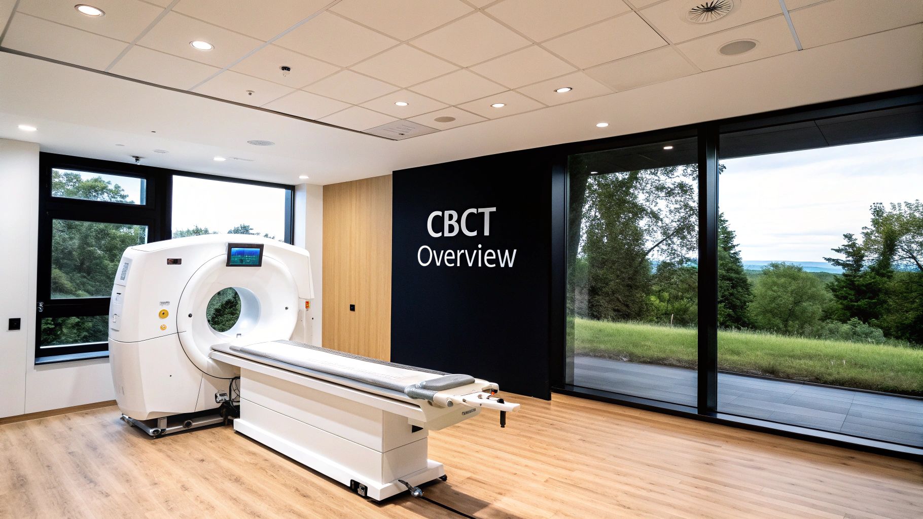
Inside CBCT Scans: The Fascinating Science Behind 3D Dental Imaging
CBCT scanners are impressive pieces of technology. In a matter of seconds, they capture hundreds of distinct 2D images. These images are then combined, like pieces of a jigsaw puzzle, to create a 3D model of the patient's anatomy. Dentists can manipulate this 3D model, rotating, zooming, and even taking virtual "slices" to view specific areas. This interactivity provides a significant improvement in diagnostic capabilities compared to traditional 2D X-rays.
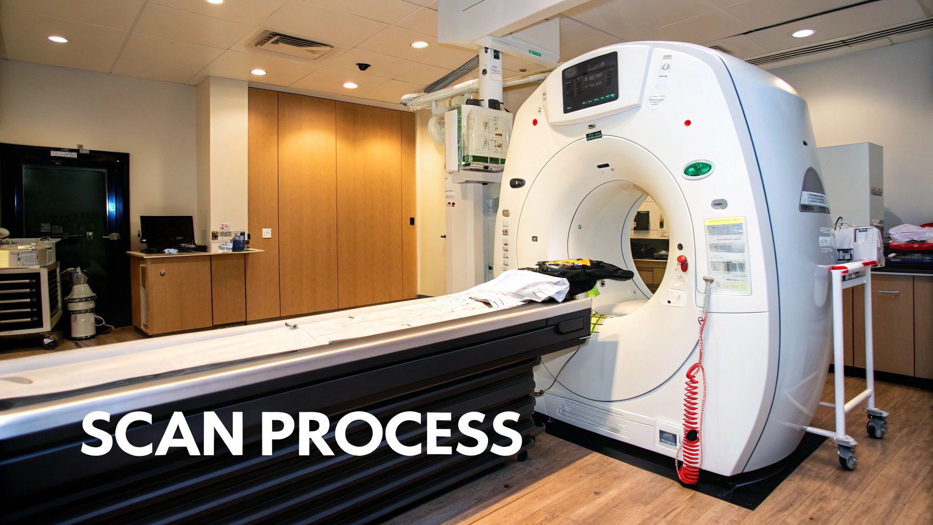
Capturing the Data: How CBCT Works
The process starts with the patient sitting comfortably inside the CBCT unit. A cone-shaped X-ray beam then rotates around the patient's head, capturing a series of images from various angles. This cone-beam geometry sets CBCT apart from medical CT scans, which use a fan-shaped beam.
The entire data acquisition process is remarkably quick, typically taking only 20-30 seconds. This speed improves patient comfort and minimizes motion blur, ensuring high-quality images. It also contributes to a more streamlined workflow for the dental practice.
Voxel Resolution and Field of View: Key Considerations
Two crucial aspects of CBCT scans are voxel resolution and field of view. Voxel resolution refers to the size of the individual 3D pixels (voxels) that compose the image. Smaller voxels provide higher resolution and reveal finer details, but require more processing power and may involve higher radiation doses.
The field of view describes the area captured by the scan. A larger field of view might seem desirable, but it can reduce the resolution. Selecting the correct field of view is a critical balancing act between diagnostic needs and radiation exposure. Local CBCT development began around 1993, spurred by the need for 3D visualization of the temporomandibular joint (TMJ). Inspired by earlier panoramic tomography, Dr. Arai's work established the foundation for this localized imaging technology. Learn more about the history of CBCT: CBCT History.
Minimizing Radiation: Advancements in CBCT Technology
A significant advancement in CBCT technology is the reduction in radiation dose. Modern CBCT units emit substantially less radiation than medical CT scans, increasing patient safety. This is particularly beneficial for children and patients requiring repeated scans.
This reduction is attributed to improvements in detector technology, refined software algorithms, and optimized scanning protocols. These innovations elevate CBCT scans to more powerful and safer diagnostic tools. The commitment to maximizing diagnostic quality while prioritizing patient safety has made CBCT scans an increasingly vital part of modern dentistry.
Why CBCT Scans Are Transforming Both Patient Care and Clinical Practice
CBCT scans are quickly becoming the preferred method for dental diagnostics, offering substantial benefits for both patients and clinicians. These advanced scans provide a wealth of information not available through traditional 2D imaging, resulting in better treatment outcomes across a variety of dental specialties.
Patient Benefits: Safety, Speed, and Accuracy
CBCT scans offer patients several key advantages. A primary benefit is the significantly reduced radiation exposure compared to traditional hospital CT scans. This is especially important for children and those requiring multiple scans.
CBCT scans also significantly shorten appointment times. The quick scan duration, often only 20-30 seconds, creates a more efficient and comfortable experience. This speed also minimizes motion blur, leading to clearer, more precise images.
Finally, the increased diagnostic accuracy of CBCT scans can eliminate unnecessary procedures. By providing a detailed view of underlying structures, dentists can make more informed treatment decisions.
One key advantage of CBCT scans is their lower radiation dose compared to traditional multi-slice CT (MSCT) scanners. Studies have shown that CBCT delivers lower organ doses, a safer option, particularly in orthognathic surgery and other dental procedures. This reduction in radiation exposure is vital for minimizing health risks associated with repeated imaging. CBCT's ability to provide detailed three-dimensional images has made it essential in dental and maxillofacial diagnostics. For further information, explore this resource: Radiation Dose in CBCT
Clinical Advantages: Enhanced Visualization and Treatment Planning
CBCT scans provide dental professionals with exceptional visualization, enabling a level of treatment planning not possible with 2D imaging. This improved visualization allows for less invasive interventions, preserving healthy tissue.
In implantology, for example, CBCT scans allow for precise implant placement, avoiding vital structures like nerves and blood vessels. This precision leads to higher implant success rates and better patient outcomes.
CBCT scans also facilitate more predictable results, even in complex cases. The detailed 3D images give a comprehensive understanding of the patient's anatomy, enabling clinicians to develop targeted treatment plans and anticipate potential challenges. This results in better long-term outcomes and greater patient satisfaction. In endodontics, CBCT scans can reveal hidden canal anatomy and subtle fractures often missed on traditional X-rays, allowing for more effective root canal treatments and preventing future complications.
The following table highlights the key differences between CBCT and other imaging modalities:
To understand the differences between CBCT and traditional imaging technologies, let's look at a comparison table.
CBCT vs. Traditional Imaging Modalities
| Imaging Modality | Radiation Dose (μSv) | Dimension | Resolution | Clinical Applications | Cost |
|---|---|---|---|---|---|
| Conventional X-ray | 5-10 | 2D | Low | Basic dental screenings | Low |
| Panoramic X-ray | 10-20 | 2D | Moderate | Overall jaw structure | Moderate |
| CBCT Scan | 20-500 | 3D | High | Implantology, Orthodontics, Oral Surgery | High |
| Medical CT Scan | 500-2000 | 3D | High | Complex medical imaging | Very High |
This table demonstrates the advantages of CBCT in terms of resolution and diagnostic capabilities, while also highlighting the significantly lower radiation dose compared to medical CT scans. The cost, although higher than 2D imaging, is justified by the enhanced precision and improved treatment outcomes.
Transforming Specialties Across the Board
The impact of CBCT scans extends across various dental specialties:
- Implantology: Precise implant placement and improved surgical planning.
- Endodontics: Identifying hidden canals and fractures for better root canal treatments.
- Orthodontics: Detailed assessment of jaw structure and tooth position for personalized orthodontic plans.
- Oral Surgery: Accurate evaluation of bone density and anatomical structures for complex procedures.
This widespread use of CBCT technology demonstrates its value in improving diagnostic accuracy and treatment outcomes across dental care. The ability to visualize structures in three dimensions allows for more informed decisions and ultimately contributes to better patient care.
CBCT Scans in Action: Game-Changing Applications Across Dentistry
CBCT (Cone Beam Computed Tomography) technology is transforming dental treatment across various specialties. Its ability to create detailed 3D images provides significant advantages over traditional 2D radiography. This leads to more accurate diagnoses, improved treatment planning, and better patient outcomes. Let's explore how CBCT scans are making a difference in specific areas of dentistry.
Implantology: Precision Placement and Enhanced Success Rates
CBCT scans are now essential for implant placement. They provide a precise 3D model of the jawbone. This allows dentists to accurately assess bone density, volume, and the location of vital anatomical structures such as nerves and blood vessels. This information is crucial for determining the right implant size and placement, enhancing osseointegration (the fusion of the implant with the bone), and reducing the risk of complications. CBCT scans enable surgeons to place implants with sub-millimeter precision. This results in higher success rates and predictable long-term outcomes.
Endodontics: Unveiling Hidden Canals and Fractures
In endodontics, CBCT scans are invaluable for identifying intricate root canal systems. This includes hidden canals and minor fractures that traditional X-rays might miss. This detailed perspective allows endodontists to thoroughly clean and seal the root canal system, boosting successful treatment rates and minimizing reinfection risks. CBCT scans also help diagnose the severity of periapical lesions (infections at the root tip). This guides treatment decisions and monitors the healing process.
Orthodontics: Beyond Tooth Alignment to Airway Analysis
Orthodontists are utilizing CBCT scans for more than just straightening teeth. The scans provide insights into the patient's airway, enabling assessment of airway size and identifying potential obstructions. This is particularly important when treating patients with sleep-disordered breathing or other airway-related concerns. By integrating airway analysis into treatment planning, orthodontists can develop comprehensive strategies that address both orthodontic and functional requirements.
Oral and Maxillofacial Surgery: Comprehensive Surgical Planning
CBCT scans are critical for complex oral and maxillofacial surgeries. They provide a complete view of the facial bones, teeth, and surrounding soft tissues, enabling surgeons to meticulously plan procedures. This precise anatomical information facilitates accurate pre-operative planning. It also minimizes intraoperative complications and promotes faster post-operative recovery. Surgeons can therefore approach complicated procedures with greater confidence, leading to better outcomes. The 1999 release of the NewTom DVT 9000, the first commercial CBCT unit—developed by Italian inventors Attilio Tacconi and Piero Mozzo—was a landmark achievement. This system offered dentists a way to acquire detailed 3D images of the maxillofacial region. This innovation transformed treatment planning by enabling accurate measurements and visualizations previously not possible with conventional radiography. Learn more: The First Dental CBCT.
Other Applications and Future Directions
The use of CBCT scans continues to expand beyond these core specialties. They are increasingly applied in periodontics to assess bone loss around teeth, in temporomandibular joint (TMJ) disorder diagnosis, and in detecting pathological conditions like cysts and tumors. The future of CBCT scans is promising, with ongoing improvements in image quality, reductions in radiation doses, and the incorporation of artificial intelligence for enhanced diagnostic capabilities. These advancements will continue to elevate patient care and broaden the applications of this valuable technology in dentistry.
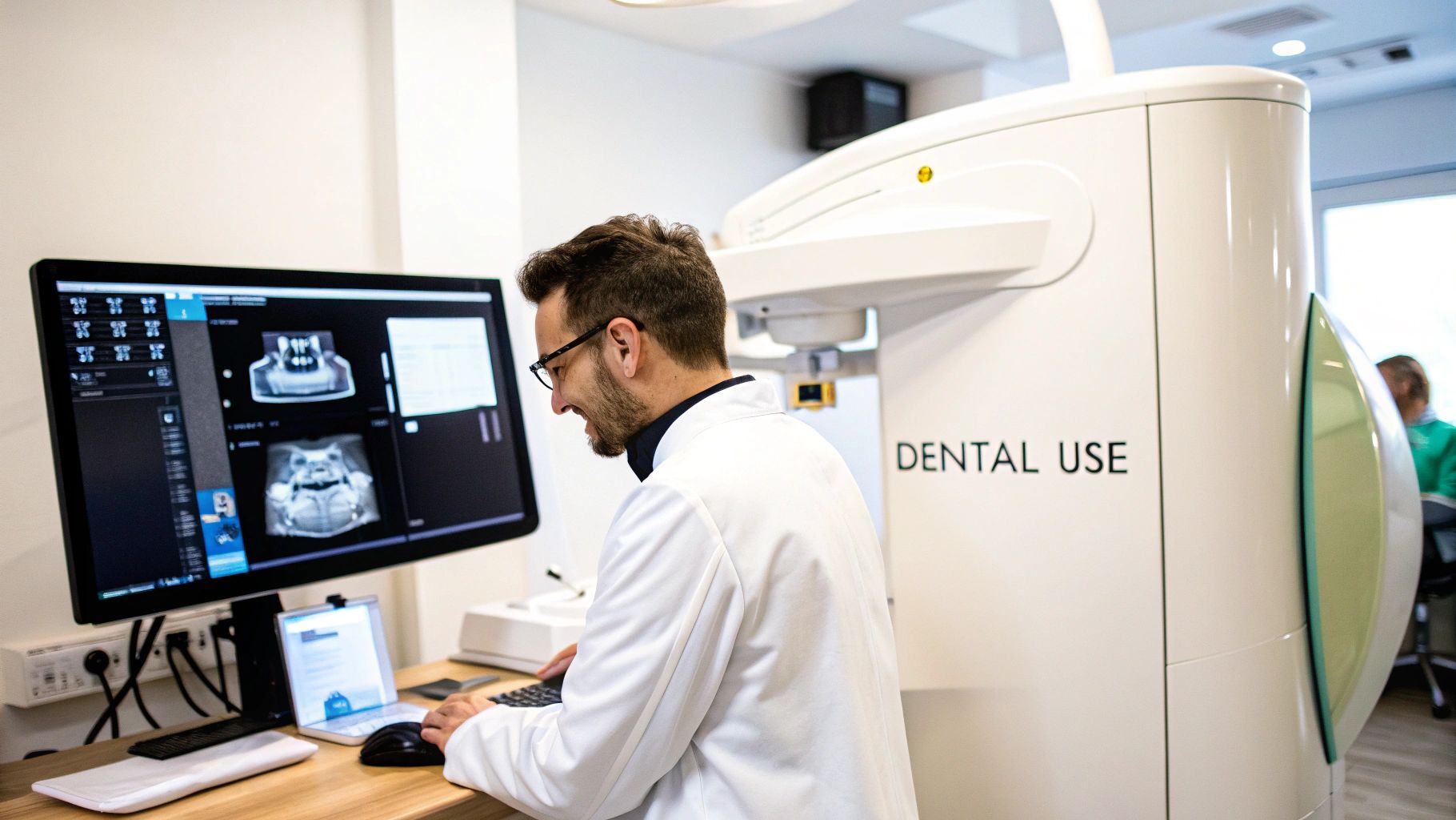
Reading CBCT Scans: What Dental Professionals Need to Know

Interpreting CBCT scans is a vital skill for dental professionals. This section offers practical advice from oral radiology experts to help you understand these detailed images. We'll explore a systematic approach to analysis, concentrating on identifying essential findings and avoiding common mistakes.
Developing a Systematic Review Pattern
A consistent review pattern is crucial for comprehensive CBCT scan interpretation. Begin by assessing the scan's overall quality, ensuring no significant artifacts are present. Then, conduct a structured examination of each anatomical region, moving methodically from one area to the next. This organized approach helps guarantee no critical details are missed.
For example, when examining the mandible, start with the condyle, progress to the ramus and body, and finish with the symphysis. This structured approach encourages thoroughness and minimizes the risk of overlooking vital findings.
Comparing the CBCT scan to any available 2D radiographs is also important. This comparison can help confirm findings and offer a more complete understanding of the patient's condition.
Distinguishing Normal Anatomy from Pathology
One of the difficulties in interpreting CBCT scans lies in differentiating normal anatomical variations from actual pathology. A deep understanding of dental anatomy is essential. Equally important is recognizing the spectrum of normal variations.
This often requires experience and continuous learning. For instance, a nutrient canal in the mandible can appear as a radiolucent line on a CBCT scan. An inexperienced clinician might mistake this for a fracture.
However, understanding normal anatomical variations allows for accurate identification as a benign finding. This knowledge is crucial for preventing misdiagnosis and unnecessary procedures.
Recognizing Imaging Artifacts
While powerful, CBCT scans can be prone to imaging artifacts. These artifacts can mimic pathology, possibly leading to an incorrect diagnosis. Common artifacts include beam hardening, scatter, and metal artifacts.
Learning to recognize these is vital for accurate interpretation. Metal restorations, for example, can cause beam hardening artifacts that manifest as dark streaks on the scan. Recognizing this helps clinicians avoid misinterpreting them as fractures or other issues.
This highlights the importance of understanding how artifacts appear on CBCT scans.
Field of View and Voxel Size: Impact on Diagnosis
The chosen field of view and voxel size significantly affect what can be reliably diagnosed on a CBCT scan. A larger field of view captures more anatomy but may reduce resolution. Conversely, a smaller field of view with a smaller voxel size provides higher resolution but covers a limited area.
Consider the diagnostic goal when selecting these parameters. Assessing the entire dentition for impacted teeth requires a larger field of view. Evaluating a small area for a subtle fracture requires a smaller field of view with higher resolution. The correct parameters are essential for accurate diagnoses.
Referral for Specialist Interpretation
While many dental professionals can interpret basic CBCT scans, complex cases often benefit from specialist interpretation. Oral and maxillofacial radiologists have advanced training in CBCT scan interpretation.
They are adept at identifying subtle findings and differentiating normal variations from pathology. Don't hesitate to refer challenging cases to a specialist. This ensures the patient receives the most accurate diagnosis and treatment plan.
This collaboration between general dentists and specialists improves patient care. Platforms like BeamReaders offer access to experienced Oral and Maxillofacial Radiologists, assisting in maximizing clinical value from CBCT scans.
The Future of CBCT Scans: Emerging Technologies Reshaping Dental Care
The field of Cone Beam Computed Tomography (CBCT) scanning is constantly evolving. New technologies promise to further enhance its capabilities and impact on dental care. These advancements will reshape how dentists diagnose, plan, and execute treatments.
Artificial Intelligence: Automating Pathology Detection
Artificial intelligence (AI) is rapidly changing dentistry. Researchers are developing AI algorithms to analyze CBCT scans, automatically detecting subtle pathologies and anomalies that might be missed by the human eye. This improves diagnostic accuracy and efficiency, enabling earlier diagnosis and intervention.
For example, AI could identify early signs of periodontal disease or even precancerous lesions, leading to timelier and more effective treatment.
Next-Generation Systems: Enhanced Image Quality and Reduced Radiation
Advancements in CBCT technology aim to deliver unprecedented image quality while minimizing radiation exposure. Next-generation systems are being developed with improved detectors and sophisticated software algorithms to achieve clearer images with less radiation.
This means patients benefit from more accurate diagnoses with lower risks, contributing to improved patient safety and diagnostic accuracy.
Augmented Reality: Visualizing CBCT Data During Procedures
Augmented reality (AR) is being integrated with CBCT data to offer surgeons real-time, 3D visualizations during procedures. Surgeons can wear specialized glasses that overlay CBCT images onto the patient's anatomy in real-time.
This technology enhances precision, especially during complex surgeries like implant placement or orthognathic surgery. It allows surgeons to navigate with greater accuracy, improving surgical outcomes and reducing complications. AR transforms static CBCT data into interactive visual guides, offering incredible potential for improved surgical precision.
Automated Treatment Planning: Simulating Outcomes with Precision
Automated treatment planning systems now use CBCT scan data to generate personalized treatment simulations. By inputting scan data into sophisticated software, dentists can preview the results of different treatment options. This allows for a more informed and collaborative decision-making process with the patient.
For example, in orthodontics, automated systems can predict tooth movement and simulate the final result of different orthodontic appliances. This enhanced predictability contributes to better patient outcomes and satisfaction.
Early Adoption and Transformative Impact
These advancements are not merely theoretical. Many dental practices are adopting these technologies. Some practices use AI-powered software for implant planning, while others use AR to guide surgical procedures.
The impact on these early adopters has been substantial, improving diagnostic accuracy, streamlining workflows, and elevating the standard of patient care. Their experiences demonstrate the real-world benefits and transformative potential of these technologies. Ready to experience the future of dental imaging? PYCAD is a leading provider of AI solutions for medical imaging. We offer a comprehensive suite of services, from data handling and model training to deployment, helping you optimize medical devices and improve diagnostic accuracy. Visit PYCAD to learn more.