
Think of it like giving a computer the same trained eye as a seasoned clinician—one that can spot subtle, almost invisible signs of disease that a person might otherwise miss. This is the heart of computer vision in medicine. It isn't about replacing doctors, but about giving them a new kind of partner, one that analyzes complex medical images with incredible speed and accuracy.
Seeing Health Through a New Lens
Computer vision is quickly becoming a fundamental tool in modern healthcare. At its core, it teaches machines how to interpret visual data—everything from MRIs and CT scans to microscopic pathology slides—and turn that information into genuine medical insights. This is already leading to earlier diagnoses, more personalized treatments, and an overall higher standard of care.
The field is seeing massive investment and growth for a reason. Valued at roughly $3.93 billion, the global computer vision market in healthcare is expected to climb to an estimated $14.39 billion by 2030, which is a compound annual growth rate of 24.3%. A big part of this boom is thanks to regulatory bodies like the FDA, which are helping to vet and approve these advanced tools for real-world clinical use. For a deeper dive into the numbers, you can explore the latest market research.
Core Areas of Medical Transformation
Computer vision's impact isn't limited to just one area; it's making waves across many medical specialties. By giving machines the ability to see and understand visual health data, we're unlocking new levels of efficiency and skill.
Here are a few key areas where this technology is already making a real difference:
- Radiology: It helps automate the detection of abnormalities in X-rays, CT scans, and MRIs. This allows radiologists to triage critical cases faster and catch faint patterns that might have been overlooked.
- Pathology: With digital analysis of tissue samples, we can achieve more consistent and objective tumor grading. This cuts down on the diagnostic variability between different pathologists.
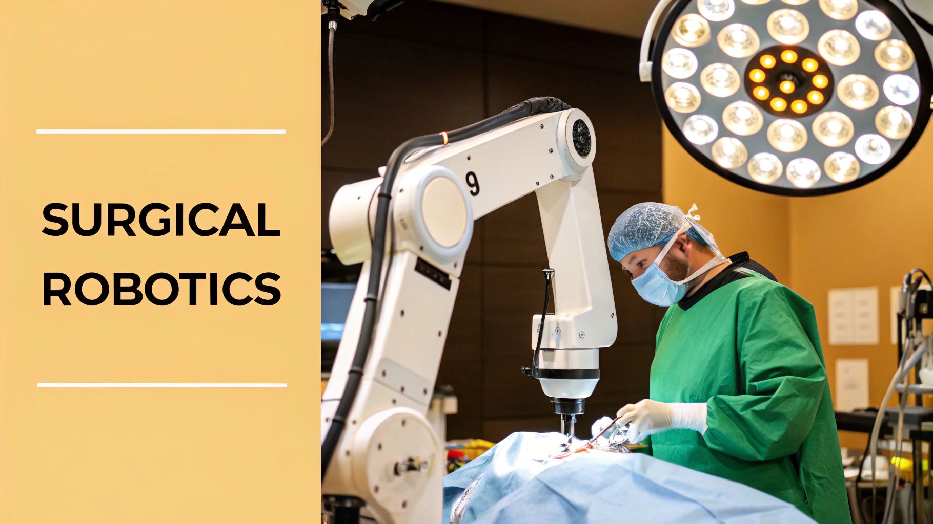
- Surgery: AI-guided robotics and augmented reality overlays are boosting surgical precision by giving surgeons real-time anatomical maps during delicate operations.
- Ophthalmology: It's now possible to screen for conditions like diabetic retinopathy from simple retinal scans, enabling early treatment that can prevent widespread vision loss.
The real power of computer vision in medicine is its role as a tireless, data-driven assistant. It can sift through thousands of images without getting fatigued, giving healthcare professionals the support they need to provide faster, more accurate, and more personalized care.
By taking over the more repetitive and data-heavy parts of medical imaging, computer vision frees up doctors and specialists. This allows them to focus on what humans do best: handling complex cases, communicating with patients, and crafting thoughtful treatment plans.
The table below gives a quick summary of how these applications are delivering real benefits across different fields of medicine.
Medical Domains Transformed by Computer Vision
| Medical Domain | Primary Application | Key Benefit |
|---|---|---|
| Radiology | Automated anomaly detection in X-rays, CTs, and MRIs | Faster diagnosis, reduced error rates, and improved workflow efficiency. |
| Pathology | Digital analysis and grading of tissue samples (histopathology) | Increased accuracy, consistency, and objective cancer staging. |
| Surgery | AI-guided robotics and real-time augmented reality overlays | Enhanced surgical precision, reduced invasiveness, and better outcomes. |
| Ophthalmology | Mass screening for conditions like diabetic retinopathy | Early detection and intervention, preventing irreversible vision loss. |
As these examples show, the goal is to create a synergy between human expertise and machine precision, ultimately elevating the quality and accessibility of care for everyone.
How AI Learns to Interpret Medical Scans
How can a machine learn to read an MRI or CT scan with the same sharp eye as a trained radiologist? The process is a lot like how a medical student learns. A student starts with textbooks to understand basic anatomy, then moves on to reviewing thousands of real-world scans to spot subtle abnormalities. AI models follow a similar path, just on a much more massive scale.
This training process is powered by deep learning, a field of AI that uses complex neural networks to uncover patterns in huge amounts of data. In medical computer vision, this data is made up of enormous, carefully organized datasets of medical images. These images are the AI's textbooks, and every single one is painstakingly labeled by human experts.
This is where annotation comes in—it’s the critical step of adding labels that tell the model what it's looking at. For a deeper dive into how this meticulous data preparation works, there are some essential image annotation tips for computer vision that offer valuable context.
The Core Tasks of Medical Vision AI
Once an AI model has its annotated dataset, it can be trained for specific tasks that mirror a clinician's diagnostic workflow. These tasks usually fall into three main categories, each one building on the last in terms of complexity and clinical value. The model isn't just "looking" at a scan; it's being taught to answer very specific questions.
These core functions are the building blocks for most medical AI systems today:
- Image Classification: This is the most basic task. The model answers a simple yes-or-no question about the entire image, like, "Does this chest X-ray show signs of pneumonia?" or "Is this tissue sample cancerous?"
- Object Detection: Taking it a step further, object detection answers the question, "Where is the problem?" The model draws a bounding box around a specific area of interest, such as identifying and highlighting a potential nodule in a lung CT scan.
- Image Segmentation: This is the most precise task of all. Segmentation answers, "What is the exact shape and size of the anomaly?" Instead of just a box, the model outlines the precise border of a tumor or lesion, pixel by pixel. This level of detail is crucial for things like surgical planning and targeting radiation therapy.
This visual breakdown shows how these advanced imaging insights come together in a real-world clinical setting.
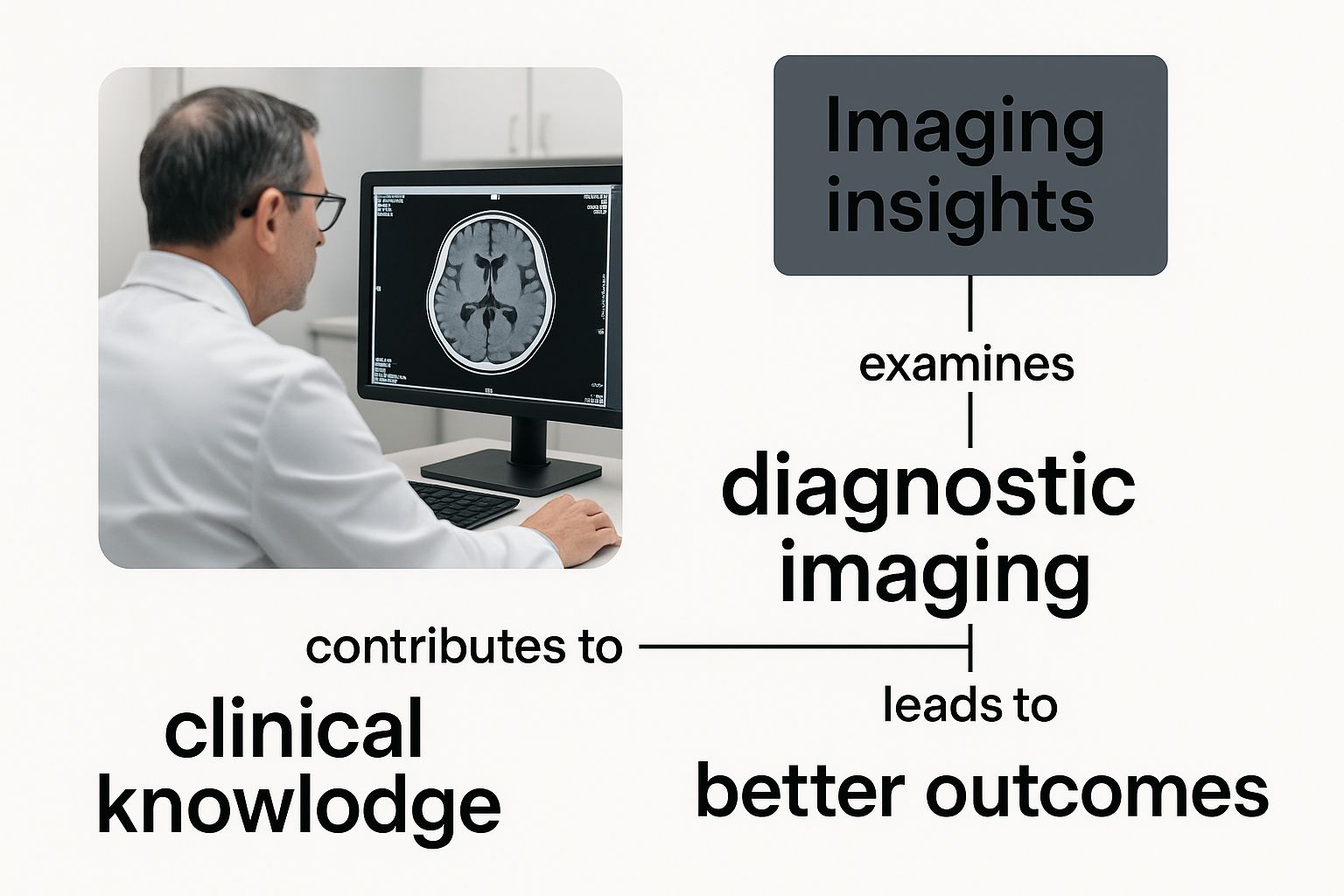
The image perfectly illustrates the synergy between human expertise and AI, where technology acts as a powerful analytical partner, supporting a medical professional’s diagnostic workflow.
Building Expertise Through Repetition
Training these models is all about iteration. An algorithm makes a prediction on an image, compares its answer to the human-provided label, and then adjusts its internal connections to correct its mistake. This cycle is repeated millions of times across the entire dataset.
Think of it like a student taking one practice exam after another. With each test, they learn from their errors and fine-tune their understanding. The AI does the same, continuously improving its ability to recognize complex patterns tied to different medical conditions.
This intense training makes it possible for the technology to handle the ever-increasing volume of medical scans we see today. From X-rays to CT scans, deep learning algorithms are improving both the speed and accuracy of diagnoses.
Through this rigorous, data-driven education, an AI model develops its own specialized form of expertise. It learns to see things that might be too subtle or time-consuming for the human eye to catch consistently, becoming an invaluable tool for medical professionals.
Real-World Impact on Diagnosis and Treatment
The true test of any technology isn't in the lab—it's in the real world. For healthcare, computer vision in medicine is making that leap, moving from research papers into the daily hustle of clinics and hospitals. This is where algorithms stop being theoretical and start acting as a trusted ally for clinicians, helping them save time, work more accurately, and ultimately, save lives.
This isn't just a distant, futuristic idea. The market itself tells the story. The global demand for computer vision in healthcare is poised for explosive growth, with a projected compound annual growth rate of 32.7%. By 2030, the market is expected to hit a staggering USD 15.6 billion. Why? Because these systems deliver tangible results, with some applications hitting diagnostic precision near 100% by catching subtle signs the human eye might overlook. You can see a full breakdown of this growth in a detailed market report.
A Second Pair of Eyes in Radiology
Picture a radiologist's day. They sift through hundreds of complex scans, their eyes trained to find minuscule, almost invisible signs of disease. The pressure is immense. Missing a tiny lung nodule on a CT scan, for example, could delay a cancer diagnosis with serious consequences. This is where computer vision steps in as a vigilant assistant.
AI algorithms, trained on vast libraries of thousands of chest CT scans, can automatically flag suspicious nodules, even those just a few millimeters across. The system doesn't make the final call; it acts more like a spotlight, drawing the radiologist's attention directly to areas that need a closer look. It’s a powerful human-AI partnership.
- The Challenge: An overwhelming volume of scans and the risk of human error from fatigue.
- The AI Solution: An automated system that pre-screens images and highlights potential trouble spots.
- The Outcome: A lower risk of missed diagnoses, faster report turnaround, and the freedom for radiologists to apply their deep expertise to the most complex cases. The result is earlier, more effective lung cancer detection.
Bringing Consistency to Pathology
Pathology is another area feeling the immediate benefits. Traditionally, grading a tumor involves a pathologist looking at a tissue slide through a microscope and making a judgment call based on what they see. This process, while highly skilled, can have slight variations from one expert to another, which can impact treatment plans.
Digital pathology, powered by computer vision, completely changes this dynamic. High-resolution scanners create digital copies of the glass slides, allowing AI models to analyze them with unwavering, objective precision. These systems can count dividing cells, measure tumor borders, and spot subtle structural patterns with a consistency that's nearly impossible to achieve by hand.
By providing a standardized, data-driven analysis of tissue, computer vision strips away a significant layer of subjectivity in cancer grading. This helps ensure a patient's diagnosis is based purely on the evidence, not on which pathologist happened to be on duty.
Preventing Blindness in Ophthalmology
Diabetic retinopathy is a major cause of blindness worldwide, but it's entirely preventable with early detection. The problem is screening the millions of diabetic patients, many of whom don't see an ophthalmologist regularly. This is a perfect job for an automated AI screener.
A patient can get a simple retinal photo taken at their primary care doctor's office. In seconds, a computer vision algorithm analyzes the image, searching for the tell-tale signs of retinopathy, like tiny aneurysms or bleeding.
The workflow is beautifully simple:
- Image Capture: A special fundus camera takes a high-resolution picture of the patient’s retina.
- AI Analysis: The algorithm scans the image for any sign of diabetic retinopathy.
- Instant Triage: The system gives an immediate result: either "no signs detected" or "referral to an ophthalmologist is recommended."
This approach makes widespread, low-cost screening a reality. It can catch the disease in its earliest stages, giving patients the chance to get treatment that can save their sight. Of course, building and deploying these advanced systems is a massive effort. To get these projects off the ground, organizations often rely on various healthcare grants to fund the essential research and infrastructure.
From flagging potential cancers to standardizing tumor grades and preventing blindness, computer vision in medicine is already making good on its promise. It's a powerful tool that enhances the skills of medical professionals, making healthcare more precise, accessible, and effective for everyone.
Overcoming Hurdles to AI Adoption in Healthcare
Bringing powerful computer vision in medicine from the lab into the real world of a busy clinic is a whole lot harder than just installing new software. The potential is massive, but the journey is filled with some serious challenges that demand smart planning and a thoughtful approach.
These aren't just technical glitches we're talking about. The biggest roadblocks involve people, established hospital routines, and a web of strict regulations. Getting it right means mastering three critical areas: the quality and privacy of your data, making sure the AI fits into how doctors already work, and clearing the high bar set by regulators. If you drop the ball on any one of these, a brilliant AI tool could end up gathering dust, never helping a single patient.
The Bedrock of Trustworthy AI: Data
At the end of the day, a medical AI model is only as good as the data it’s trained on. It’s a simple concept. You can't train a medical student to become a world-class radiologist by showing them a handful of blurry, mislabeled X-rays from one small town. The same exact principle holds true for AI. Your data has to be high-quality, diverse, and gathered ethically.
A huge piece of this puzzle is locking down patient data security. For anyone wanting to dig deeper into the specifics, this guide on improving patient data security in healthcare IT provides excellent context on what it takes to protect this highly sensitive information—a non-negotiable for any AI deployment.
A model trained on a non-diverse dataset will inevitably develop biases. If it only learns from data representing one demographic, its accuracy will likely drop when used on patients from different backgrounds, potentially leading to diagnostic errors and health inequities.
This is exactly why getting the data right is so fundamental. It's more than just grabbing images. It involves a painstaking process of anonymization to meet privacy laws like HIPAA, getting expert clinicians to annotate the data for accuracy, and building a dataset that truly mirrors the diverse patient population you serve.
Integrating AI into Clinical Workflows
Here’s a hard truth: a genius AI tool that forces a doctor to change their entire routine is a tool that simply won't get used. For AI to make a real impact, it has to slide seamlessly into the hospital’s existing world, especially the Electronic Health Record (EHR) and Picture Archiving and Communication Systems (PACS).
This integration is a massive technical and operational lift. The whole point is to make the AI's insights feel like a natural part of the software clinicians already use every single day. A radiologist, for instance, should see an AI-flagged nodule as an intuitive overlay on their standard imaging viewer, not have to open a separate, clunky application.
To make this complex process more manageable, it helps to follow a structured plan. The checklist below outlines the essential steps for healthcare institutions aiming to bring computer vision tools on board.
Implementation Checklist for Medical AI
| Phase | Key Action Item | Primary Consideration |
|---|---|---|
| Data Governance | Establish clear data access and usage policies. | Ensuring patient privacy and HIPAA compliance. |
| Workflow Analysis | Map out the current clinical process to identify pain points. | Minimizing disruption and maximizing efficiency for staff. |
| System Integration | Develop or procure tools that connect with existing EHR/PACS. | Achieving seamless data flow and a unified user interface. |
| User Training | Train all clinical staff on how to use and interpret AI results. | Building trust and ensuring the tool is used correctly. |
Without this kind of practical, on-the-ground planning, even the most promising technology can become a source of frustration, adding to a clinician's workload instead of lightening it.
Navigating the Regulatory Landscape
Finally, medical AI isn't the Wild West. Before any AI system can touch a patient for diagnosis or treatment, it has to pass a tough evaluation by regulatory bodies like the U.S. Food and Drug Administration (FDA). This process exists for one reason: to make sure the technology is both safe and effective.
Getting that green light is a marathon, not a sprint. Developers have to submit mountains of documentation and clinical proof showing their model works as promised in different situations. They must prove its accuracy, its reliability, and spell out the exact clinical scenarios where it’s meant to be used.
The FDA acts as a crucial gatekeeper, protecting patients from unproven tech while still making room for genuine innovation. This demanding oversight is what builds trust, assuring doctors and patients that any AI tool in use has met the highest possible standards. Without that regulatory seal of approval, even the most advanced computer vision in medicine would stay stuck in the lab.
The Next Frontier of Medical AI Perception
We've seen what computer vision can do in medicine today, and it's already impressive. But the real excitement lies in what's coming next. We're on the verge of moving past simply finding and labeling problems and into a new age of predictive, highly personalized, and proactive healthcare. This isn't science fiction; it's the next evolution of AI perception.
The technology moving from research labs into clinics right now won't just change diagnostics—it's set to redefine the entire patient experience. The focus is shifting from reacting to what's already happened to forecasting what might happen next. This gives clinicians a powerful advantage: the ability to step in before a condition takes a turn for the worse.
From Reactive to Predictive Analytics
Think about this: what if an AI could not only spot a tumor but also predict its growth pattern over the next six months? That's the core idea behind predictive analytics in medical imaging. By training computer vision models on sequences of scans over time, they can start to recognize the incredibly subtle visual cues of how a disease progresses.
This completely changes the game, allowing doctors to be proactive instead of reactive. For example, after a patient has a stroke, an AI could analyze their initial brain scan to forecast the most likely areas of long-term functional damage. Armed with that knowledge, therapists could design a hyper-targeted rehab plan from the very first day.
At its heart, this is about turning a medical image into a sort of crystal ball. If we can teach an AI to understand the visual dynamics of a disease, we can start anticipating its next move and plan our interventions with incredible foresight.
This approach is a massive breakthrough for managing chronic illnesses like multiple sclerosis or degenerative arthritis, where success depends on tracking tiny changes over many years.
Personalized Medicine and Real-Time Feedback
Another game-changing area is the development of truly personalized medicine. Right now, many treatments are based on what works for a broad average of patients. The future is about using computer vision to see how an individual's body and their specific disease are responding to treatment, right now.
Imagine an oncologist using AI to analyze a patient's weekly CT scans during chemotherapy. The system could measure microscopic changes in a tumor's size, density, and even its blood supply, offering immediate insight into whether the treatment is effective. If the tumor isn't shrinking as it should, the oncologist could pivot to a new strategy weeks sooner than current methods allow.
This creates a powerful feedback loop for patient care:
- Administer Therapy: The patient is given a specific drug or radiation treatment.
- Analyze Imaging: Computer vision models evaluate the biological response on a granular level.
- Adjust Strategy: Clinicians use this real-time data to tweak the plan for the best possible outcome.
AI-Guided Surgical Precision
The operating room is also getting a major tech upgrade. With AI-guided surgery, computer vision essentially gives surgeons a form of "x-ray vision" during a procedure. The system can project a 3D map of critical structures—like major blood vessels or nerve bundles—directly onto the surgeon’s field of view.
It's like having a GPS for navigating the human body. This augmented reality overlay, which updates in real-time, helps surgeons move through delicate anatomy with much more confidence. The result is a lower risk of accidental damage and better outcomes for the patient, especially in fields like neurosurgery or complex cancer removal where every millimeter counts.
The Power of Multimodal AI
Perhaps the most profound shift will come from multimodal AI. This is where we stop thinking only about images. The next generation of systems will integrate insights from computer vision with all kinds of other patient data to build a complete, 360-degree picture of their health.
A single model could analyze a patient's chest X-ray while also factoring in their genetic markers, recent lab results, and the clinical notes in their electronic health record. By weaving together these different data streams, the AI can spot complex patterns that would be invisible when looking at any single source. This fusion of information is what will unlock a future of truly deep, predictive, and precise care.
The Human and AI Partnership in Healthcare
Whenever the topic of computer vision in medicine comes up, one question almost always follows: will AI replace doctors? The short answer is no. The real goal isn’t replacement, but empowerment.
Think of it this way: the AI isn't an autonomous expert, but a brilliant co-pilot sitting alongside a human clinician. It’s built to handle the very things humans struggle with, like sifting through thousands of scans without getting tired or spotting minuscule patterns the naked eye might miss. This frees up the doctor to do what they do best: practice medicine.
Augmenting Human Expertise, Not Replacing It
The most exciting progress in medical AI is happening right where machine precision and human judgment meet. The AI does the heavy lifting—the tedious, data-intensive analysis—while the clinician applies their years of experience, critical thinking, and empathy to make the final call. It's a powerful combination.
By taking on the pixel-by-pixel analysis, computer vision gives doctors more time to actually be with their patients. This partnership is building a future where healthcare is more precise, proactive, and ultimately, more human.
The true value of medical AI isn't its ability to work alone, but its power to amplify a human expert's skills. It's a tool that makes good doctors even better, faster, and more effective at their jobs.
This collaborative approach is fundamental for building trust. It ensures technology serves as a tool for better, human-led medicine, not as a replacement for it.
How The Partnership Creates Better Outcomes
This human-AI collaboration directly tackles some of modern healthcare's biggest hurdles. Clinicians are drowning in data, and AI gives them a way to manage the flood.
This partnership brings a few key advantages:
- Reduces Cognitive Load: The AI acts as a first-pass filter, pre-screening images and flagging potential areas of concern. This helps doctors focus their attention where it matters most, cutting down on fatigue and the chance of an oversight.
- Enhances Diagnostic Confidence: When an AI's analysis confirms a doctor's own suspicion, it adds an extra layer of confidence. And when it flags something unexpected, it triggers a valuable second look that might have otherwise been missed.
- Frees Up Time for Patient Interaction: Automating repetitive analytical work gives doctors back their most valuable resource: time. That time can be spent talking with patients, building trust, and carefully explaining treatment options.
The Clinician-in-the-Loop Model
This dynamic is often called a “clinician-in-the-loop” system. In this setup, the AI might make a recommendation or highlight a potential anomaly, but a human expert is always required to validate the finding and make the final clinical decision. This model is absolutely critical for patient safety and accountability.
It keeps the technology in its proper role as a powerful assistant. The AI delivers data-driven insights, but the nuanced, context-aware decisions stay firmly in the hands of the trained medical professional. It’s this balance that’s creating a safer, more efficient, and more human-centric healthcare system for us all.
Frequently Asked Questions
As computer vision finds its way into clinics and hospitals, it naturally raises some big questions. People want to know what this means for doctors, how reliable the technology really is, and what ethical guardrails are in place. Getting a handle on these issues is key to understanding how this technology is being thoughtfully woven into modern medicine.
Will AI Replace Radiologists and Doctors?
This is probably the most common question out there, and it’s a valid one. The best way to think about medical AI isn't as a replacement, but as a powerful collaborator. It’s like giving a doctor a super-powered assistant that can sift through immense amounts of data at incredible speeds. This frees up the human expert to focus on what they do best: complex problem-solving, patient interaction, and exercising clinical judgment.
AI is fantastic at spotting patterns that might be invisible to the human eye, but it doesn't have a doctor's intuition, real-world experience, or ethical compass. The future isn't about AI taking over; it's about a "clinician-in-the-loop" approach. The AI presents its findings, but the doctor makes the final call. This partnership elevates a clinician's abilities, it doesn't erase them.
The goal of computer vision in medicine is to empower clinicians, not to replace them. By automating repetitive analytical work, it frees up doctors to spend more time with patients, not pixels.
How Accurate Are Medical AI Models?
Accuracy is the bottom line, and it’s not a simple one-size-fits-all answer. An AI model's performance really depends on the task at hand, the quality of the data it was trained on, and how complex the medical condition is.
That said, for specific, well-defined jobs—like spotting signs of diabetic retinopathy in eye scans or flagging suspicious lung nodules on a CT—the best models can perform on par with, and sometimes even better than, human specialists.
Getting an AI model into a clinical setting is a tough process. They are rigorously tested on huge datasets they've never seen before, with performance measured by metrics like sensitivity and specificity. Regulatory bodies like the FDA demand extensive clinical proof that a tool is both safe and effective before it ever gets near a patient.
What Are the Main Ethical Concerns?
Putting such powerful technology into healthcare means we have to talk about ethics. These aren't afterthoughts; they are central to building trust with doctors and patients alike.
There are three major areas we have to get right:
- Algorithmic Bias: This is a huge one. If an AI is trained primarily on data from one demographic, it might not work as well for others. This could accidentally worsen health disparities. Building fair AI means starting with diverse, representative datasets.
- Data Privacy: Patient data is incredibly sensitive. We have to be uncompromising about security and anonymization, following strict rules like HIPAA. This is non-negotiable.
- Accountability: If an AI-assisted diagnosis is wrong, who's responsible? Is it the developer, the hospital, or the clinician? We need clear guidelines for accountability to ensure everyone feels safe using these tools.
At PYCAD, we navigate these complex technical and ethical waters every day. We build robust AI solutions for medical imaging, from data annotation to model deployment, helping organizations integrate computer vision safely and effectively. Find out how we can help your project at https://pycad.co.


