When you look in the mirror, you're seeing yourself in the coronal plane. Coronal CT images give doctors that same unique, "face-on" view of your internal anatomy, slicing the body from front to back to reveal crucial diagnostic details that other perspectives might hide.
It’s one of the most fundamental tools in medical imaging, helping specialists piece together complex anatomical puzzles.
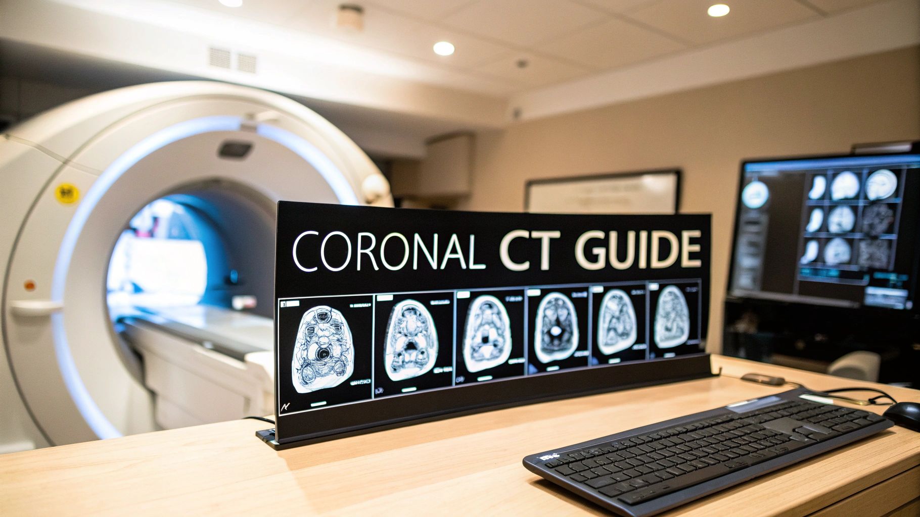
Decoding the Coronal View in Medical Scans
Think of how you'd slice a loaf of bread. Slicing from top to bottom gives you axial slices, and slicing from the side gives you sagittal slices. But if you were to slice it from the front crust all the way to the back, you’d be creating coronal slices. That's precisely what coronal CT images show—a frontal, or anterior-to-posterior, look at your anatomy.
This "face-on" view isn't a separate scan. Instead, it's a clever digital reconstruction. The CT scanner first takes hundreds of incredibly thin axial images. Then, powerful software stacks and reassembles this data to generate the coronal view, letting doctors scroll through the body as if they were flipping through pages of a book from front to back.
Why This Perspective Matters
The coronal plane is so powerful because it shows the side-to-side and up-and-down relationships between organs in a way that’s incredibly intuitive. It’s perfect for getting a clear picture of symmetry and alignment, which is often the first step in spotting a problem.
Here’s where the coronal perspective really shines:
- Anatomical Context: It lays out exactly how organs like the liver, spleen, and kidneys are positioned next to each other in the abdomen.
- Symmetry Assessment: It's the ideal view for comparing the left and right sides of the body, which is vital for evaluating structures like the sinuses, eye sockets, and lungs.
- Spinal Alignment: For conditions like scoliosis, the coronal view gives a direct look at the spine's lateral curvature, making it indispensable for proper assessment.
Think of it this way: an axial view shows one "floor" of the body at a time, and a sagittal view shows it from the side. The coronal view, however, provides a complete, front-to-back blueprint.
This unique viewpoint helps radiologists and clinicians build a full three-dimensional mental map of a patient's condition. By combining what they learn from the coronal, axial, and sagittal planes, they can pinpoint abnormalities, plan surgeries, and track treatment with much greater confidence and precision.
How a Scan Becomes a Coronal Image
A coronal CT image doesn't just appear on a radiologist's screen fully formed. It's the end product of a brilliant process that starts with the scanner acquiring hundreds, sometimes thousands, of ultra-thin images in the axial plane—think of these as individual cross-sectional slices.
Imagine taking a massive stack of transparent Lego bricks. Each thin brick is a single slice of the body. On its own, one brick doesn't tell you much. But stack them all together, and you have a complete, three-dimensional block of data. This initial data grab is the foundation for everything that comes next.
The infographic below breaks down the fundamental steps of this acquisition process.
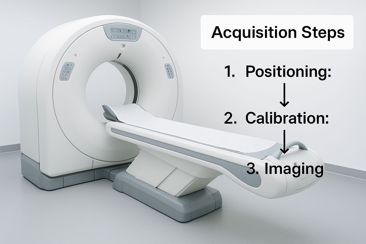
As you can see, getting high-quality coronal views all starts with capturing precise data during that first pass.
The Power of Digital Reslicing
With that 3D dataset captured, the real magic begins. It's a technique called Multiplanar Reformation (MPR). The software essentially takes that digital stack of "Lego bricks" and carves it up in a whole new direction. Instead of looking at the stack from the top down (the axial view), the computer slices through it from front to back, generating those crystal-clear coronal images.
The best part? This is all done digitally. No new scans, no extra radiation for the patient. Radiologists can then scroll through these newly created coronal slices just like the original axial ones, finally getting that essential "face-on" perspective.
The ability to create coronal CT images evolved from groundbreaking developments in computed tomography. Following the first clinical CT exam on October 1, 1971, the shift from single-slice to multi-detector scanners in the 1990s dramatically increased scan speeds, paving the way for detailed reconstructions. You can discover the full history of these CT advancements and their impact.
Why Thinner Slices Matter Most
The final quality of those coronal images hinges directly on the technical decisions made during the initial axial scan. And the single most important factor? Slice thickness.
Think back to our Lego analogy. Using thinner initial slices is like building your model with more, much thinner bricks. The result is a far smoother, more detailed final structure.
- Thinner Slices (e.g., 0.625 mm): These give you more data points to work with. The payoff is sharp, high-resolution coronal images with almost no distortion, which is absolutely critical for looking at tiny, intricate structures like the bones of the inner ear or face.
- Thicker Slices (e.g., 5 mm): With less data packed in, the reconstructed images can look blocky or jagged—an effect we call a stair-step artifact. This can easily hide subtle problems and make a confident diagnosis much harder.
At the end of the day, the goal is always to capture the most detailed raw data possible. This gives the software the best possible material to work with, ensuring the final coronal CT images are clear, accurate, and truly useful for diagnosis.
When Are Coronal CT Images Most Useful?
While every CT plane offers a unique piece of the diagnostic puzzle, some clinical questions are best answered with a "face-on" view. This is where coronal CT images truly shine. Think of it as looking at the body as if you were standing directly in front of the patient. This perspective is fantastic for understanding the vertical and side-to-side relationships between anatomical structures, making it an essential tool in many medical fields. It’s often the key that unlocks the final diagnosis.

The rise of the coronal view goes hand-in-hand with the incredible advancements in CT technology. As scanning exploded—from just 3 million exams worldwide by 1980 to more than 68 million a year by 2005—so did the demand for more comprehensive imaging. Faster, more powerful scanners made it possible to generate crisp, detailed coronal reconstructions as a standard part of many exams. This evolution cemented its importance in specialties from cardiology to orthopedics. You can get a great overview of how CT technology evolved over the last half-century to see how far we've come.
Assessing Sinuses and Facial Structures
When it comes to the paranasal sinuses, the coronal plane is the undisputed champion. Imagine trying to understand a multi-story building by looking at individual floor plans (the axial view). You’d miss how the rooms and stairwells stack up. The coronal view gives you that complete vertical picture, perfectly laying out the sinus cavities and their delicate drainage pathways.
This perspective is critical for a few key tasks:
- Diagnosing Sinusitis: It gives a crystal-clear look at fluid levels and inflammation inside the sinuses.
- Pinpointing Obstructions: Clinicians can easily spot blockages in the ostiomeatal complex—a crucial drainage area that's often the culprit in chronic sinusitis.
- Evaluating Facial Fractures: For trauma patients, nothing beats the coronal view for assessing fractures of the orbital floor (the bone at the bottom of the eye socket) and other complex facial injuries.
Mapping Abdominal and Pelvic Anatomy
In the tightly packed real estate of the abdomen and pelvis, knowing how organs are situated is everything. The coronal view acts like a clear anatomical map, showing how organs like the liver, spleen, and kidneys are positioned relative to one another from top to bottom.
This is especially powerful in oncology for tumor staging. A coronal image can reveal the full vertical spread of a tumor, answering critical questions like whether it has grown from a kidney into the adrenal gland just above it. This information is absolutely vital for staging the cancer accurately and planning the right surgical strategy.
A seasoned radiologist once told me that trying to read an abdominal scan without a coronal view is like navigating a city with only a street-level map. You can see what's next to you, but you have no concept of the towering skyscrapers. The coronal view gives you that all-important perspective from above.
Evaluating Spinal Alignment and Curvature
For spinal problems, particularly scoliosis, the coronal view is a must-have. By definition, scoliosis is a side-to-side curve of the spine. While other views provide context, the coronal plane gives the most direct and intuitive look at this abnormal curvature.
Orthopedic surgeons lean heavily on coronal CT images to:
- Measure the Cobb Angle: This is the universal measurement for quantifying how severe a scoliotic curve is.
- Assess Spinal Balance: The view helps them see if the head is properly centered over the pelvis.
- Plan Surgical Correction: Surgeons use these images as a blueprint for placing rods and screws during spinal fusion surgery, with the goal of restoring proper alignment.
The following table provides a snapshot of how different specialties rely on the coronal perspective for specific diagnostic challenges.
Key Clinical Uses of Coronal CT Views by Specialty
| Medical Specialty | Primary Application of Coronal View | Diagnostic Advantage |
|---|---|---|
| Otolaryngology (ENT) | Evaluating chronic sinusitis and nasal polyps | Clearly visualizes the ostiomeatal complex and sinus drainage pathways, which are often hidden in other views. |
| Trauma/Emergency | Assessing complex facial and orbital floor fractures | Provides an unparalleled view of vertical bone displacement and alignment, crucial for surgical planning. |
| Oncology | Staging abdominal and pelvic tumors | Shows the cranio-caudal (head-to-tail) extent of a tumor and its relationship to adjacent organs, like liver-diaphragm interface. |
| Orthopedics | Measuring scoliosis and assessing spinal balance | Offers the most direct and accurate method for calculating the Cobb angle and planning corrective surgery. |
| Urology | Evaluating kidneys, ureters, and bladder (CT Urogram) | Maps the entire urinary tract in a single view, making it ideal for detecting stones, strictures, or tumors. |
From tracing the intricate pathways of the sinuses to planning a life-saving cancer operation, the coronal view consistently delivers a layer of diagnostic insight that is simply indispensable.
How to Interpret Coronal CT Images
Reading a coronal CT scan is a bit like learning to read a complex map of the human body. You need to know the landmarks, of course, but you also need a strategy to navigate from point A to point B without getting lost. The most fundamental technique is to simply scroll through the image slices sequentially, moving from the front of the body (anterior) to the back (posterior).
As you scroll, you’re not just looking at random pictures; you're following specific structures slice by slice. This front-to-back journey helps you build a complete 3D picture in your mind. You can trace the entire path of a major blood vessel, follow the loops of the bowel, or get a feel for the true size and shape of organs like the liver and spleen. This systematic review is your best defense against missing a subtle abnormality that’s hiding in plain sight.
Mastering the Art of Windowing
One of the most powerful tools at your disposal is windowing. Think of it like adjusting the contrast and brightness on a photo to make certain details pop. A CT scanner captures an incredible range of tissue densities—far more than the human eye can see all at once. Windowing is how we filter this massive amount of data to focus on specific types of tissue.
You’ll use different presets to optimize the view for whatever you’re looking for:
- Bone Window: This setting cranks up the contrast to make bone appear bright white and incredibly sharp. It’s perfect for spotting a hairline fracture or subtle bone lesion. Everything else, like soft tissue, just fades into a uniform gray.
- Soft Tissue Window: Here, the settings are tuned to highlight the subtle differences between organs. You can distinguish the liver from the kidneys and spot inflammation or a tumor within the muscle.
- Lung Window: This preset is designed to visualize the delicate, air-filled structures of the lungs. It makes it possible to find tiny nodules or the faint signs of pneumonia that would be invisible otherwise.
A classic rookie mistake is trying to evaluate a structure in the wrong window. For example, you can't confidently rule out a rib fracture if you're still in a soft tissue window. Always remember to switch to the appropriate setting for the anatomy you’re examining.
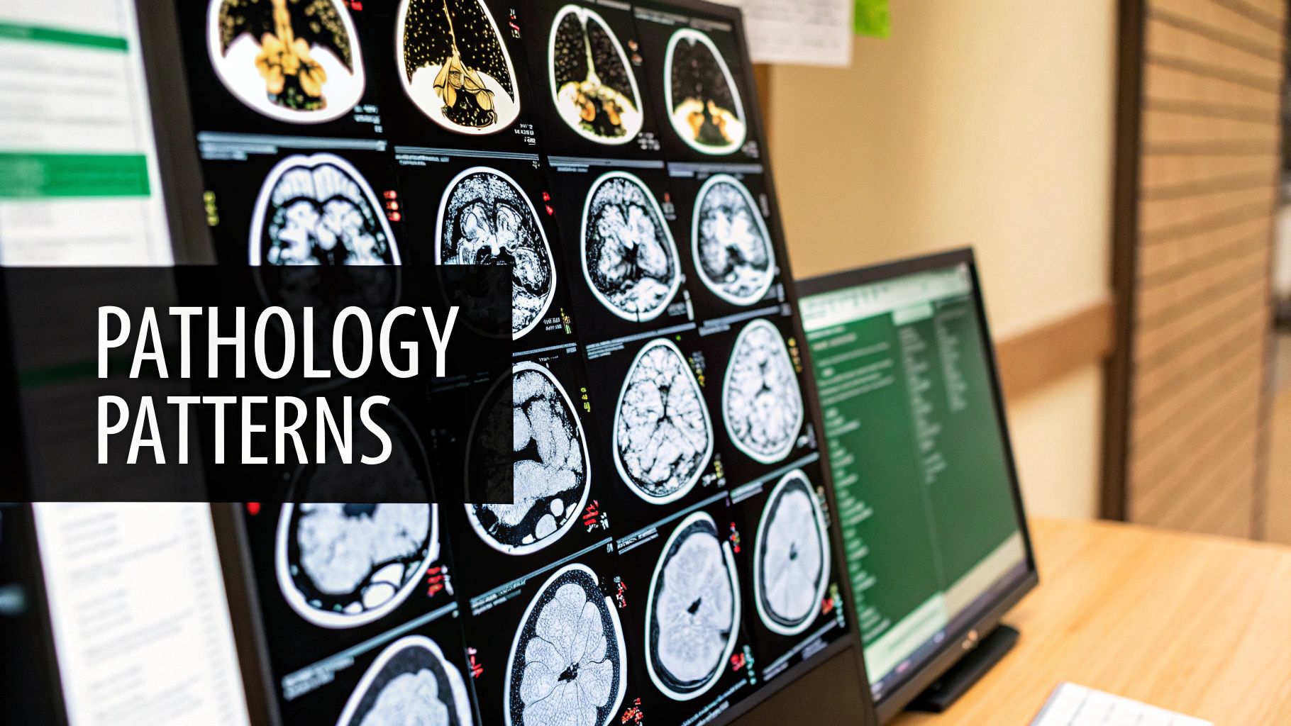
Spotting Artifacts on Coronal Images
Because coronal images are usually reconstructed from the original axial slices, they can sometimes have visual glitches called artifacts. Learning to recognize these is crucial so you don’t mistake one for a real medical problem. A common one is the "stair-step" artifact, which makes the edges of organs look jagged or blocky. This usually means the original axial slices were on the thicker side, giving the software less data to work with when creating a smooth reconstruction.
Another artifact you'll see all the time is caused by metal, like surgical clips or dental fillings. These dense objects can create bright streaks that shoot across the image, completely obscuring the anatomy behind them. Knowing what causes these artifacts helps you mentally look past them to see the true picture underneath.
The Power of Cross-Referencing Planes
If there’s one golden rule in CT interpretation, it’s this: never rely on a single plane. Any finding that looks suspicious on a coronal view must be checked on the axial and sagittal images. This cross-referencing is your best fact-checking tool.
For instance, something that looks like a small nodule in the liver on a coronal slice might just be a blood vessel seen in cross-section. A quick switch to the axial view will often clear it right up. Combining all three views is how you build a true 3D understanding, allowing you to confirm the size, shape, and location of any potential finding with confidence. This multiplanar approach is the absolute cornerstone of an accurate diagnosis.
The Role of AI in Coronal Image Analysis
Artificial intelligence is no longer just a futuristic idea in radiology; it's becoming a practical, everyday tool, especially for analyzing coronal CT images. It’s a bit like how your phone’s photo app instantly finds and tags faces. Medical AI works on a similar, though far more sophisticated, principle. It’s trained on thousands of existing scans, learning to identify specific organs, bones, and blood vessels with incredible accuracy.
This advanced pattern recognition allows AI algorithms to handle certain tasks with remarkable speed and precision. For example, an algorithm can automatically segment organs on a coronal view—essentially "coloring in" the liver, kidneys, and spleen. This not only saves radiologists precious time but also provides a consistent, measurable foundation for tasks like tracking organ volume or tumor size.
Enhancing Diagnostic Precision
Beyond just identifying structures, AI is exceptionally good at flagging subtle abnormalities that the human eye might miss, particularly during a long and demanding shift. By highlighting suspicious areas—a tiny lung nodule, a hairline fracture, or the faint, early signs of inflammation—AI acts as a diligent second pair of eyes. This collaborative dynamic helps reduce diagnostic errors and can lead to spotting diseases much earlier.
Here are a few key areas where AI is already making a real difference:
- Automated Nodule Detection: AI tools can systematically scan hundreds of coronal lung images, flagging potential nodules for the radiologist to review.
- Fracture Identification: In trauma cases, algorithms can rapidly screen coronal views of the spine or pelvis for fractures, helping to prioritize the most urgent situations.
- Quantitative Analysis: AI can precisely measure a tumor's volume or track subtle changes in an organ's size over time, providing objective data to gauge how well a treatment is working.
This ability to process massive amounts of visual data isn't unique to medicine. For those curious about the technology's reach, you can find broader discussions on AI's impact across many other fields.
The Future of AI in Radiology
Let's be clear: the goal of AI in medical imaging isn’t to replace radiologists. It's to empower them. By taking over the repetitive and time-consuming tasks, AI frees up specialists to concentrate on complex interpretations, patient consultations, and multidisciplinary teamwork. It’s a partnership that pairs human expertise with machine efficiency.
The integration of AI into the review of coronal CT images is creating a powerful synergy. The machine handles the high-volume data processing, while the human expert provides the critical context, clinical judgment, and diagnostic conclusion.
As these algorithms continue to improve, they will weave themselves even more seamlessly into the daily workflow. The future of radiology likely involves AI-driven tools that can pre-process scans, highlight key findings on coronal CT images, and perhaps even draft preliminary reports for review. This human-AI collaboration points toward a future where diagnoses are faster, more accurate, and ultimately lead to better outcomes for patients.
Common Questions About Coronal CT Images
Diving into medical imaging can feel like learning a new language, especially with terms like coronal CT images. Let's clear up some of the most common questions about how these essential views are made and why they matter so much.
Is a Coronal CT Scan a Separate Procedure?
Nope, not at all. Think of it this way: a coronal image isn't a new scan, but a different way of looking at the data from the original CT scan.
First, the scanner captures a detailed series of thin axial (or cross-sectional) images—imagine slices of a loaf of bread. Then, powerful computer software digitally "reslices" that data from a different angle, creating the front-to-back coronal view. This all happens behind the scenes, giving your doctor multiple perspectives from a single, efficient scan without you spending any extra time in the machine.
Do Coronal Images Require More Radiation?
This is a great question and a common concern. The answer is a clear no. Because coronal CT images are built by software after the scan is over, they don't add a single bit of radiation exposure.
The total radiation dose is set during that initial data capture. The magic of modern CT is its ability to generate coronal, sagittal, and even 3D views from that one set of data. This approach maximizes the diagnostic information doctors can get while sticking to the ALARA principle—As Low As Reasonably Achievable—for radiation safety.
The ability to create multiple image planes from a single scan is one of the biggest wins for modern CT. It gives us a complete diagnostic picture without increasing a patient's radiation dose for each new view.
Why Do Doctors Need the Coronal View?
Some parts of the body just make more sense when you look at them from the front. The coronal view gives doctors that intuitive, head-on perspective that can reveal anatomical relationships other views might hide or distort.
It’s often the go-to view for specific situations:
- Evaluating Sinuses: It's the gold standard for tracing the complex drainage pathways of the paranasal sinuses.
- Checking Spinal Alignment: When looking for scoliosis, nothing beats the coronal view for a direct look at the spine's side-to-side curve.
- Mapping Tumors: It helps oncologists see how a tumor might be spreading up or down through the abdomen and pelvis, which is critical for staging.
- Assessing Joints: In trauma cases like facial fractures, this view is perfect for understanding bone alignment and joint integrity.
In short, the coronal plane offers a unique and often indispensable angle for getting the diagnosis right.
Is the Quality of All Coronal Images the Same?
Definitely not. The final quality of a reconstructed coronal image is only as good as the data it was built from. It all goes back to the settings used during the original axial scan.
The single most important factor? The thickness of the original slices. Thinner initial slices provide more raw data for the computer, resulting in sharp, high-resolution coronal images with minimal distortion. On the flip side, if the original slices are too thick, the reconstructed coronal CT images can look blurry or pixelated, almost like a low-resolution photo. That "blocky" appearance can hide subtle problems and limit how useful the scan is.
At PYCAD, we focus on applying artificial intelligence to pull deeper insights from complex medical images. Our tools are built to help medical device companies and researchers improve diagnostic accuracy and make workflows more efficient. Discover how our AI expertise can advance your medical imaging projects.


