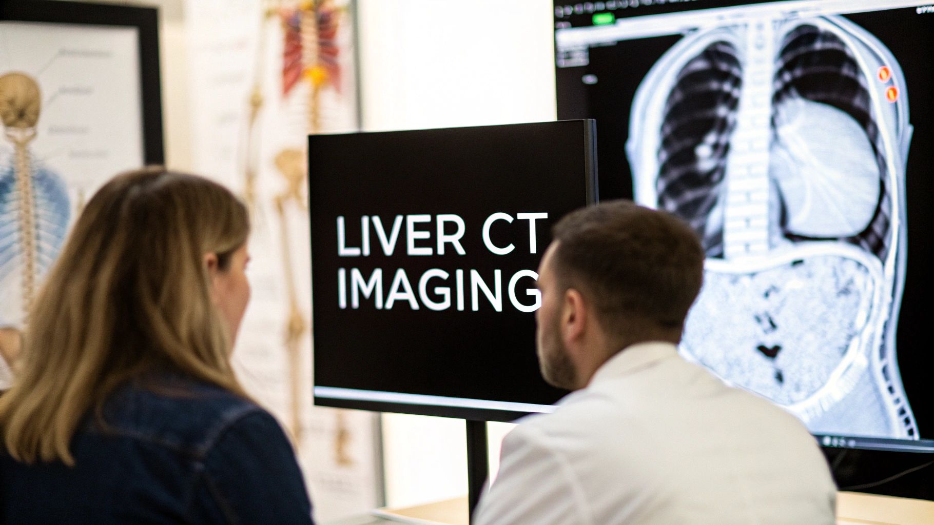
Mastering Liver Segment Visualization on CT
Visualizing the liver's complex structure is essential for accurate diagnoses and treatment plans. While appearing as a single organ, the liver is divided into eight functional segments. Each segment has its own blood supply and biliary drainage. Accurately identifying these segments on a CT scan is critical in modern hepatology.
This understanding allows healthcare professionals to precisely locate lesions. It also allows them to plan surgical resections and monitor treatment responses effectively.
Understanding The Importance of Liver Segment Identification
Why is segment identification so crucial? Think of the liver as a city with distinct districts. Each district has its own vital services. Knowing the exact location of a problem in one district allows for efficient resource allocation.
Similarly, precise segment identification on a CT scan allows for targeted interventions. During surgery, understanding segment boundaries allows surgeons to remove only affected tissue. This precision preserves healthy liver function, minimizes complications, and improves patient outcomes. Accurate segment identification also guides treatment strategies for liver diseases.
Challenges and Solutions In CT Liver Segment Visualization
CT scans are fundamental for visualizing liver segments, but interpretation can be challenging. It's like navigating a complex city map with overlapping roads and hidden landmarks. The Couinaud classification system, based on vascular anatomy, provides a crucial roadmap for navigating this complexity. This system promotes clear communication among radiologists and surgeons regarding liver abnormalities.
However, even with the Couinaud system, accurately defining segments on a CT scan remains difficult. Scans can sometimes misattribute liver portions to incorrect segments, particularly in central zones. Studies show that in marginal liver areas, approximately 17.3% of the area on axial CT scans can be incorrectly assigned. This error increases to about 51.6% in central zones. Learn more about these statistics.
This difficulty highlights the need for careful analysis and the use of additional tools. Meticulous analysis, coupled with advanced visualization techniques, is key for precise segmentation.
By understanding the challenges and utilizing advanced techniques, healthcare professionals can enhance accuracy. This ultimately leads to better patient care through improved diagnoses, surgical planning, and treatment outcomes.
Navigating the Couinaud Classification System
Understanding advanced liver anatomy requires familiarity with the Couinaud classification system. This system is crucial for effective communication between radiologists and surgeons regarding liver anatomy. It provides a standardized method for identifying specific liver segments, essential for CT imaging and surgical planning.
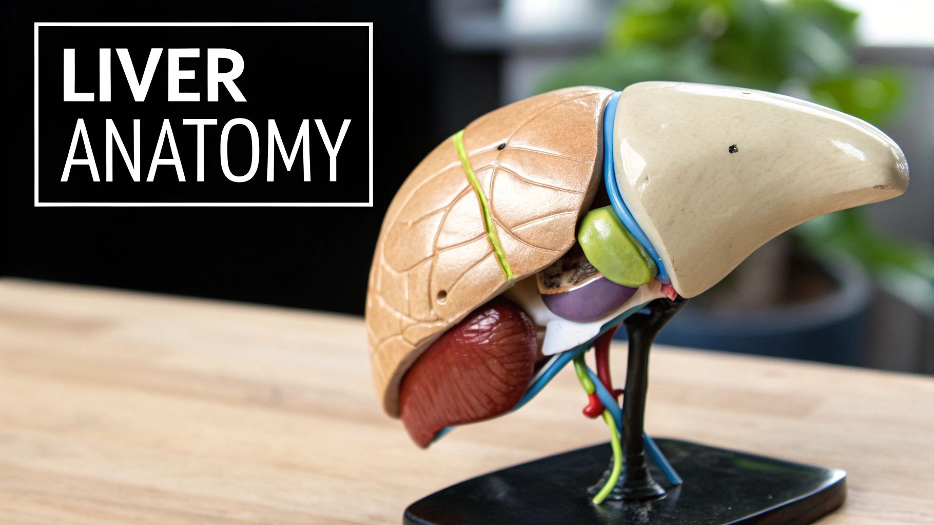
Understanding The Eight Liver Segments
The liver, though a single organ, is functionally divided into eight independent segments. This segmentation is based on the distribution of key structures: the portal vein, hepatic artery, and bile ducts. Each segment has its own blood supply and biliary drainage, much like apartments in a building have separate utilities. This independent functionality allows for precise, targeted interventions.
This segmented architecture allows surgeons to resect diseased portions without compromising healthy liver tissue. This precision has significantly improved surgical planning and execution, enabling complex procedures with greater confidence. The Couinaud classification, established by Claude Couinaud in 1957, forms the basis of this segmented approach. Learn more about the Couinaud Classification.
Identifying Key Anatomical Landmarks
Accurate identification of liver segments on CT scans depends on recognizing key anatomical landmarks. These landmarks serve as critical guides for navigating the liver's complex structure. The middle hepatic vein separates the liver into right and left lobes. The right and left portal veins, along with their branches, further define the segment boundaries.
Precisely locating these structures on CT scans is fundamental for accurate segment identification. This accuracy is essential for both diagnosis and the development of effective treatment plans.
Common Anatomical Variations and Expert Strategies
While the Couinaud system offers a standardized framework, anatomical variations are frequently encountered. Individual livers can exhibit unique characteristics, making segment identification challenging. Variations can include extra or missing vessels, or altered branching patterns.
Skilled radiologists utilize specific strategies to address these variations. Careful examination of CT images in multiple planes, combined with extensive anatomical knowledge, are essential. Meticulous tracing of vascular structures, correlated with expected segment boundaries, allows for accurate identification even in complex cases. This expertise is vital for ensuring diagnostic accuracy and optimal treatment planning.
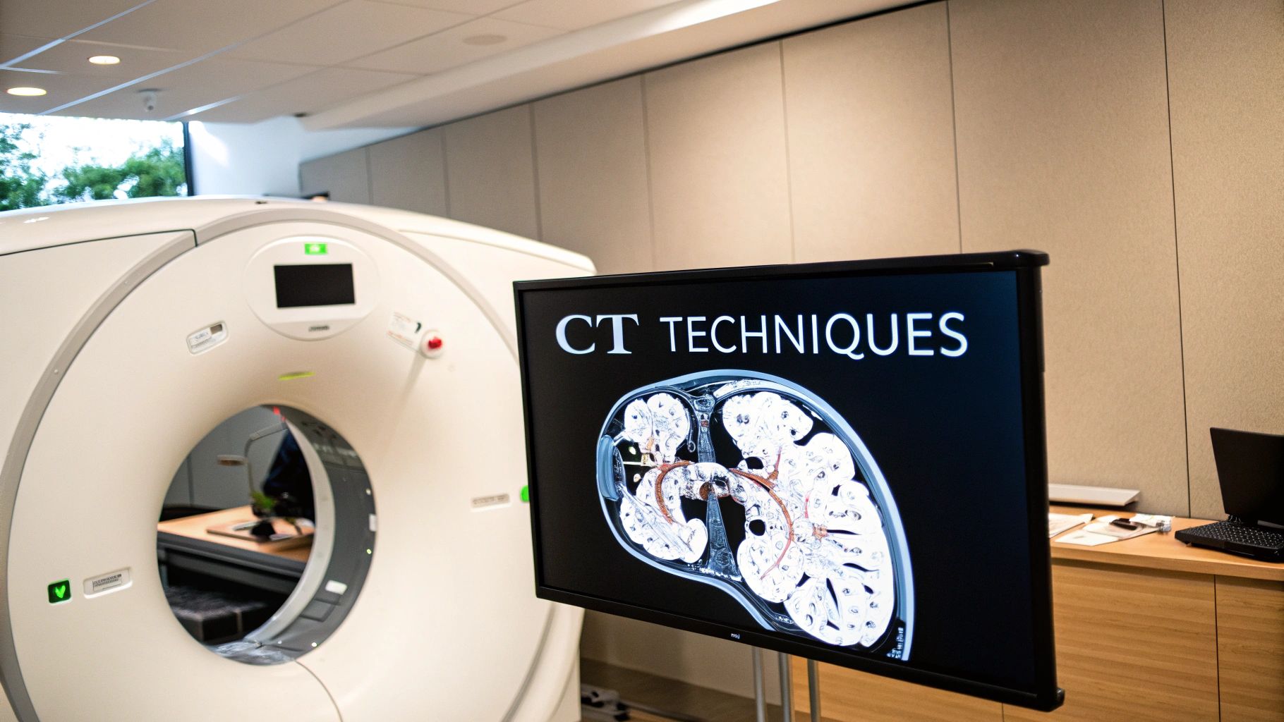
Improving CT Imaging Techniques
Research continues to advance CT imaging techniques for enhanced visualization of liver segments. Advanced software now allows for 3D reconstructions of the liver from CT data. These 3D models provide an intuitive and interactive method for visualizing the segments, their relationships, and the location of any lesions. This improved visualization is especially valuable for surgical planning, enabling precise mapping and minimizing potential complications. Furthermore, the development of AI-powered segmentation tools holds promise for automated and even more precise segment identification in the future.
CT Protocols That Reveal Hidden Liver Segments
Optimizing CT protocols is essential for clear visualization of liver segments. This clarity forms the basis for accurate diagnoses and effective treatment plans. This section explores the technical aspects of CT protocols, enhancing liver segment visualization.
Fine-Tuning Acquisition Parameters for Optimal Results
Visualizing the complex network of liver segments requires precise CT acquisition parameters. These parameters act like the settings on a camera, directly influencing the clarity and detail of the resulting image. Key parameters include slice thickness, reconstruction kernel, and contrast administration.
Thin slices provide better differentiation of small structures. Specific reconstruction kernels optimize image contrast. For example, a sharper kernel might be preferred for visualizing vascular structures. A smoother kernel might be chosen to assess parenchymal changes.
The timing of contrast administration is especially critical. Different phases of contrast enhancement highlight specific aspects of liver anatomy. This highlights the importance of multiphase CT imaging.
Multiphase Imaging: Capturing Dynamic Contrast Enhancement
Multiphase CT imaging involves acquiring images at different times after contrast injection. This captures the dynamic flow of contrast through the liver. The most important phases for segment visualization are the arterial phase, portal venous phase, and delayed phase.
The arterial phase, captured approximately 20-30 seconds after injection, highlights the hepatic arteries. The portal venous phase, imaged around 60-70 seconds post-injection, best visualizes the portal veins. The delayed phase, acquired several minutes later, helps characterize certain lesions and assess the biliary system.
Managing Challenging Patients and Anatomical Variations
Certain patient characteristics can make clear visualization challenging. Patients who struggle to hold their breath can introduce motion artifacts that blur the images. Patients with anatomical variations can deviate from the standard Couinaud segmentation.
In these cases, adapting the scanning protocol is key. Shorter scan times can help reduce motion artifacts. 3D imaging and post-processing reconstructions can further enhance visualization in challenging cases, compensating for anatomical variations and enabling precise segmentation even when traditional landmarks are obscured.
The table below summarizes key parameters and timings for each phase of liver segment imaging:
CT Protocol Parameters for Liver Segment Imaging
Optimal scanning parameters and contrast timing for clear visualization of liver segments
| Parameter | Arterial Phase | Portal Venous Phase | Delayed Phase |
|---|---|---|---|
| Time after injection | 20-30 seconds | 60-70 seconds | 3-5 minutes |
| Key structures visualized | Hepatic arteries | Portal veins | Parenchyma, biliary system |
| Clinical significance | Detecting arterial lesions | Segmental anatomy, portal vein thrombosis | Characterizing lesions, biliary obstruction |
By tailoring CT protocols to individual patient needs and using advanced imaging techniques, radiologists can achieve optimal visualization of liver segments, leading to improved diagnostic accuracy and more informed treatment decisions.
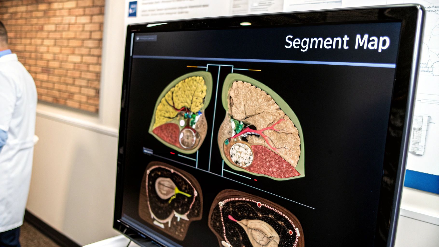
3D Reconstruction: Bringing Liver Segments To Life

Advanced visualization techniques are transforming how medical professionals understand and interact with CT scans of liver segments. Moving beyond traditional 2D images, 3D reconstructions offer a more intuitive and comprehensive view of the liver's complex anatomy. This leads to improved identification of liver segments and more effective communication among healthcare providers.
Transforming CT Data Into Interactive 3D Models
Leading institutions are using post-processing techniques to convert standard CT scan data into interactive 3D models. These models provide a dynamic representation of the liver. Healthcare providers can rotate, zoom, and even dissect the virtual organ to visualize segment boundaries and their relationships to surrounding structures. This ability to manipulate and explore the 3D model is particularly useful for complex cases, facilitating a deeper understanding of the liver's unique anatomy in each patient.
Efficient Workflows For 3D Reconstruction
Creating these 3D reconstructions isn't necessarily a time-consuming process. Modern software tools offer efficient workflows for generating meaningful 3D models from CT scan data. These streamlined processes allow radiologists and other healthcare professionals to incorporate 3D visualization into their routine practice without significantly increasing their workload. This accessibility makes 3D reconstruction a practical tool for enhancing clinical decision-making.
Implementation Strategies For All Centers
The advantages of 3D reconstruction are within reach for centers of all sizes and resource levels. While some advanced software packages may require specialized hardware, there are also cost-effective solutions for facilities with limited budgets. This means that even smaller centers can implement 3D visualization techniques, improving their capabilities in liver segment identification and surgical planning.
Improving Surgical Planning Accuracy
3D reconstructions of liver segments greatly improve the accuracy of surgical planning. By providing surgeons with a detailed, interactive model of the liver, these reconstructions allow for precise pre-operative planning. Surgeons can virtually simulate procedures, identify critical structures, and determine optimal resection margins with increased confidence. This improved visualization translates to greater precision in the operating room, ultimately leading to better patient outcomes.
Reducing Operative Complications
The increased precision offered by 3D models also contributes to a reduction in operative complications. By enabling surgeons to more accurately plan and execute procedures, these models minimize the risk of damaging vital structures during surgery. This results in fewer complications, faster recovery times, and shorter hospital stays for patients undergoing liver surgery.
Enhancing Interdisciplinary Communication
3D reconstructions are invaluable for interdisciplinary communication. They provide a common visual language that facilitates clear communication between radiologists, surgeons, and other members of the healthcare team. This enhanced communication ensures that everyone involved in a patient's care shares a consistent understanding of the liver's anatomy. This shared understanding contributes to more informed decision-making and improved patient care. For example, a radiologist can clearly explain the extent of a tumor to a surgeon using a 3D model, facilitating a collaborative approach to treatment. These advancements, including AI-driven tools like those developed by NVIDIA, are pushing the boundaries of what's possible with CT liver segment analysis and 3D reconstruction.
Surgical Planning With CT Liver Segments
Precise segment identification is crucial for successful liver surgery. Hepatobiliary surgical teams rely on CT scans of liver segments to navigate complex procedures, including resections, transplantations, and interventional radiology procedures. This detailed anatomical understanding plays a vital role in surgical planning and directly impacts patient outcomes.
CT Segmentation and Surgical Workflow
The process begins with a high-quality CT scan, optimized for clear visualization of the liver segments. Radiologists, leveraging their expertise and anatomical classification systems like the Couinaud classification, meticulously identify and label each segment on the CT images. This segmented data is then shared with the surgical team. This collaborative workflow between radiology and surgery is essential for effective preoperative planning. This detailed mapping allows surgeons to create a precise surgical roadmap. This roadmap guides critical decisions regarding incision placement, resection boundaries, and the extent of tissue removal.
Segment-Based Volumetry and Remnant Liver Viability
A critical aspect of surgical planning is determining the viability of the remnant liver, the portion that remains after resection. Accurate segment-based volumetry, enabled by CT segmentation, allows surgeons to calculate the volume of the remaining liver tissue. This precise measurement helps ensure sufficient liver function post-operatively.
Insufficient remnant liver volume can lead to postoperative liver failure, a severe complication. Accurate volumetry helps mitigate this risk.
Impact on Surgical Approaches and Resection Boundaries
Precise CT segment mapping directly influences surgical approaches and resection boundaries. Consider a right hepatectomy, a procedure typically involving segments 5 through 8. Understanding the precise location and volume of these segments allows for a tailored surgical approach. This precision optimizes access while minimizing disruption to surrounding tissues.
A left hepatectomy typically involves segments 2 through 4, and sometimes segment 1. Accurate CT segmentation ensures precise identification of these segments, enabling surgeons to define clear resection boundaries. This minimizes the risk of resecting healthy tissue or leaving behind diseased tissue.
To illustrate the relationship between surgical procedures and the liver segments involved, let's examine the following table:
Surgical Procedures and Required Liver Segments: Common hepatobiliary surgical procedures and the liver segments involved
| Surgical Procedure | Segments Involved | Key Considerations |
|---|---|---|
| Right Hepatectomy | 5-8 | Remnant liver volume in the left lobe, vascular variations |
| Left Hepatectomy | 2-4 (sometimes 1) | Middle hepatic vein location, proximity to major vessels |
| Segmentectomy | Specific segment | Precise identification of the target segment, minimizing resection of adjacent segments |
| Living Donor Liver Transplantation | Specific segments | Ensuring adequate graft volume for both recipient and donor |
This table highlights the critical role of segment identification in various procedures. The key considerations emphasize the importance of precise planning based on individual patient anatomy.
Case Studies and Patient Outcomes
Numerous case studies demonstrate the positive impact of precise CT segment mapping on patient outcomes. Improved accuracy of resection margins, facilitated by 3D reconstructed CT images, has been linked to reduced blood loss and shorter hospital stays. These outcomes underscore the importance of integrating CT liver segment analysis into surgical planning.
Multidisciplinary Collaboration and Surgical Success
Multidisciplinary collaboration between radiologists, surgeons, and other healthcare professionals is crucial for successful liver surgery. Open communication and a shared understanding of the segmented imaging data promote a team-based approach to surgical planning. NVIDIA is among the companies developing AI-driven tools that are further enhancing the precision and efficiency of CT liver segment analysis. This continuous advancement in imaging technologies translates anatomical knowledge into practical surgical applications, ultimately improving outcomes for patients undergoing liver surgery.
Overcoming Challenges in CT Liver Segment Identification
Accurately identifying liver segments on CT scans presents several challenges for radiologists. These difficulties can stem from anatomical variations, pathological distortions caused by diseases, and inherent limitations in CT technology. Let's explore these challenges and effective strategies to overcome them.
Anatomical Variations
A significant hurdle is the inherent variability in human liver anatomy. While the Couinaud system offers a standardized framework, individual livers often deviate from this ideal. Accessory hepatic veins, variations in portal vein branching, and differences in segment shape and size can all complicate segment identification. For instance, an accessory right hepatic vein can be mistaken for the fissure for segment VIII. Similarly, an early bifurcation of the right portal vein can create ambiguity between segments V and VIII.
Pathological Distortions
Diseases like cirrhosis and tumors can distort normal liver architecture. Cirrhosis, with its characteristic fibrosis and nodular regeneration, can obscure normal landmarks such as hepatic veins and portal vein branches. Think of it like a map where the roads (vessels) are twisted and obscured by an earthquake (cirrhosis). Large tumors can compress or displace liver segments, making it difficult to define their original boundaries. This is similar to a landslide (tumor) reshaping a mountain (liver). In these situations, relying solely on standard anatomical landmarks can be misleading.
Technical Limitations
CT technology has limitations that can affect segment delineation. Partial volume averaging, where different tissues within a single voxel are averaged, can blur the boundaries between segments. Motion artifacts, caused by patient breathing, can also reduce image quality. These limitations underscore the importance of optimized CT protocols.
Strategies for Accurate Segment Identification
Despite these challenges, several strategies can improve the accuracy of CT liver segment identification.
-
Multiplanar Reformats: Examining CT images in axial, coronal, and sagittal planes provides a more comprehensive 3D understanding of liver anatomy, helping to clarify ambiguous findings.
-
Thin-Slice CT: Acquiring thinner slices reduces partial volume averaging and improves the visualization of small structures, allowing for more precise delineation of segment boundaries.
-
Contrast Enhancement: Using intravenous contrast agents highlights vascular structures, crucial for segment identification. Multiphasic imaging during arterial, portal venous, and delayed phases further enhances vascular visualization.
-
3D Reconstructions: 3D reconstructions significantly aid in understanding the spatial relationships between segments. These models allow for interactive manipulation and visualization of the liver, offering a clearer view of the segmental anatomy.
-
Correlation with Other Modalities: In complex cases, comparing CT findings with other imaging modalities like MRI or ultrasound can provide valuable insights and improve diagnostic confidence.
-
Expertise and Experience: A radiologist's expertise and experience remain paramount. Familiarity with anatomical variations, pathological distortions, and CT imaging principles is essential for navigating complex cases.
By integrating these strategies, radiologists can maintain diagnostic accuracy even in challenging anatomical scenarios. Precise segment identification is critical for accurate diagnosis, effective treatment planning, and successful surgical interventions. Companies like NVIDIA are advancing liver segment analysis with innovative tools, showcasing the ongoing evolution of CT technology and its potential to revolutionize liver visualization and understanding.
The Future of CT Liver Segment Visualization
The field of CT liver segment visualization is constantly advancing, offering increasing precision and clinical impact. Several key developments are shaping this future, promising to change how we identify, analyze, and interact with liver anatomy.
Artificial Intelligence: Refining Liver Analysis
Artificial Intelligence (AI) is playing a growing role in CT liver segment visualization. AI acts like a specialized assistant, analyzing large datasets, recognizing subtle patterns, and automating difficult tasks. Algorithms are being developed to automatically segment the liver, accurately outlining the boundaries between segments. This automation saves radiologists time and improves precision, minimizing the variability of manual segmentation. This is especially helpful in complex cases involving anatomical variations or pathological distortions. AI-powered tools, like NVIDIA's VISTA-3D model, can segment over 100 organs and numerous diseases from CT scans, providing a more thorough and efficient analysis. Learn more about NVIDIA's advancements in medical imaging.
Augmented Reality: Connecting Radiology and the Operating Room
Augmented Reality (AR) is another exciting advancement bridging radiology and surgery. AR overlays digital information onto the real world, letting surgeons visualize CT liver segments in real-time during procedures. Imagine surgeons wearing specialized glasses projecting a 3D model of the segmented and labeled liver directly onto the patient's abdomen. This real-time visualization improves surgical precision, guiding surgeons through complex anatomy and preventing damage to vital structures. This enhanced precision can result in better patient outcomes and shorter recovery periods.
Advances in Contrast and Functional Imaging
Improvements in contrast agents and functional imaging techniques are also leading to better liver segment delineation. New contrast agents are designed to specifically highlight liver segments on CT scans. This is similar to using a dye that emphasizes different map sections, making them easier to see. Functional imaging techniques, like perfusion CT, offer details about blood flow within the liver. This metabolic data complements anatomical imaging, giving a more complete view of liver function and helping identify diseased segments. Perfusion CT offers valuable insights for diagnosis and treatment planning.
Enhanced Precision and Accessibility: The Future of Liver Imaging
These innovations point to a future of increased precision, accessibility, and clinical impact for CT liver segment visualization. AI-powered automation, AR-assisted surgery, and enhanced imaging techniques empower healthcare professionals to make more informed decisions, improving patient care. The greater precision in segment identification enables more targeted interventions, including surgical resection, localized drug delivery, or radiation therapy. These advancements also promise to make sophisticated imaging techniques more accessible to a wider range of healthcare providers, benefiting patients everywhere.
Ready to explore the future of medical imaging? PYCAD offers leading AI solutions transforming healthcare. From automated segmentation to advanced visualization, PYCAD gives medical professionals the tools they need to improve diagnostic accuracy and patient outcomes. Discover how PYCAD can revolutionize your medical imaging workflow.


