The Evolution of CT Scan Readers: From Hours to Seconds
The journey of the CT scan reader reflects a constant drive for innovation in patient care. Early CT scanners, while revolutionary for their time, posed significant logistical hurdles. Imagine waiting hours for a single head scan, followed by days of image processing. This was the reality of CT technology in the 1970s.
The development of Computed Tomography (CT) scans, starting with Godfrey Hounsfield's invention in 1972, has profoundly changed medical imaging. Initially, these scanners were limited to head imaging, with scans taking hours and reconstruction spanning days.
The first patient CT scan, performed in 1971 by James Ambrose and Godfrey Hounsfield, ushered in a new era of diagnostic imaging. Explore this topic further. By 1980, CT scanners were more widely accessible. Today, there are approximately 6,000 CT scanners in the U.S. and over 30,000 worldwide.
The Rise of Multi-Slice Scanning
The initial slow process of CT scanning drove rapid technological advancements. A key development was the multi-slice scanner. These machines could capture multiple image slices simultaneously, significantly reducing scan times.
This improvement brought several benefits. Shorter scan times meant patients spent less time holding their breath, minimizing motion artifacts and improving image quality. This resulted in clearer, more diagnostically useful images.
Faster Scans, Clearer Images
The progress didn't stop with multi-slice scanning. Technology continued to advance, increasing the number of slices acquired per rotation. From 4-slice scanners to 16, 64, and beyond, each generation brought improvements in speed and resolution.
CT scan readers could now capture highly detailed images of internal structures much faster. This enabled imaging of dynamic processes like blood flow and organ function, expanding the diagnostic capabilities of CT.
The Impact on Diagnosis and Treatment
Modern CT scan readers, with their speed and precision, have transformed clinical practice. Procedures that once took hours or days are now completed in seconds. This allows for quick diagnoses and treatment in critical situations like stroke or trauma.
The ability to acquire high-quality images rapidly also improves diagnostic accuracy. This empowers physicians to make more informed treatment decisions, ultimately leading to better patient outcomes.
Today, CT scan readers, some enhanced by companies like PYCAD, can deliver crucial diagnostic information within minutes. This is a dramatic change from the time-consuming procedures of the past. Ongoing advancements promise even faster, clearer, and more insightful images, solidifying the CT scanner's crucial role in modern medicine.
Inside Modern CT Scan Readers: The Science of Seeing
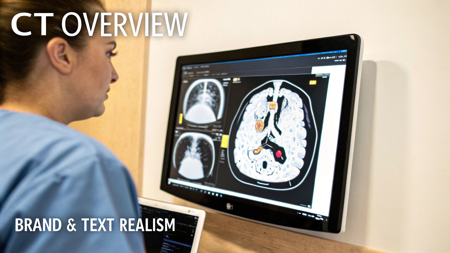
Modern CT scan readers provide highly detailed cross-sectional images of the body. These complex machines allow physicians to see internal structures with remarkable precision. This advanced visualization relies on the interplay of several key components.
The X-Ray and Detector Duo
At the core of every CT scanner are two essential parts: the X-ray tube and the detectors. The X-ray tube emits a focused beam of X-rays that rotates around the patient. As these X-rays travel through the body, they are absorbed or weakened at varying rates based on tissue density. Denser structures, like bone, absorb more X-rays than less dense tissues, such as lung tissue.
On the opposite side, an array of detectors measures the X-rays that pass through. These detectors convert the X-ray energy into electrical signals, creating the raw data. Sophisticated computer algorithms then process this data to construct the final CT images.
Reconstruction: Building the Image
Image reconstruction is the process of transforming this raw data into a usable medical image. This involves intricate mathematical calculations that analyze the X-ray attenuation patterns measured by the detectors. The final images represent cross-sectional "slices" of the body.
Advancements in reconstruction algorithms and increased computing power have dramatically reduced image processing time. This allows for near real-time imaging in certain applications, which has proven especially useful in interventional radiology. Physicians can guide procedures with live CT images.
From Slices to Volumes: The Multi-Slice Revolution
The development of multi-slice technology marked a significant advancement in CT scanning. By the early 2000s, 64-slice scanners were becoming common. Learn more about this milestone in CT scanning. This technology enabled faster scan times and higher resolution images.
For instance, modern multi-slice CT systems can scan an entire chest in just five to ten seconds. This speed is crucial in emergency situations where rapid diagnosis is essential.
Let's take a look at how CT scanner technology has progressed through the years:
The following table, "Evolution of CT Scan Reader Technology", illustrates the advancements in CT scanners over time. It shows the dramatic improvement in speed, the number of slices acquired per rotation, and the image resolution.
| Time Period | Slices per Rotation | Scan Time | Resolution | Key Applications |
|---|---|---|---|---|
| Early Generations (1970s-1980s) | 1 | Minutes | Relatively low | Head imaging |
| Single-Slice (1990s) | 1 | ~30 seconds | Improved | Body imaging |
| Multi-Slice (Early 2000s) | 4-16 | <10 seconds | Significantly improved | Cardiac, angiography |
| Modern Multi-Slice (Present) | 64-128+ | <5 seconds | High resolution, sub-millimeter | Interventional radiology, whole-body scans |
This table highlights the remarkable evolution of CT technology. From single-slice scanners taking minutes to acquire images, we've advanced to systems capturing hundreds of slices in mere seconds. This leads to faster diagnoses and more detailed information.
Contrast Enhancement: Highlighting Structures
A contrast agent, usually iodine-based, is frequently used before a CT scan. It absorbs X-rays more effectively than surrounding tissues, highlighting specific structures like blood vessels and organs in the CT images. This makes it easier for physicians to identify subtle anomalies that might be missed otherwise.
Scanning Modes: Tailoring the Approach
CT scanners offer a range of scanning modes to meet various clinical needs. These modes optimize the balance between image quality, radiation dose, and scan time. Spiral CT, for example, allows for continuous data acquisition, making it ideal for quickly scanning larger body regions. Specialized modes also exist for cardiac imaging, angiography, and other applications. Choosing the right mode ensures the most diagnostically valuable information for each patient.
CT Scan Readers in Action: Transforming Clinical Practice

CT scan readers have become indispensable tools in modern healthcare, offering rapid and precise diagnostic capabilities. Their transformative impact is evident across various medical specialties, reshaping how physicians approach patient care. From the urgency of the emergency room to the complexities of oncology, CT scanners provide crucial insights within minutes.
Emergency Medicine: Rapid Diagnosis
In emergency medicine, time is critical. CT scan readers enable physicians to swiftly evaluate trauma injuries, pinpoint internal bleeding, and diagnose strokes. This speed is paramount for initiating prompt interventions and optimizing patient outcomes. For instance, a CT scan can quickly detect a brain hemorrhage in a suspected stroke patient, allowing for immediate and appropriate treatment.
Oncology: Precision Treatment
CT scan readers play a vital role in oncology, assisting with cancer staging and treatment planning. The detailed images offer valuable information regarding tumor size, location, and extent of spread. This data allows oncologists to determine the most effective course of treatment, whether surgery, chemotherapy, or radiation therapy. Furthermore, CT scans are instrumental in monitoring treatment response and detecting potential recurrence.
Neurology: Insights Into the Nervous System
Neurologists utilize CT scan readers to visualize the brain and spinal cord, facilitating the diagnosis of conditions such as tumors, infections, and vascular abnormalities. The detailed cross-sectional images provide valuable insights into complex neurological issues, guiding treatment strategies. Moreover, these scans can identify areas of brain damage following a stroke, crucial for assessing the extent of the injury.
Cardiology: Visualizing the Heart's Structure
In cardiology, CT scan readers offer essential information about the heart’s structure and function. They are employed to assess coronary artery disease, evaluate heart valve function, and diagnose congenital heart defects. This information is vital for accurate diagnoses and personalized treatment plans. A cardiac CT scan, for example, can reveal blockages in the coronary arteries.
CT Scan Usage: A Dramatic Increase
The adoption of CT technology has grown significantly, reflecting its diagnostic power and continuous advancements. By 1980, approximately three million CT examinations were performed. This number surged to over 68 million annually by 2005. Currently, more than 80 million CT scans are ordered annually in the U.S. alone, underscoring their vital role in diagnosing conditions like heart disease, tumors, and fractures. Innovations like dual-energy CT, introduced in 2006, further enhance the technology's capabilities, enabling the identification of specific materials within the body and expanding diagnostic possibilities. Find more detailed statistics here.
Collaboration: Enhanced Patient Care
The effective utilization of CT scan readers hinges on collaboration between radiologists and clinicians. They work together to determine the most appropriate scanning protocols based on the patient's specific diagnostic needs. The resulting images directly inform treatment decisions, significantly improving patient outcomes. This collaborative approach ensures patients receive the most suitable and effective care.
AI-Powered CT Scan Readers: Beyond Human Vision
The medical field is constantly evolving, with technology playing a critical role. Artificial intelligence is now partnering with CT scan reader technology, pushing the boundaries of diagnostic imaging. This collaboration between human expertise and AI is changing patient care.
Enhancing Detection and Automating Analysis
AI algorithms are proving invaluable in enhancing the capabilities of CT scan readers. They are improving detection rates for subtle abnormalities that might be overlooked by the human eye. AI can also automate routine analyses, freeing up CT scan readers to focus on complex cases.
This automation results in faster turnaround times for patients and more efficient use of resources. Companies like PYCAD are at the forefront of integrating AI into medical imaging.
Identifying Subtleties and Improving Accuracy
A key advantage of AI-powered CT scan readers is their ability to identify subtle abnormalities. These nuances might be missed by even the most experienced CT scan reader. This increased sensitivity leads to earlier diagnosis and improves treatment success rates.
AI also contributes to improved diagnostic accuracy by minimizing human error and providing consistent interpretations of CT scans.
The Evolving Role of the Radiologist
The integration of AI is changing the workflow of CT scan readers. AI algorithms are handling more routine tasks, allowing radiologists to focus on complex analyses and patient interaction. This shift creates a synergy where human expertise and AI optimize diagnostic precision.
Addressing Challenges and Looking Ahead
While AI offers numerous benefits, there are challenges. Validating AI algorithms and ensuring their reliability is crucial for safe and effective implementation.
As this technology matures, AI-powered CT scan readers have the potential to become the standard of care, improving diagnostics and patient outcomes. Forward-thinking healthcare providers are already integrating AI into their practices.
Real-World Impact and Case Studies
The positive impact of AI-augmented CT scan readers is evident in real-world applications. Several case studies demonstrate improved diagnostic accuracy and efficiency. This real-world evidence builds confidence in the technology and highlights its value. The future of CT scanning promises continued improvement, further optimizing the role of the CT scan reader.
Selecting the Ideal CT Scan Reader: A Decision Guide
Choosing the right CT scan reader is a critical decision for any healthcare facility. It's a multifaceted process, demanding careful evaluation of everything from technological advancements to budget limitations. This guide will help you navigate the selection process and empower you to make a well-informed decision.
Key Considerations for CT Scan Reader Selection
The perfect CT scan reader will align seamlessly with your unique clinical requirements, patient demographics, and operational objectives. Several crucial factors should guide your assessment:
-
Slice Capabilities: The number of slices a scanner can acquire simultaneously directly impacts both scan speed and the resulting image quality. A higher slice count facilitates faster scans and thinner slices. This translates to more detailed images, an especially important feature for cardiac and neurological imaging.
-
Reconstruction Speeds: Quick image reconstruction is fundamental for a streamlined workflow. This speed becomes particularly critical in emergency situations where rapid diagnosis is of the essence.
-
Radiation Dose Management: Minimizing patient radiation exposure is paramount in modern healthcare. Look for CT systems equipped with advanced dose modulation features. These features allow for optimized image quality while simultaneously reducing radiation levels.
-
Specialized Clinical Applications: Carefully consider your facility's particular needs. Do you require specialized applications such as cardiac CT, neuro CT, or interventional radiology capabilities? Ensure the chosen system is equipped to support these critical functionalities.
-
Integration with Existing Systems: Seamless integration with your hospital's Picture Archiving and Communication System (PACS) and other IT infrastructure is essential. This integration ensures efficient data management and contributes to a smoother overall workflow.
Evaluating Systems and Balancing Costs
Choosing a CT scan reader involves a practical approach. You must balance desired performance with existing budgetary realities. Begin by thoroughly evaluating available systems, focusing on your pre-identified needs. A comparison table, like the one below, can help you objectively assess the different options.
To help you compare available options, the table below summarizes key features of different CT Scanners available.
| System Type | Slice Count | Scan Speed | Special Features | Ideal Applications | Approximate Cost Range |
|---|---|---|---|---|---|
| Entry-Level | 16-64 | Moderate | Basic dose modulation | Routine body imaging | Lower |
| Mid-Range | 64-128 | Fast | Advanced dose modulation, some specialized applications | General radiology, some cardiac and neuro | Mid-Range |
| High-End | 128+ | Very Fast | Advanced dose modulation, comprehensive specialized applications, AI capabilities | Cardiac, neuro, interventional radiology, research | Higher |
This table ("CT Scan Reader System Comparison") provides a framework for comparing key features and functionalities across different systems. Remember to request quotes from multiple vendors and carefully consider long-term costs such as maintenance and service agreements.
Vendor Evaluation and Training
Partnering with a reputable vendor is essential for long-term success. Seek out vendors with a proven track record of reliability, coupled with excellent customer support and comprehensive training programs. Providing adequate training for your staff is essential to maximize system utilization, ensuring optimal image quality and, ultimately, patient safety.
Maintenance and Integration
Planning for ongoing maintenance is vital for sustained system performance. Thoroughly investigate available service agreements and preventive maintenance plans offered by the vendor. Finally, seamless integration with your existing healthcare IT systems is crucial to maximize efficiency and optimize workflow.
By carefully considering these factors, you can confidently select the ideal CT scan reader tailored to your facility’s specific needs and contribute to delivering excellent patient care. Choosing the right system is a strategic investment in the future of your practice.
The Future of CT Scan Readers: Beyond Today's Limits
The field of CT scanning is constantly evolving. From its very beginning, the technology has seen remarkable progress. But what's next for the CT scan reader? Several promising developments are on the horizon, ready to further transform diagnostic imaging.
Photon-Counting Detectors: A Quantum Leap
One of the most exciting advancements is the development of photon-counting detectors. Unlike conventional detectors that measure the total energy deposited by X-rays, these detectors count individual X-ray photons and measure their energy. This leads to more precise measurements, better image quality, and a lower radiation dose for patients. Think of a camera that not only captures an image but also analyzes the color spectrum of each pixel—photon-counting detectors bring similar capabilities to X-ray imaging. This richer data creates new possibilities for differentiating and characterizing materials, resulting in more accurate diagnoses.
Ultra-High Resolution: Seeing the Unseen
Another key area of research is ultra-high-resolution imaging. New CT systems are being developed to visualize structures at the microscopic level, offering an unprecedented level of detail. This is particularly important in fields like oncology, where the ability to see tiny tumors or precancerous lesions could have a major impact on early detection and treatment. Imagine being able to see the intricate details of a cell through a high-powered microscope—ultra-high-resolution CT scans are working towards that same level of detail for medical imaging.
Dose Reduction: Minimizing Risks
With CT scanning becoming more common, minimizing radiation exposure is critical. Improved dose reduction techniques, including AI-driven optimization algorithms and advanced shielding materials, are currently being developed. These methods aim to maintain or even enhance image quality while substantially lowering the radiation dose patients receive. This is particularly important for children and patients requiring repeat CT scans.
AI Integration: Enhancing Human Expertise
The integration of artificial intelligence (AI) continues to reshape CT scan reading. AI algorithms are being created to automate image analysis, identify subtle anomalies, and personalize treatment plans. This is especially relevant for image-guided procedures in interventional radiology, where AI can improve both precision and workflow. As AI and machine learning progress, we can anticipate even more advanced applications of these technologies in CT scanning.
Regulatory Adaptation and Clinical Practice
As these advancements are implemented, regulatory frameworks will need to adapt to ensure patient safety and appropriate use of these new technologies. This includes developing new validation methods for AI-driven tools and updating guidelines for radiation dose monitoring. These technologies will also reshape clinical practices. Radiologists and technicians will require specialized training to operate the new equipment and interpret the data generated by these next-generation CT scanners.
For healthcare leaders, understanding the future of CT scan readers is crucial for strategic planning. Staying informed about these emerging technologies ensures that institutions are prepared for the next wave of innovation, allowing them to offer the best possible care to their patients.
Ready to explore the potential of AI in medical imaging? Discover how PYCAD can enhance your CT scan readers and transform your diagnostic capabilities.