
Transforming Medical Imaging: The CT Scan to 3D Model Evolution
The journey from two-dimensional CT scan images to interactive 3D models has significantly changed medical visualization. Initially, CT scans offered a series of cross-sectional "slices," much like looking at individual slices of bread. While these 2D images provided information, grasping the complex relationships between anatomical structures proved challenging. Visualizing the "whole loaf," or a complete organ in 3D, required physicians to perform substantial mental reconstruction.
This visualization challenge evolved with the arrival of 3D modeling from CT scan data. Creating these models has dramatically impacted how medical professionals diagnose, plan surgeries, and educate patients. For example, surgeons can now manipulate a virtual replica of a patient's heart.
This allows examination from all angles and facilitates planning complex procedures with remarkable precision. It's a considerable advancement from relying only on 2D images. The increasing accessibility of these tools further extends benefits to a broader range of healthcare environments.
The shift to interactive 3D models was not immediate. The development of CT scan to 3D model technology involved overcoming substantial computational obstacles. In the mid-1980s, reconstructing a relatively small number of CT slices—around 50-60—into a 3D image could require over 24 hours to generate just 84 images. Technologies like the Pixar Image Computer were innovative at the time, but the process remained labor-intensive and time-consuming. Since then, hardware and software advancements have drastically reduced costs and processing times. This makes high-resolution 3D models readily available for medical procedures and educational resources. Explore this topic further.
Impact of Faster Processing
The considerable improvements in processing speed have been essential for expanding the clinical use of 3D models. What once took days now takes minutes, thanks to the continuous development of computer processing power and algorithm efficiency. This speed allows 3D models to be generated quickly for time-critical situations. This includes pre-operative planning and even intraoperative use during surgical procedures. Increased speed has also reduced the cost, making them a practical choice for more hospitals and clinics.
Democratization of 3D Modeling
The increasing accessibility of CT scan to 3D model technology is another key aspect of its development. High computational requirements previously restricted access to these visualization tools. Primarily, larger institutions with significant resources could utilize them. As hardware and software become more affordable and powerful, smaller hospitals and clinics can now leverage these tools.
This wider accessibility expands the potential benefits of 3D modeling to a larger patient population. Patients can now potentially benefit from improved diagnostic and treatment capabilities regardless of location or medical facility size. The ongoing development of cloud-based platforms further promotes this democratization, enabling broader collaboration and access to specialized expertise.
CT Scan to 3D Model: Your Complete Step-by-Step Conversion Guide

Transforming CT scan data into a tangible 3D model involves a multi-stage process, with each step crucial for the final model's accuracy and usability. This guide outlines the essential steps in creating effective 3D models from CT scans.
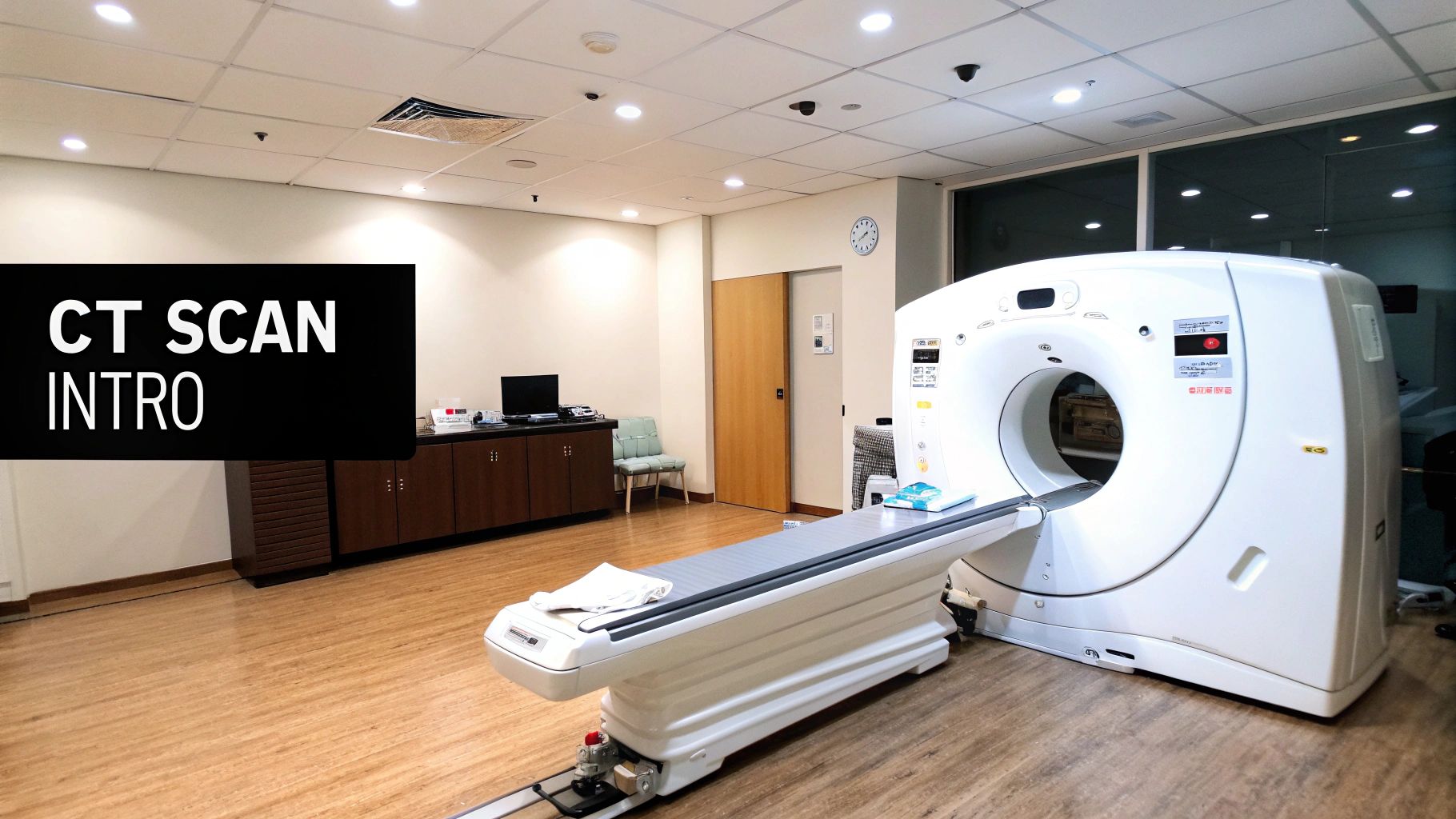
Data Acquisition: The Foundation of Your 3D Model
The process begins with acquiring high-quality CT scan data. Imaging professionals carefully calibrate the CT scanner, optimizing parameters like slice thickness and resolution. These settings influence the detail captured, ultimately impacting the 3D model's precision.
A thinner slice thickness provides more data, leading to a higher-resolution model. However, this also increases the dataset's size, increasing processing demands. Meticulous data acquisition is paramount, as this initial stage sets the foundation for a successful conversion.
Segmentation: Isolating Anatomical Structures
After acquiring the CT scan data, the next crucial step is segmentation. This process isolates specific anatomical structures within the dataset. It's akin to digitally sculpting the desired organ or bone from surrounding tissue.
This isolation uses various techniques, most notably thresholding. Thresholding defines a range of Hounsfield units, representing tissue density, differentiating between tissues.
Segmentation Approaches: Automatic, Semi-Automatic, and Manual
Three primary segmentation approaches exist: automatic, semi-automatic, and manual. Automatic segmentation uses algorithms to isolate structures based on predefined parameters. It's faster but may need manual refinement.
Semi-automatic segmentation combines automated processes with user input, balancing speed and control. Manual segmentation, the most time-consuming but often most accurate, relies on trained professionals who delineate structures slice by slice. The chosen approach depends on the clinical scenario, desired detail, and resources.
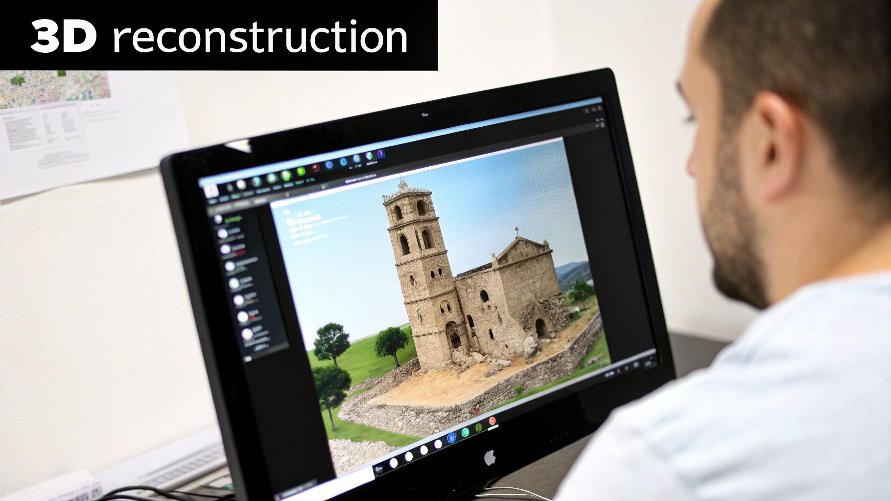
From Segmentation to 3D Model: Building Your Virtual Replica
Post-segmentation, the isolated data is converted into a 3D model by creating a mesh. This network of interconnected triangles or polygons represents the segmented structure's surface. The mesh is refined and textured to create a realistic anatomical representation.
Cardiovascular CT imaging is well-suited for 3D printing because of its high-resolution images. However, factors like voxel isotropy and motion artifacts from ungated imaging can affect quality. ECG gating is crucial for precise anatomical models, especially with small structures like coronary arteries. These models have diverse applications, from surgical planning to education. Learn more about the conversion process.
Software Selection: Choosing the Right Tools for the Job
Choosing the right software for CT scan to 3D model conversion is essential. Consider features, ease of use, system compatibility, and cost. Many popular options offer specialized tools for medical image processing and 3D model creation.
Some cater to specific medical specialties like cardiovascular or orthopedic imaging, providing tailored tools and functionalities. Selecting software that aligns with your specific needs is vital for maximizing efficiency and optimizing results.
To help you choose the right software, we've compiled a comparison table:
CT to 3D Model Software Comparison
Comparison of popular software solutions for converting CT scan data to 3D models, highlighting key features, learning curve, and price points.
| Software | Key Features | Learning Curve | Price Range | Best For |
|---|---|---|---|---|
| 3D Slicer | Open-source, multi-platform, extensive image processing tools, 3D visualization and modeling | Steep | Free | Research, education, advanced users |
| Mimics | Powerful segmentation and modeling tools, CAD integration, specialized modules | Steep | High | Medical device design, bioengineering |
| ITK-SNAP | User-friendly interface, focused on segmentation, basic 3D visualization | Moderate | Free | Clinical applications, segmentation tasks |
| Blender | Open-source, versatile 3D modeling software, can import medical image data | Steep | Free | Artistic modeling, animation, requires scripting for advanced medical applications |
| Meshmixer | Free, user-friendly, focuses on mesh manipulation and repair | Moderate | Free | Mesh editing, preparing models for 3D printing |
This table highlights the varied features and price points available, allowing you to choose software that best meets your needs. Consider your technical expertise and project requirements when selecting.
Precision Matters: Ensuring CT Scan to 3D Model Accuracy
Accuracy is paramount when converting CT scans into 3D models, especially for medical applications. In these situations, the models can directly impact patient care. Several factors contribute to the fidelity of the final 3D model, starting with the initial CT scan parameters. This requires a thorough understanding of the balance between resolution and processing demands.
CT Resolution and Slice Thickness: Balancing Detail and Processing
The resolution of the CT scan and the thickness of each slice significantly affect the level of detail captured. Think of it like building with LEGOs. Thinner bricks (like thinner CT slices) allow for more intricate details but require more bricks and more building time (like increased processing power). This results in higher resolution and a more detailed 3D model, but potentially longer processing times.
Conversely, thicker slices are like larger LEGO bricks. They create a simpler structure that's faster to assemble but lacks finer details. Finding the right balance between detail and processing time is crucial for an effective 3D model.
Segmentation Thresholds: Defining Anatomical Boundaries
Segmentation, the process of isolating anatomical structures, is critical for accurate CT scan to 3D model conversion. This is done by setting appropriate segmentation thresholds. These thresholds act like boundaries in the CT data, based on tissue densities. They determine which data is included in the final model.
For example, during bone segmentation, the threshold differentiates bone tissue from surrounding soft tissue. Incorrect thresholds can lead to missing parts of the bone or the inclusion of unwanted soft tissue. This can compromise the accuracy and usefulness of the 3D model.
This is especially important for surgical planning, where even small inaccuracies can have significant consequences. Maintaining accuracy is a key challenge, particularly when integrating data from different sources. Aligning 3D anatomical models with radiological templates requires precise segmentation and registration techniques to minimize discrepancies.
Studies show that the choice of threshold values significantly impacts the final 3D reconstruction, with mean discrepancies ranging from 0.004 mm to 0.141 mm depending on the method. Achieving optimal alignment is crucial for surgical planning, where accurate models are essential for successful outcomes. Read the full research here.
Validation and Quality Control: Ensuring Model Integrity
Validation protocols are essential for verifying the dimensional accuracy of the final 3D model. This often involves comparing the model to the original CT scan data, quantifying any deviations, and minimizing them. This process acts as a quality control check, ensuring the model accurately reflects the source data.
This rigorous process ensures the 3D model is a faithful representation of the patient's anatomy. This precision is paramount for clinical applications where reliability is non-negotiable. This level of accuracy makes these models suitable for critical uses where precise anatomical details are needed.
AI Revolution: Transforming CT Scan to 3D Model Workflows
Artificial intelligence is changing how medical professionals convert CT scans into 3D models. Processes that once took days are now possible in minutes. This increased speed is largely due to the automation of complex tasks, especially within the segmentation phase.
Automating Segmentation With Deep Learning
Traditionally, segmentation was a manual process requiring significant effort from trained professionals. Now, deep learning algorithms are automating this once labor-intensive task. These algorithms are trained to identify anatomical structures within CT scan data. This not only saves valuable time but also improves the accuracy and consistency of 3D model creation.
Specialized Neural Networks for Medical Imaging
Specialized neural network architectures are key to this advancement in medical imaging. These networks are adept at processing medical images and can identify subtle tissue variations that might be missed by even the most experienced human eye. This ability to discern fine details leads to greater precision in 3D models, particularly for complex anatomical structures.
For example, imagine needing a 3D model of a complicated bone fracture. AI can analyze the CT scan, precisely identifying the fractured fragments and their exact locations. This detailed information allows surgeons to develop targeted surgical plans specific to the fracture pattern.
The use of AI in reconstructing CT scans into 3D models has also yielded impressive results. The DiffusionBlend framework allows for the efficient creation of high-quality 3D CT images from sparse-view scans, reducing the need for high levels of X-ray exposure. This framework uses diffusion models to learn spatial correlations between image slices, significantly improving both the speed and quality of reconstruction. This technique is particularly valuable, as it enhances patient safety by minimizing radiation and accelerates the diagnostic process. Discover more insights about this framework.
Integrating AI into Clinical Workflows
AI tools are designed for seamless integration into existing clinical workflows, encouraging wider adoption within healthcare settings. This integration enhances both the efficiency and accuracy of CT scan to 3D model conversion.
Benefits of AI-Driven Conversion
- Increased Speed: Faster processing translates to quicker turnaround times for time-sensitive procedures like surgical planning.
- Improved Accuracy: AI algorithms are capable of identifying subtle details, potentially exceeding human capabilities.
- Reduced Radiation Exposure: Techniques like sparse-view reconstruction minimize the amount of radiation a patient is exposed to.
- Enhanced Consistency: Automation reduces variability, resulting in more dependable 3D models.
Current Limitations and Future Potential
While AI offers substantial potential, it's important to acknowledge current limitations. These include the need for extensive, high-quality datasets for training and the ongoing challenge of validating AI-generated models. However, continuous research and development are tackling these challenges. The future of AI in medical imaging is promising, holding the potential to further improve patient care across numerous medical specialties.
Clinical Impact: How CT Scan to 3D Model Technology Saves Lives
CT scan to 3D model technology represents a significant advancement in patient care. This technology empowers medical professionals to perform complex procedures, improves patient understanding, and is revolutionizing medical education.
Enhanced Surgical Planning and Execution
One of the most impactful applications of 3D modeling is in pre-operative planning. Surgeons can now visualize a patient's anatomy in 3D, allowing for a more precise and personalized surgical approach. This detailed visualization helps surgeons anticipate potential challenges and optimize surgical strategies.
For example, orthopedic surgeons can practice complex reconstructions on a 3D printed model before the actual surgery. This practice can increase precision and potentially reduce operating times. The benefits extend beyond orthopedics. Neurosurgeons use these models to navigate the intricate structures of the brain, minimizing invasiveness and improving outcomes.
Maxillofacial specialists also use patient-specific 3D models for complex facial reconstructions. The ability to visualize and manipulate a tangible model of a patient's skull before surgery enables more accurate pre-operative planning, particularly in facial trauma cases.
Improved Patient Communication and Informed Consent
3D models are invaluable tools for patient education and informed consent. A physical model of their own anatomy helps patients understand their condition and the planned surgical procedure. This tangible representation empowers patients to ask informed questions, bridging the communication gap and fostering a more patient-centered approach to healthcare.
This increased understanding can also reduce patient anxiety, making the healthcare journey less daunting. Over 70 million CT scans are performed annually in the United States, highlighting their importance in medical diagnostics. Google Research's CT Foundation simplifies 3D CT model development, improving diagnostic efficiency and research.
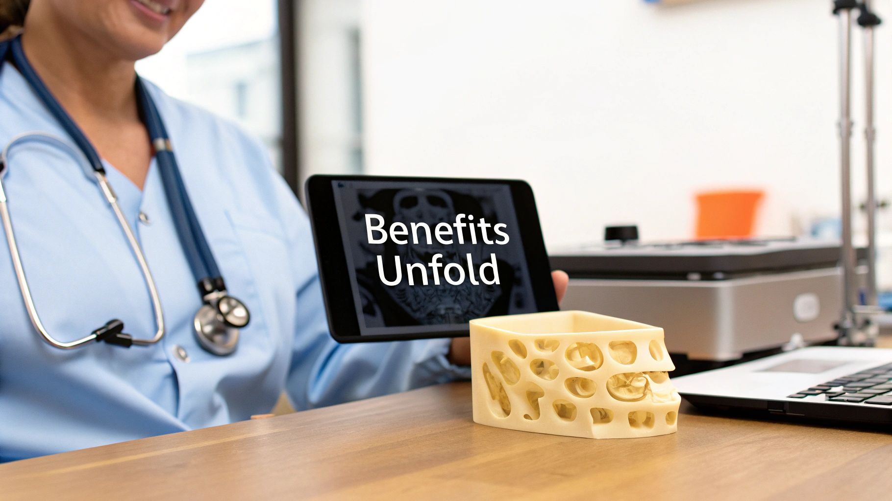
Personalized Implants and Prosthetics
CT scan to 3D model technology significantly benefits the field of patient-specific implants and prosthetics. These models enable the creation of implants and prosthetics perfectly tailored to an individual's anatomy.
This personalization improves fit, function, and patient satisfaction. This represents a substantial advancement, offering solutions that are both functional and improve patients' quality of life.
Transforming Medical Education
Beyond patient care, 3D models are transforming medical education. Students can interact with detailed anatomical representations, gaining a deeper understanding of complex structures and their relationships. This interactive learning enhances comprehension compared to traditional 2D images.
The ability to visualize and manipulate 3D models strengthens students' grasp of spatial relationships within the body, a critical skill for many medical disciplines. This technology equips future medical professionals with a more comprehensive understanding of human anatomy, ultimately contributing to better patient outcomes.
The Future of CT Scan to 3D Model Technology
The future of CT scan to 3D model technology is brimming with exciting innovations. These advancements promise to reshape how these models are created and utilized, focusing on improved speed, accuracy, and accessibility across various medical specialties.
Real-Time Model Generation
Imagine a 3D model of a fractured bone generated instantly during an ER visit. This is the potential of real-time model generation. This technology could drastically change emergency medicine and surgical decision-making, allowing for immediate assessments and treatment plans.
In trauma situations, real-time 3D models could provide surgeons with instant insight into the extent of injuries. This would enable faster, more informed decisions during critical moments, leading to more positive patient outcomes.
Immersive Visualization with Augmented and Virtual Reality
Augmented and virtual reality (AR/VR) are transforming surgical interaction with 3D models. These technologies create immersive visualization environments, allowing surgeons to virtually "step inside" a patient's anatomy.
This pre-operative planning capability is invaluable. Surgeons can examine structures, explore surgical approaches, and gain a more thorough understanding of the patient's anatomy. This leads to improved surgical precision and potentially better results.
Multi-Material 3D Printing
3D printing continues to evolve, with multi-material printing offering significant advantages in surgical training. These printers create models with tissue-specific properties, allowing surgeons to realistically simulate procedures.
A surgeon can practice a complex procedure on a model that mimics the feel of bone and muscle. This tactile feedback enhances surgical training and improves preparedness for real-world operations.
Cloud-Based Collaboration
Cloud computing is breaking down geographical barriers. Cloud-based platforms enable medical teams worldwide to access and interact with 3D models concurrently. This fosters international consultations, knowledge sharing, and global medical education.
A surgeon in New York can consult with a specialist in London using the same 3D model. They can discuss strategies and collaboratively determine the best approach. This global collaboration is transforming medical communication and raising the standard of care.
Clinical Outcomes and Future Directions
To illustrate the tangible benefits, let's examine some statistical data demonstrating how 3D models enhance surgical outcomes across different medical specialties. The following table shows some key improvements achieved using this technology:
| Medical Specialty | Procedure Type | Reduction in OR Time | Complication Reduction | Patient Satisfaction |
|---|---|---|---|---|
| Orthopedics | Joint Replacement | 15% | 10% | 20% |
| Neurosurgery | Tumor Resection | 20% | 12% | 15% |
| Cardiology | Valve Repair | 10% | 8% | 18% |
As you can see, the integration of 3D models leads to significant improvements across several key metrics, including a reduction in operating room time, a decrease in complications, and increased patient satisfaction.
These advancements highlight the significant potential of CT scan to 3D model technology. Continued innovation promises even more impactful healthcare applications.
Ready to explore the future of medical imaging? PYCAD specializes in integrating artificial intelligence into medical imaging workflows. Learn more about how PYCAD can empower your medical device with advanced AI capabilities.