Viewing Medical Images Made Easy
Navigating the intricacies of medical imaging demands tools that are both powerful and user-friendly. DICOM (Digital Imaging and Communications in Medicine) is the established standard for medical images. However, accessing and interpreting these files can be difficult without specialized software. From quick consultations to extensive research, the right DICOM viewer can significantly impact efficiency and the accuracy of diagnoses. This article explores seven leading DICOM viewers, outlining their features, functionalities, and pricing to help you make an informed choice.
Choosing the right DICOM viewer is essential for a variety of professionals, including those developing medical devices, conducting research, managing hospital IT infrastructure, or launching medtech startups. Key factors to consider include platform compatibility (Windows, macOS, Linux, web-based), advanced features such as 3D rendering and image manipulation tools, integration with existing systems like PACS and HIS, and budgetary limitations.
An effective DICOM viewer provides seamless navigation, clear image display, robust measurement tools, and customizable workflows. These features ultimately enhance collaboration and improve patient care. Some viewers offer free versions with limited functionality, while others require subscriptions or one-time purchases to unlock premium features. Technical aspects, like hardware requirements and the potential for cloud deployment, should also be considered.
By the end of this article, you'll have a solid understanding of the strengths and weaknesses of each viewer. This will empower you to choose the ideal tool to fulfill your specific medical imaging needs.
1. Horos
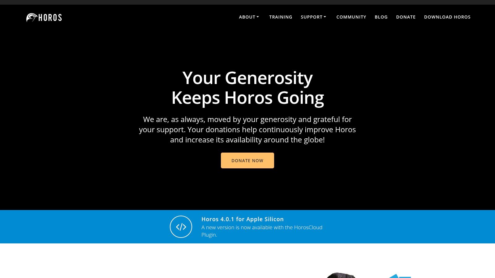
Horos is a powerful, free, and open-source DICOM viewer designed for macOS. Built upon the foundation of OsiriX, it presents a compelling alternative to commercial viewers. This makes it particularly attractive to researchers, academic institutions, and medtech startups seeking robust functionality without the high cost. For macOS users, it’s a top contender for daily DICOM image viewing and analysis.
Horos boasts a comprehensive feature set, rivaling many commercial options. It offers advanced 3D and 4D rendering capabilities, including volume rendering, maximum intensity projection (MIP), and surface rendering. These features are crucial for visualizing complex anatomical structures and pathologies. This makes Horos a valuable tool for medical device manufacturers developing and testing new imaging technologies. Healthcare technology companies integrating DICOM visualization into their platforms also benefit from its capabilities.
Beyond visualization, Horos facilitates in-depth image analysis. It provides tools such as multiplanar reconstruction (MPR), thick slab viewing, and a suite of ROI measurement and annotation tools. These functionalities are essential for medical researchers and scientists conducting quantitative image analysis. They allow for precise measurements and detailed annotations for research publications and presentations. Hospital and clinic IT departments can also use Horos for reviewing medical images and collaborating with physicians.
Key Features
- Advanced 3D/4D Rendering: Explore intricate anatomical details with volume rendering, MIP, and surface rendering.
- Complete DICOM Support: View a wide array of DICOM formats and utilize query/retrieve functionality for seamless data access.
- Multiplanar Reconstruction (MPR) and Thick Slab Viewing: Reconstruct images in any plane and visualize thicker slices for enhanced diagnostic capabilities.
- ROI Measurements and Annotation Tools: Perform precise measurements and add annotations directly to images for detailed analysis and reporting.
- DICOM PACS Server Integration: Integrate with existing PACS infrastructure for streamlined workflow and data management.
Pros and Cons
Here’s a quick look at the advantages and disadvantages of using Horos:
Pros:
- Free and Open-Source: Eliminates licensing costs, increasing accessibility.
- Robust Feature Set: Provides a wide range of tools comparable to paid software.
- Excellent 3D Visualization: Enables detailed exploration of complex anatomical structures.
- Active Community Support: Benefit from a helpful community of users and developers.
Cons:
- macOS Only: Limited to the macOS operating system.
- Resource-Intensive: Large datasets may require high-end hardware.
- Steeper Learning Curve: Mastering advanced features takes time and effort.
- Limited Technical Support: Primarily relies on community resources.
Getting Started with Horos
Website: https://horosproject.org/
Implementation Tips:
- Download the latest version from the official Horos website.
- Ensure your macOS system meets the minimum hardware requirements.
- Familiarize yourself with the user interface and navigation.
- Explore the available documentation and online tutorials.
- Engage with the Horos community for support and guidance.
Horos offers a compelling balance of power and accessibility. While the macOS exclusivity and potential resource demands are factors to consider, its rich feature set and open-source nature make it valuable. For those working within the Apple ecosystem, Horos offers a strong alternative to commercial DICOM viewers.
2. RadiAnt DICOM Viewer
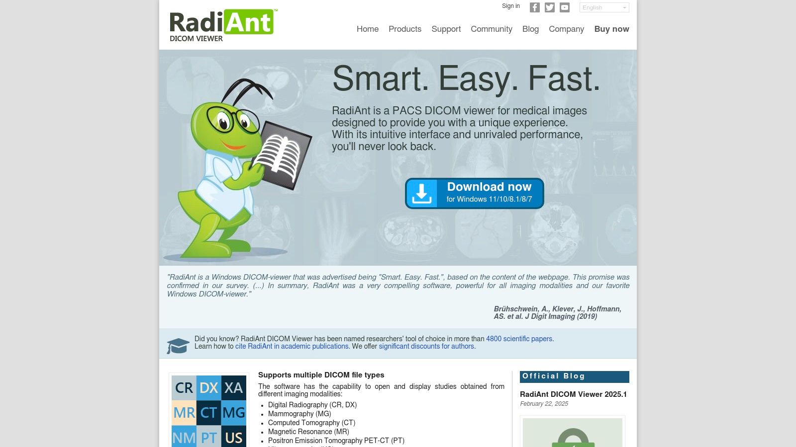
RadiAnt DICOM Viewer is a powerful, lightweight DICOM viewer designed for Windows. It caters specifically to radiologists and medical professionals who need a fast and efficient solution for their daily clinical workflow. Its intuitive interface and focus on speed make it an attractive option for those prioritizing rapid image loading and seamless navigation.
This viewer excels in situations demanding quick image review, such as during patient consultations or emergency procedures. The mouse-driven navigation and measurement tools are designed for ease of use. This minimizes the learning curve for new users and allows experienced professionals to work with maximum efficiency. Essential features like MPR (multiplanar reconstruction) provide valuable diagnostic insights. The ability to view and import DICOM CD/DVDs simplifies data management within clinical settings.
RadiAnt also supports 2D/3D image processing with MIP, MinIP, and volume rendering, offering advanced visualization for complex cases. The integrated PACS connectivity, using DICOM query/retrieve, streamlines workflow by enabling direct access to patient images from the PACS server.
Use Cases Across the Medical Industry
For medical device manufacturers, healthcare technology companies, and medtech startups, RadiAnt can be a valuable tool. It’s particularly useful for testing and demonstrating imaging software and hardware compatibility. Its low system requirements and ease of implementation make it perfect for rapid integration into testing environments. Hospital and clinic IT departments will appreciate the straightforward deployment and minimal training needed for staff.
Academic institutions focused on medical imaging can use the free version for educational purposes. Researchers and scientists can explore the paid version for its image processing capabilities. However, its research-focused functionality may be less extensive than dedicated research platforms like OsiriX MD or 3D Slicer. DICOM communication and transfer companies can use RadiAnt as a client-side viewer for verifying successful data transfer and integrity.
Features, Benefits, and Drawbacks
Key Features and Benefits:
- Speed and Efficiency: RadiAnt offers exceptionally fast image loading and smooth navigation, crucial for busy clinical environments.
- User-Friendly Interface: The intuitive design requires minimal training, enabling rapid adoption by medical staff.
- Essential Tools: The viewer provides core DICOM viewing features, including MPR, measurement tools, and 2D/3D processing.
- PACS Connectivity: Direct access to patient data via DICOM query/retrieve simplifies workflow integration.
- Affordable Pricing: RadiAnt offers both a free version for basic viewing and a paid version for advanced functionalities.
Pros:
- Exceptionally fast image loading and fluid navigation.
- User-friendly interface requiring minimal training.
- Affordable pricing, including a free version.
- Low system requirements.
Cons:
- Windows-only platform.
- Some advanced features are exclusive to the paid version.
- Fewer comprehensive tools than enterprise solutions like OsiriX MD or 3D Slicer.
- Limited research-oriented functionality.
Implementation and Evaluation
Website: https://www.radiantviewer.com/
To quickly evaluate RadiAnt’s capabilities, download and install the free version. The paid version offers a trial period, allowing thorough assessment of the advanced features before purchasing. Contact the vendor for pricing and licensing details. While system requirements are low, ensure your hardware meets the recommended specifications for optimal performance.
RadiAnt DICOM Viewer balances speed, functionality, and affordability. This makes it a practical choice for various medical imaging applications. It’s particularly well-suited for clinical settings prioritizing efficiency and ease of use. However, users needing advanced research tools or cross-platform compatibility should consider more comprehensive alternatives.
3. OsiriX MD
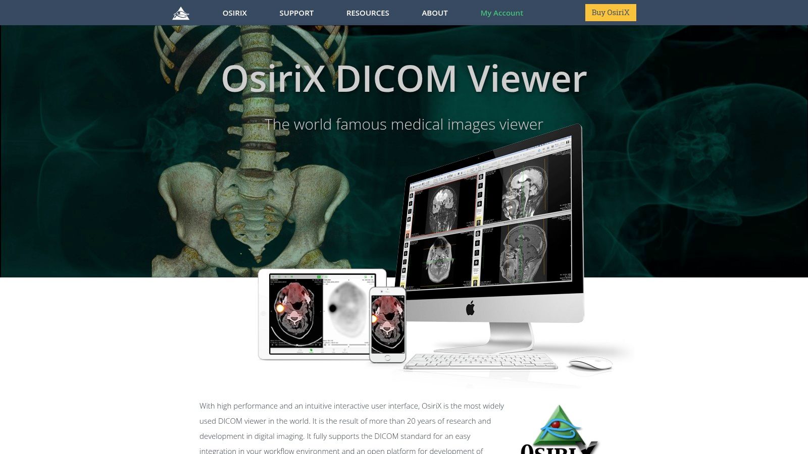
OsiriX MD stands out as the leading FDA-cleared DICOM viewer for macOS. It's designed specifically for the demanding world of diagnostic imaging in professional medical environments. While other viewers might offer basic DICOM viewing, OsiriX MD provides advanced tools. These include 3D/4D rendering, image fusion, precise measurements, and comprehensive DICOM networking.
This robust platform caters to a diverse range of users. Clinicians can make critical diagnostic decisions with confidence, while medical researchers can explore intricate datasets. It's a powerful solution for anyone working with medical imaging.
OsiriX MD also serves as a valuable tool for medical device manufacturers and healthcare technology companies. They can utilize it for testing and validating their imaging devices and software. Its FDA clearance and comprehensive feature set offer a reliable environment for assessing image quality and performance, ensuring compatibility.
Key Features and Benefits
-
FDA-Cleared for Diagnostic Imaging (510k): OsiriX MD's 510(k) clearance from the FDA sets it apart. Unlike many free or low-cost viewers, it can be used for primary diagnosis, making it a crucial tool for clinicians and researchers needing a legally compliant and trustworthy diagnostic tool.
-
Advanced 3D/4D Rendering and Fusion Techniques: These features enable detailed visualization and analysis of complex anatomical structures. This is particularly beneficial in specialties like radiology, cardiology, and oncology, where precise imaging is paramount.
-
Sophisticated Measurement and Analysis Tools: OsiriX MD offers a wide range of tools for accurate measurements, region of interest analysis, and quantitative image assessment. This supports both clinical diagnosis and research applications, offering valuable insights into patient data.
-
Complete DICOM Networking Capabilities: The software integrates seamlessly with PACS systems and other DICOM devices, enabling efficient image sharing and streamlined workflow integration within healthcare settings.
-
Extensive Plugin Architecture for Customization: Users can extend OsiriX MD's functionality with specialized plugins tailored to specific needs, adding a layer of personalization to the platform.
Pros and Cons
Pros:
- FDA cleared for primary diagnosis: This ensures legal compliance and reliability.
- Comprehensive feature set: A wide array of advanced tools is available for visualization, analysis, and processing.
- Regular updates: The software stays current with the latest advancements in medical imaging through continuous updates and new clinical tools.
- Professional technical support: Dedicated support is provided for troubleshooting and assistance.
Cons:
- Cost: At $699 for a standard license, it's a significant investment.
- macOS only: This limits accessibility for users on other operating systems.
- Resource intensive: Demanding tasks like 3D rendering may require a powerful Mac configuration.
- Learning curve: The extensive feature set can be initially challenging to master.
Getting Started with OsiriX MD
- System Requirements: Ensure your Mac meets the minimum system requirements.
- Performance: A dedicated high-performance Mac is recommended for optimal performance, especially with large datasets or complex 3D rendering.
- Training: Utilize the available tutorials and documentation to learn the software's features.
- Plugins: Explore the plugin architecture to add functionality based on your specific needs.
Website: https://www.osirix-viewer.com/
While the cost and macOS exclusivity may be drawbacks, OsiriX MD's comprehensive features, FDA clearance, and advanced capabilities make it a powerful tool. For professionals requiring a top-tier diagnostic imaging solution, OsiriX MD's focus on advanced visualization, measurement, and analysis justifies its premium price. It provides a level of functionality and performance that sets it apart from basic viewers.
4. MicroDicom
MicroDicom is a free and accessible DICOM viewer known for its simplicity and speed. It's an ideal choice for educational purposes, quick image reviews, or situations with limited hardware resources. Medical students, researchers performing quick image analysis, or clinicians seeking a lightweight viewer outside their primary PACS system will appreciate its user-friendly design. Even medtech startups can utilize MicroDicom for basic image validation during proof-of-concept projects.
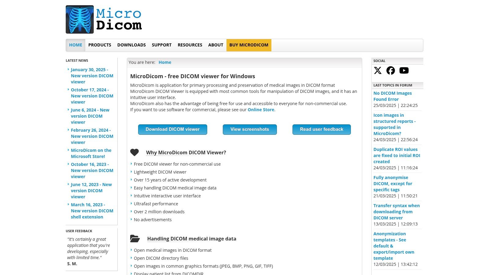
MicroDicom prioritizes core DICOM viewing functionalities, avoiding the complexity of advanced features often found in more comprehensive, and often costly, viewers. This minimalist approach leads to a small file size and minimal system requirements. As a result, it runs smoothly even on older or less powerful hardware, a significant advantage for environments with limited IT budgets or for fieldwork using portable devices.
Key Features
- Basic Measurement and Annotation: MicroDicom offers tools to measure length, angle, and area, and allows text annotation for image analysis.
- Windowing and Image Manipulation: Users can adjust brightness and contrast, and apply preset windowing levels to optimize image visualization for various tissues and structures.
- DICOM Directory Scanning and Thumbnail Previews: Navigate DICOM folders and preview images quickly with generated thumbnails.
- Histogram Analysis and Pixel Data Viewing: Examine pixel value distribution and inspect raw pixel data for in-depth image analysis.
- Export to Common Image Formats: Save images in JPEG, BMP, or TIFF format for presentations, reports, or sharing.
Pros
- Completely Free: MicroDicom is free to download and use, making it an excellent option for budget-conscious users.
- Small Footprint: Minimal system requirements and small file size ensure quick installation and efficient performance.
- Simple Interface: The intuitive interface is easy to learn, even for those new to DICOM viewers.
- Fast Startup and Image Loading: Experience quick startup and rapid image loading for improved workflow efficiency.
Cons
- Limited Advanced Visualization: MicroDicom lacks advanced features like 3D rendering, MIP, and MPR, making it unsuitable for complex clinical diagnoses or research requiring these capabilities.
- Minimal PACS Connectivity: Limited PACS integration restricts its use in complex clinical workflows reliant on PACS communication. It is best suited for standalone image review.
- Less Suitable for Complex Clinical Workflows: Its simplicity makes it less ideal for high-volume clinical environments or those requiring advanced image analysis tools.
Website: https://www.microdicom.com/
Implementation Tips
- Download the correct version for your Windows operating system from the official website.
- Installation is simple and requires minimal user input.
- While the interface is intuitive, the website offers documentation and tutorials for users new to DICOM viewing software.
MicroDicom fills a specific need by providing a free, lightweight, and accessible DICOM viewer. While not suited for complex clinical scenarios requiring advanced visualization and PACS integration, its simplicity and speed make it valuable for education, basic research, and quick image review. It's a good alternative to complex viewers when resources are limited or basic functionality is all that's required.
5. 3D Slicer
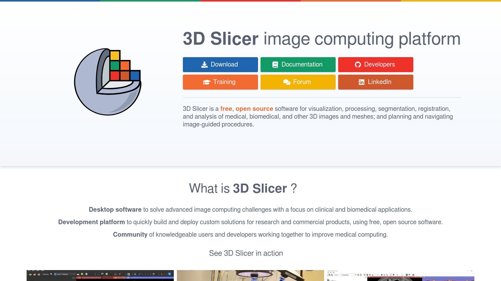
3D Slicer is a leading open-source software package for advanced visualization and analysis of DICOM images. Unlike basic DICOM viewers that offer limited functionality, 3D Slicer provides a comprehensive platform for research, development, and education in medical imaging. It goes beyond simple viewing, offering powerful tools for segmentation, registration, 3D modeling, and even surgical planning.
This makes it particularly valuable for organizations and individuals working on medical image analysis algorithms, surgical simulations, and image-guided interventions. 3D Slicer offers an extensive toolkit to manipulate and visualize DICOM data, exceeding the capabilities of standard viewers. Medical device manufacturers and healthcare technology companies can use 3D Slicer for prototyping new devices and algorithms, while researchers can leverage its powerful tools for detailed image analysis.
Key Features and Benefits
-
Advanced Segmentation and Registration Tools: 3D Slicer provides a robust set of tools for image segmentation, allowing users to isolate specific anatomical structures or regions of interest. Its registration capabilities enable accurate alignment of multiple images, crucial for tasks like image fusion and longitudinal studies.
-
Extensive 3D Visualization and Volume Rendering: Explore and visualize complex 3D medical image datasets with ease. The rendering engine facilitates interactive exploration and manipulation of volume data, providing valuable insights into anatomical structures.
-
Cross-Platform Compatibility: Available on Windows, macOS, and Linux, ensuring accessibility across various operating systems commonly used in research and development.
-
Extensible Platform with Numerous Specialized Modules: 3D Slicer's modular architecture is a key strength. Extend its functionality through a vast library of extensions addressing specific needs, such as imaging modalities, analysis techniques, or integration with surgical navigation systems. This adaptability makes it suitable for a wide array of research and clinical workflows.
-
Support for Various Image Formats Beyond DICOM: Although primarily focused on DICOM, 3D Slicer supports other medical image formats, offering flexibility and integration with diverse data sources.
Pros
-
Free and Open-Source: No licensing fees encourage widespread adoption and contribute to a vibrant, supportive community.
-
Powerful Research Capabilities: Provides tools for advanced image processing typically not found in basic DICOM viewers.
-
Cross-Platform Availability: Facilitates collaboration across different operating systems.
-
Extensive Plugin Ecosystem: Allows users to customize the platform according to specific research requirements.
Cons
-
Steep Learning Curve: Mastering the advanced features requires time and effort.
-
Not FDA-Cleared for Clinical Diagnosis: Primarily designed for research and not intended for direct diagnostic use in clinical settings.
-
Complex Interface: The numerous features can initially feel overwhelming for new users.
-
High Computing Resource Demands: Advanced visualization and processing tasks may necessitate a powerful computer with sufficient RAM and a dedicated graphics card.
Implementation/Setup Tips
-
Download the correct version for your operating system from the official website.
-
Explore the available online documentation and tutorials.
-
Start with simpler modules before exploring the more advanced features.
-
Utilize the community forums for support and guidance.
3D Slicer is an invaluable tool for researchers, developers, and educators seeking a powerful, free platform for advanced DICOM processing and visualization. While its complexity may present a challenge initially, the extensive capabilities make it an essential resource for advancing medical imaging innovation.
6. Syngo.via (Siemens Healthineers)
Syngo.via from Siemens Healthineers is an enterprise-grade clinical imaging software solution. It's designed for advanced visualization, analysis, and diagnostic reading across various medical imaging modalities. Its comprehensive features and integration capabilities make it a popular choice for large healthcare institutions. However, its complexity and cost may be less suitable for smaller practices. Syngo.via stands out due to its robust features, AI integration, and focus on streamlined clinical workflows within a hospital setting.
Practical Applications and Use Cases
Syngo.via excels in complex diagnostic situations. In oncology, its advanced visualization tools allow for precise tumor segmentation and volumetric analysis, assisting in treatment planning and monitoring. In cardiology, dedicated workflows enable efficient assessment of cardiac function and blood flow.
Neurologists can use Syngo.via for detailed brain imaging analysis, including diffusion tensor imaging (DTI) for visualizing white matter tracts. The platform's multi-modality capabilities allow clinicians to correlate findings from different imaging exams (e.g., CT, MR, PET) for a more complete patient overview.
Key Features and Benefits
-
Specialized Clinical Workflows: Optimized workflows for specific specialties (e.g., cardiology, oncology, neurology) improve efficiency and standardize reporting.
-
Advanced Visualization and Quantification: Tools for 3D rendering, multiplanar reformatting (MPR), maximum intensity projection (MIP), and volumetric analysis empower in-depth image interpretation.
-
AI-Powered Assistance: Integration with Siemens' AI-Rad Companion provides automated image analysis, lesion detection, and quantification, helping radiologists make faster, more accurate diagnoses.
-
Seamless Integration: Syngo.via integrates with RIS/PACS and EMR systems, facilitating efficient data sharing and workflow optimization.
-
Comprehensive Modality Support: Supports all major imaging modalities, including CT, MR, PET, SPECT, ultrasound, and X-ray.
Pros
-
Enterprise-Grade Reliability and Support: Backed by Siemens Healthineers, Syngo.via offers strong support and maintenance.
-
Streamlined Clinical Workflows: Optimized workflows improve efficiency and reduce report turnaround times.
-
Advanced AI Integration: AI-powered tools enhance diagnostic accuracy and speed.
-
Comprehensive Training and Implementation Support: Siemens provides training and support for successful implementation and user adoption.
Cons
-
Very Expensive Enterprise Pricing Model: Syngo.via's pricing is designed for larger institutions and can be expensive for smaller practices. Contact Siemens for pricing details.
-
Requires Significant IT Infrastructure: Implementation requires substantial IT resources, including powerful servers and dedicated network bandwidth.
-
Can be Complex to Implement and Maintain: The software's complexity can make implementation and maintenance challenging.
-
Vendor Lock-in with Siemens Ecosystem: Deep integration can lead to vendor lock-in with Siemens.
Implementation/Setup Tips
-
Thorough Planning: Engage with Siemens Healthineers early to assess your needs and develop an implementation strategy.
-
Dedicated IT Resources: Allocate sufficient IT resources for installation, configuration, and maintenance.
-
Comprehensive Training: Ensure all users receive adequate training.
-
Ongoing Optimization: Regularly evaluate and optimize workflows for continued efficiency.
Comparison With Similar Tools
Syngo.via competes with platforms like 3D Slicer (open-source), MIM Software's MIMics, and Philips IntelliSpace Portal. While 3D Slicer is free and flexible, it may not have the same enterprise-grade support and integration. MIMics focuses on image processing and 3D modeling. IntelliSpace Portal is Philips' counterpart, offering similar advanced visualization and analysis capabilities.
Website: https://www.siemens-healthineers.com/medical-imaging-it/advanced-visualization-solutions/syngo-via
7. Weasis
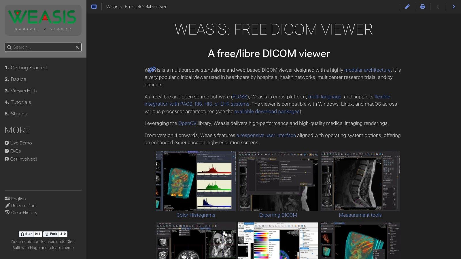
Weasis secures a spot on this list because of its effective combination of open-source availability, standards compliance, and web integration capabilities. It's a truly versatile DICOM viewer, suitable for a wide variety of uses, from individual researchers to expansive healthcare organizations. While it may not have the sleek, polished interface of some commercial viewers, its flexibility and cost-effectiveness make it a powerful option.
Weasis is especially appealing to organizations seeking a DICOM viewer that seamlessly integrates into their current web infrastructure. This makes it a superb choice for teleradiology setups, facilitating remote consultations and efficient image sharing. Its cross-platform compatibility further enhances this versatility, allowing for deployment across Windows, macOS, and Linux systems. For medical device manufacturers and medtech startups, Weasis presents a solid base for building and testing DICOM-compatible products without the burden of licensing fees. Academic institutions and medical researchers can benefit from its open-source nature for educational purposes and contribute to its ongoing development.
Key Features and Benefits
- Cross-Platform Compatibility: Deployable on Windows, macOS, and Linux, offering flexibility for varied environments.
- Web-Based Deployment: Integrates seamlessly with web-based healthcare systems, simplifying teleradiology and remote image access.
- MPR and Basic 3D Visualization: Provides essential visualization tools for medical image analysis.
- Integration with Major PACS/VNA Systems: Connects with existing hospital infrastructure for streamlined, efficient workflows.
- Customizable Interface and Plugins: Allows users to tailor the viewer to their unique needs and extend its functionality.
- Free and Open-Source: Eliminates licensing costs and fosters community development.
- Standards-Compliant: Ensures smooth interoperability with other DICOM-compliant systems.
Pros
- Free and open-source, offering a cost-effective solution.
- Excellent cross-platform compatibility for wide deployment.
- Designed for web integration and teleradiology applications.
- Strong adherence to DICOM standards ensures interoperability.
Cons
- User interface may appear less refined than commercial options.
- Advanced visualization features are somewhat limited.
- Requires a Java Runtime Environment, which may involve additional setup.
- Documentation can be technically dense and less user-friendly than some commercial products.
Technical Requirements
A Java Runtime Environment (JRE) is required to run Weasis.
Implementation/Setup Tips
- Download the Weasis distribution from the official website.
- Ensure a compatible JRE is installed on your system.
- For web integration, review the documentation for specific instructions on deploying Weasis within your web server environment. A dedicated server is recommended for optimal performance when managing a large volume of medical images.
Comparison with Similar Tools
Commercial viewers like OsiriX MD offer more advanced visualization tools and a more refined user experience, but Weasis provides a compelling free and open-source alternative. Horos, another open-source option, is macOS-specific, whereas Weasis supports a wider range of operating systems. For organizations prioritizing web integration and cost-effectiveness, Weasis presents a valuable choice.
Website: https://nroduit.github.io/en/
Top 7 DICOM Viewers: Feature Comparison
| Solution | Core Features ★ | Experience & Support 🏆 | Value Proposition 💰 | Target Audience 👥 |
|---|---|---|---|---|
| Horos | Advanced 3D/4D rendering, full DICOM support, MPR, ROI tools | User-friendly interface; active community; steeper learning for advanced use | Free & open-source | Radiologists, Physicians, Researchers |
| RadiAnt DICOM Viewer | 2D/3D imaging, MPR, PACS connectivity | Intuitive, fast performance; minimal training required | Affordable with a free version option | Clinicians, Radiologists (Windows) |
| OsiriX MD | FDA-cleared diagnostic imaging, advanced 3D/4D, extensive plugins | Comprehensive features; professional technical support; resource intensive | Premium diagnostic solution | Medical Professionals (macOS) |
| MicroDicom | Basic DICOM viewing, annotations, export to common formats | Simple, lightweight, fast startup | Completely free; minimal system requirements | Students, Researchers, Clinicians |
| 3D Slicer | Advanced segmentation, registration, 3D visualization, modular design | Highly powerful with a steep learning curve; extensive plugin ecosystem | Free & open-source with rich research capabilities | Researchers, Advanced Users, Multi-platform |
| Syngo.via | Specialized clinical workflows, AI-powered tools, multi-modality support | Enterprise-grade reliability; robust support; complex integration | High-end pricing with premium features | Large Healthcare Institutions |
| Weasis | Cross-platform compatibility, web-based deployment, customizable UI | Standards-compliant; integration-ready; less polished interface | Free & open-source solution | Healthcare Systems, Teleradiology Applications |
Choosing the Right DICOM Viewer
Selecting the right DICOM viewer is a critical decision for any healthcare professional or organization working with medical images. With a variety of options available, including Horos, RadiAnt DICOM Viewer, OsiriX MD, MicroDicom, 3D Slicer, Syngo.via, and Weasis, understanding your specific needs is paramount. Making an informed choice ensures efficient and effective image analysis.
Several factors play a key role in choosing the ideal DICOM viewer:
-
Platform Compatibility: Consider your operating system (Windows, macOS, Linux) and whether you need a desktop, web, or mobile solution. Choosing a viewer that seamlessly integrates with your existing infrastructure is essential.
-
Feature Set: DICOM viewers offer a wide range of capabilities, from basic image viewing and manipulation to advanced features like 3D reconstruction, segmentation, and AI integration. Evaluate your workflow requirements and prioritize the features you need most.
-
Budget: DICOM viewers can range from free and open-source options to premium commercial products. Setting a budget early on helps narrow your choices and ensures you find a cost-effective solution.
-
Technical Expertise: Some viewers demand more technical knowledge for installation and configuration. Consider your team's technical skills to ensure a smooth implementation and effective usage.
-
Integration and Compatibility: Assess the viewer’s compatibility with your current PACS, RIS, and other healthcare IT systems. Seamless integration is crucial for optimized workflows and data exchange.
-
Implementation and Getting Started: A straightforward deployment process and readily available support resources are essential for a smooth transition. Look for viewers with comprehensive documentation and readily accessible support channels.
Key Takeaways
-
The right DICOM viewer streamlines medical image analysis, enhancing efficiency and diagnostic accuracy.
-
Consider factors like platform compatibility, features, budget, technical skills needed, and integration requirements when making your decision.
-
Careful evaluation will help you find a viewer that perfectly aligns with your workflow and resource availability.
Integrating AI into medical imaging demands a comprehensive platform that goes beyond basic viewing capabilities. PYCAD offers a robust solution for medical device manufacturers, healthcare technology companies, and research institutions seeking to incorporate cutting-edge AI into their imaging workflows. PYCAD supports data annotation, anonymization, deploying trained models as APIs, and creating MVP UIs. This enables optimized medical devices, increasing diagnostic accuracy and operational efficiency. Learn more and explore PYCAD solutions at https://pycad.co.
