
The Critical Foundation of Heart Segmentation

Heart segmentation is the process of isolating specific heart regions within medical images. This crucial step allows healthcare professionals to analyze and understand the heart's structure and function with greater precision. This precise mapping is essential for accurate diagnosis, treatment planning, and monitoring various cardiovascular diseases.
For example, knowing the exact dimensions of the left ventricle allows doctors to calculate the ejection fraction, a key indicator of heart health. This measurement provides critical insights into the heart's ability to pump blood effectively.
Challenges In Heart Segmentation
The heart's continuous motion presents a significant challenge for effective segmentation. The subtle differences in tissue density, combined with the constant beating, create a complex imaging environment. Respiratory movement adds another layer of complexity, making it challenging to acquire clear, consistent images.
These combined factors create motion artifacts and blur, hindering accurate segmentation. The resulting distortions can obscure important details and make it difficult to define the boundaries of different heart structures.
Moreover, segmenting cardiac structures like the left ventricle (LV) and right ventricle (RV) from Magnetic Resonance Imaging (MRI) can be challenging due to poor contrast between the ventricular blood pool and myocardium. There's also significant shape variability between patients.
This variability has led to the development of sophisticated models, often using AI-based approaches like deep learning, to automate segmentation. These advanced techniques aim to improve the accuracy of clinical indices, such as ventricular volumes. As of 2022, over 200 studies have focused on cardiac MRI segmentation, with short-axis images being the most common modality. Explore this topic further here.
Why MRI Is The Gold Standard
Despite these complexities, Magnetic Resonance Imaging (MRI) remains the preferred method for heart segmentation. MRI offers superior soft tissue contrast compared to other imaging techniques, allowing for detailed visualization of heart structures.
This enhanced clarity is crucial for accurately defining the boundaries between different heart chambers and the surrounding tissues. This precise delineation forms the basis for accurate measurements and subsequent analyses.
The Impact of Accurate Heart Segmentation
Accurate heart segmentation significantly impacts patient care. It allows physicians to measure ventricular volumes, assess wall thickness, and identify areas of scar tissue with increased confidence. This precise anatomical information empowers doctors to make more informed treatment decisions.
Furthermore, accurate segmentation enables effective monitoring of treatment efficacy over time. This ability to track changes provides valuable insights into the progression of cardiovascular disease and allows for adjustments to treatment plans as needed. Ultimately, accurate heart segmentation contributes to improved patient outcomes and supports a more personalized approach to cardiovascular care. This personalized approach is increasingly important for managing complex conditions and tailoring treatment strategies to individual patient needs.
How AI Transformed Heart Segmentation Overnight
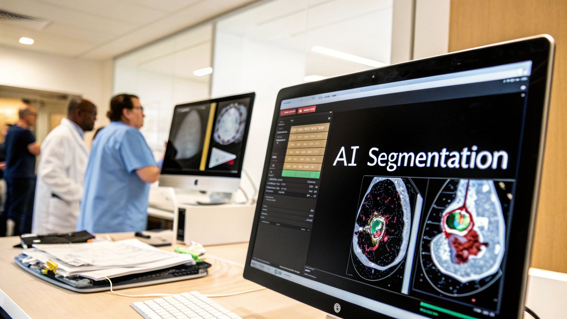
Building upon existing heart segmentation techniques, Artificial Intelligence has significantly changed how we analyze cardiac images. Time-consuming manual processes are now automated, leading to improvements in both speed and accuracy. This shift has dramatically impacted cardiovascular care.
The Rise of Deep Learning in Heart Segmentation
Deep learning algorithms excel at identifying complex patterns within images, making them well-suited for tasks like heart segmentation. These algorithms can differentiate subtle variations in tissue density and accommodate the heart's continuous motion, improving on traditional methods. This increased precision leads to more accurate diagnoses.
Deep learning models like U-Net, with its encoder-decoder architecture, are especially effective. U-Net captures contextual information and fine details critical for accurate segmentation. Attention mechanisms, inspired by human visual focus, further refine the model’s ability to isolate target structures.
Deep learning's efficient extraction and utilization of complex image features have solidified its role in cardiac image analysis. Studies reveal that the majority of AI-based cardiac MRI segmentation research employs deep learning, particularly for ventricle segmentation. Learn more about this research. The widespread adoption of AI highlights its potential to reshape cardiology.
Overcoming Data Challenges
Training deep learning models requires large datasets. However, acquiring extensive, labeled cardiac datasets is challenging. This scarcity has driven innovative approaches to maximize the use of available data.
One approach is data augmentation, modifying existing images to create synthetic variations. These variations expose the model to broader anatomical variability, increasing its robustness. Another is transfer learning, fine-tuning a model pre-trained on a larger, related dataset (like general medical images) for heart segmentation. This utilizes existing knowledge and reduces the need for massive specialized datasets.
The Impact on Speed and Accuracy
To illustrate the key differences between AI-powered and traditional methods, let's examine a comparison table:
To illustrate the key differences between AI-powered and traditional methods, let's examine a comparison table:
AI vs. Traditional Heart Segmentation: The Numbers
Real-world performance comparison between AI-powered and conventional heart segmentation approaches
| Method Type | Accuracy (Dice Score) | Processing Time | Manual Intervention Needed | Handling of Anatomical Variability |
|---|---|---|---|---|
| Traditional (Manual) | ~0.80 – 0.90 | Hours | High | Limited |
| AI-Powered (Deep Learning) | ~0.90 – 0.95 | Seconds/Minutes | Minimal | High |
This table highlights the advantages of AI-based methods, showcasing improved accuracy and drastically reduced processing times. While manual methods typically achieve Dice scores between 0.80 and 0.90 and take hours to process, AI-powered methods achieve higher scores (0.90-0.95) in seconds or minutes. AI’s superior handling of anatomical variability is also a significant advantage.
The benefits of AI in heart segmentation are clear. Processing times have decreased dramatically, from hours to mere seconds, enabling faster diagnoses and treatment decisions. Accuracy has also improved substantially due to deep learning's ability to handle anatomical differences between patients. The Dice Similarity Coefficient (DSC), a standard metric for segmentation accuracy, demonstrates this improvement with high scores for certain algorithms. This metric compares automated segmentations with manual ground truths, showcasing the power of AI. This combined speed and accuracy empowers medical professionals to make better informed decisions, potentially leading to improved patient outcomes.
Breaking Through Cardiac MRI's Toughest Challenges
Even with advancements in artificial intelligence, accurately segmenting the heart in medical imaging remains a significant challenge. This section explores these persistent hurdles and discusses why they continue to pose difficulties for even the most advanced algorithms.
Motion Artifacts: A Moving Target for Segmentation
The heart's constant motion introduces motion artifacts into MRI scans. These artifacts manifest as blurring or distortions, making it difficult for AI systems to accurately define the boundaries of the heart's various structures.
For instance, attempting to segment the left ventricle during its contraction can lead to inaccurate measurements of both its volume and wall thickness. Respiratory motion further complicates matters.
As the patient breathes, the heart shifts position, introducing additional motion artifacts. This combined motion, from both the beating heart and respiration, makes precise heart segmentation a complex task.
Pathological Hearts: Unique Anatomical Puzzles
Pathological hearts, affected by disease or structural abnormalities, present further segmentation challenges. Scar tissue, commonly found after a heart attack, can have a similar image intensity to healthy tissue, making it hard for algorithms to differentiate between the two.
Unusual heart shapes, stemming from congenital defects or other conditions, also complicate segmentation. Standard models struggle to accurately segment hearts with irregular anatomies. Implanted devices, such as pacemakers or artificial valves, add yet another layer of complexity.
These devices create artifacts in MRI images, obscuring clear views of the heart's structure and interfering with segmentation algorithms.
Innovative Solutions: Combining Physics and AI
Researchers are developing innovative solutions to overcome these challenges. One promising avenue combines physics-based modeling with deep learning. Physics-based models incorporate existing knowledge of cardiac anatomy and physiology, providing constraints for the segmentation process.
This integration helps guide the AI algorithms, improving their accuracy and robustness, particularly in the presence of motion artifacts and anatomical variations. These models can learn to compensate for both breathing motions and the heart's contractions, ultimately producing more precise segmentation results.
Preprocessing Techniques: Laying the Groundwork for Success
Robust preprocessing techniques are essential for enhanced heart segmentation outcomes. These techniques aim to reduce noise and improve image quality before segmentation begins. Motion correction algorithms, for example, can mitigate the impact of breathing and heart motion on image clarity.
Image normalization techniques help standardize image intensities across different scans and patients. This standardization enables AI models to learn more effectively and generalize across diverse datasets. By optimizing image quality, preprocessing lays the foundation for more accurate and reliable segmentations.
Just as an artist prepares a canvas before painting, proper preprocessing allows the AI to perform at its best. This careful preparation can significantly improve the final results, leading to more accurate and dependable segmentations.
Beyond Ventricles: Mapping the Whole Heart in 3D

While ventricular segmentation has seen significant progress, the field of heart segmentation is expanding to include the entire heart in 3D. This involves mapping complex structures like the atria, valves, and coronary vessels in detail. This comprehensive view provides a more complete understanding of how the heart functions.
The Atria: A Different Segmentation Challenge
The atria, with their thin walls, present a unique challenge for heart segmentation. Unlike the thicker ventricles, the atria require specialized imaging and analytical techniques. The subtle variations in atrial wall thickness can be difficult to detect, requiring highly sensitive algorithms.
Conventional segmentation methods may not be enough for accurate atrial analysis. New approaches, often using AI-powered image enhancement and refined algorithms, are needed to capture the intricate details of these structures.
Valve Segmentation: Transforming Surgical Planning
Accurate heart segmentation of cardiac valves is transforming surgical planning. Detailed 3D models of valves allow surgeons to visualize and assess valve function with remarkable precision before surgery. This enhanced visualization leads to more accurate diagnoses and more informed treatment decisions.
Valve segmentation also helps create patient-specific surgical plans. Surgeons can simulate different surgical approaches and anticipate potential complications, resulting in better surgical outcomes. This personalized approach is particularly valuable in complex valve repair or replacement cases. Researchers like Dr. Alison Pouch at the University of Pennsylvania are exploring the use of 3D segmentation and modeling to improve the surgical treatment of valve regurgitation.
Mapping Coronary Arteries: Revolutionizing Heart Disease Prevention
Mapping the coronary arteries, the heart's blood supply network, holds tremendous potential for improving heart disease prevention. Precise segmentation of these vessels allows for early detection of blockages or narrowing, even before symptoms appear.
This early detection allows for timely intervention and preventative measures, which significantly impacts patient outcomes. By identifying at-risk individuals before major cardiac events, heart segmentation contributes to proactive healthcare and better long-term cardiac health. Accurate mapping of coronary arteries can also guide minimally invasive procedures, reducing patient recovery time and improving their quality of life.
Multi-Structure Models: Creating Digital Twins
Multi-structure heart segmentation models are pushing the boundaries of cardiac imaging. These models can identify numerous cardiac components simultaneously, creating comprehensive digital twins of individual hearts. These digital replicas offer a complete 3D representation of the heart's anatomy, allowing clinicians to visualize and analyze its function in detail. The use of 3D MRI in medical imaging has expanded beyond cardiac applications to areas like breast imaging and bone segmentation. Learn more about abnormal 3D MRI here. This detailed mapping is particularly useful for diagnosing and planning treatment for complex congenital heart defects, where traditional imaging can be insufficient. These detailed models provide important insights for personalized medicine, allowing for tailored treatment strategies based on a patient’s unique heart structure and function. These advancements promise to greatly improve diagnosis, treatment planning, and patient outcomes in the future.
Heart Segmentation That's Changing Patient Outcomes
Heart segmentation is rapidly changing how cardiologists diagnose and treat heart conditions. Previously a time-intensive process, advancements in Artificial Intelligence (AI) have made heart segmentation much more efficient. This technology is significantly impacting patient care across a wide range of cardiovascular conditions.
Automated Chamber Analysis: Early Detection of Heart Failure
Heart segmentation allows for the automated analysis of the heart's chambers. This enables early detection of even subtle changes that may indicate heart failure, often before patients experience any noticeable symptoms. This early detection is vital for timely intervention and effective disease management.
For instance, AI-driven segmentation can precisely measure changes in ventricular volume and wall thickness, key indicators of declining heart function. These precise measurements can often reveal early signs of heart failure months before traditional diagnostic methods. This provides a critical window of opportunity for intervention.
Myocardial Mapping: Transforming Cardiomyopathy Treatment
Myocardial mapping, facilitated by precise heart segmentation, is transforming cardiomyopathy treatment plans. By creating a clear visualization of the heart muscle, segmentation allows physicians to pinpoint areas of damage or scarring.
This detailed information is essential for determining the most appropriate treatment strategy for each individual patient. This granular level of detail empowers cardiologists to make better informed treatment decisions, ultimately leading to improved patient outcomes.
Virtual Surgical Planning: Improving Outcomes for Congenital Heart Defects
Virtual surgical planning based on patient-specific heart segmentation is dramatically improving outcomes for individuals with congenital heart defects. By creating a virtual 3D model of the patient's heart, surgeons can meticulously plan and simulate the procedure beforehand.
This pre-operative planning allows surgeons to anticipate potential challenges and optimize their surgical approach, thereby reducing surgical risks and complications. This technology is especially valuable in cases involving complex congenital heart defects.
Emerging Applications in Electrophysiology
Heart segmentation is finding growing applications in electrophysiology. By providing detailed anatomical information, segmentation helps guide complex ablation procedures for treating heart rhythm disorders. Precise mapping of the heart’s electrical pathways enables targeted interventions.
This targeted approach maximizes the effectiveness of the procedure while minimizing the risk of complications. The enhanced precision offered by heart segmentation contributes to safer and more effective treatment of heart rhythm abnormalities.
Fusion with Other Imaging Modalities: A Comprehensive Cardiac Assessment
Integrating heart segmentation with other imaging modalities like CT and echocardiography allows for a more comprehensive cardiac assessment. Combining information from multiple sources provides a holistic view of both heart structure and function.
For example, integrating segmentation data from MRI with perfusion information from nuclear medicine scans can provide a detailed picture of blood flow within the heart. This complete picture helps identify areas with reduced blood supply, enabling more accurate diagnosis and treatment planning.
To illustrate the impact of heart segmentation on patient care, let's examine some real-world clinical applications where this technology is transforming diagnosis and treatment.
The following table details the specific cardiovascular conditions, the segmentation focus, the clinical benefits, and the impact on treatment decisions:
| Cardiovascular Condition | Segmentation Focus | Clinical Benefits | Impact on Treatment Decisions |
|---|---|---|---|
| Heart Failure | Ventricular Volume, Wall Thickness | Early detection of functional decline | Timely initiation of medication and lifestyle changes |
| Cardiomyopathy | Myocardial Scarring, Tissue Characterization | Assessment of disease extent and severity | Guidance for device implantation (e.g., ICDs) or surgical interventions |
| Congenital Heart Defects | 3D Heart Modeling | Pre-operative planning, simulation of surgical repair | Optimized surgical approach, reduced risk of complications |
| Heart Rhythm Disorders (e.g., Atrial Fibrillation) | Atrial Anatomy, Electrical Pathways | Guidance for catheter ablation procedures | Improved targeting of ablation sites, increased procedural success rates |
| Coronary Artery Disease | Coronary Vessel Mapping | Early detection of stenosis or blockages | Timely intervention with angioplasty or bypass surgery |
As demonstrated in the table above, heart segmentation provides valuable insights into a variety of cardiovascular conditions, impacting the entire spectrum of care from diagnosis to treatment planning.
These advancements highlight the increasingly important role of heart segmentation in improving patient outcomes. The ability to visualize, analyze, and understand the complexities of individual heart structures empowers physicians to make more accurate diagnoses, personalize treatment strategies, and ultimately improve the quality of life for their patients. As AI algorithms continue to advance, heart segmentation will undoubtedly remain a cornerstone of modern cardiology, driving further improvements in cardiovascular care.
The Future of Heart Segmentation Is Already Here
Heart segmentation is constantly evolving. Exciting advancements are emerging from research labs worldwide, promising to reshape cardiac imaging in the coming years and offering more precise and comprehensive insights into heart health.
Federated Learning: Global Collaboration, Enhanced Privacy
One key advancement is federated learning. This approach allows for global collaboration on segmentation models while maintaining patient privacy. Imagine multiple hospitals around the world training a shared AI model without directly exchanging sensitive patient data.
This collaborative training is possible by sharing model updates instead of raw data. This results in more robust and generalizable models, benefitting from diverse datasets while upholding patient confidentiality.
Multimodal Fusion: Combining the Strengths of Multiple Imaging Techniques
Multimodal fusion represents another important development. This technique combines the strengths of different imaging modalities, such as MRI, CT, and echocardiography. By integrating data from these varied sources, clinicians can gain a more holistic view of heart structure and function.
For instance, combining the detailed anatomical information from MRI with CT's ability to visualize calcifications offers a more complete view of a patient’s cardiovascular health. This integrated approach improves diagnostic accuracy and treatment planning.
Dynamic Segmentation: Capturing the Heart in Motion
Traditional heart segmentation methods analyze static images. However, the heart is constantly in motion. Dynamic segmentation addresses this by capturing the heart's movements throughout the cardiac cycle.
Tracking this motion yields insights into heart function that static images cannot provide. Dynamic segmentation allows for a better assessment of heart performance under different conditions. This detailed analysis can contribute to earlier and more accurate diagnoses.
Tissue Characterization: Moving Beyond Structure
Emerging heart segmentation techniques go beyond outlining anatomical structures. These techniques focus on tissue characterization, identifying subtle differences within tissues. This allows for the detection of fibrosis, inflammation, and perfusion defects, even before structural changes are visible.
This ability to detect early signs of disease is promising for preventative care. By identifying these subtle tissue changes, clinicians can intervene earlier, potentially preventing irreversible damage. Early detection is vital for improving patient outcomes.
From Research to Reality: Overcoming Challenges
Implementing these innovations in routine clinical practice faces some obstacles. Regulatory approval is a crucial step, ensuring new techniques meet strict safety and efficacy standards.
Integrating these technologies into existing hospital workflows presents practical challenges and requires planning and training. These challenges highlight the need for continued research, collaboration, and communication between researchers, clinicians, and regulatory bodies. These combined efforts are crucial to translate these advancements into patient benefits.
Ready to experience the future of cardiac imaging? PYCAD offers comprehensive AI solutions for heart segmentation, from data handling to model deployment. Visit PYCAD to learn more about how we can help you enhance diagnostic accuracy and improve patient care.


