
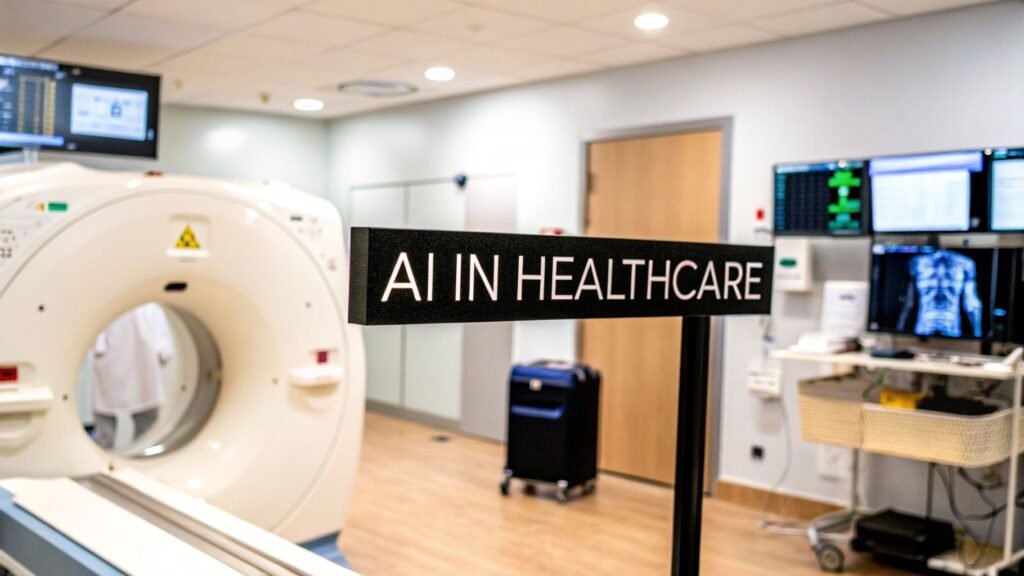
Artificial intelligence in healthcare is software that finds patterns in medical data — images, notes, labs, waveforms, claims — and returns a score, a mark, or a short list a clinician then uses or ignores. It is not a robot doctor. It does not sign the note.
The same job gets sold under different nouns: benefits, solutions, implementation, “how to implement AI.” Those are keyword wraps of this page. Diagnosis as a specialty topic is a different article. So is explainable AI (why a model flagged a pixel).
What the stack actually is
Four names, one pipeline. A model is trained on labeled examples, then run on a new case. The output is a suggestion. The clinician keeps or drops it.
| Name | Input | Typical job |
|---|---|---|
| Machine learning | Tabular records, labs, vitals | Risk scores, triage, readmission flags |
| Natural language processing | Notes, reports, claims text | Extract findings; draft or code from prose |
| Deep learning | Pixels or sequences at scale | The image-era name for the same training loop |
| Computer vision | X-ray, CT, MRI, OCT, slides | Marks on a scan — CAD-style second reader |
Imaging is the part that shipped. A convolutional net sees a labeled study (chest X-ray + “nodule / no nodule”) and later marks candidates on a new one. That workflow has been called computer-aided diagnosis for decades. The generic diagnosis explainer is AI for medical diagnosis — a different job from this page.
Two published image results that are actually cited, not market decks:
- De Fauw et al., Nature Medicine 2018 — a deep-learning OCT referral system matched expert ophthalmologists across dozens of retinal conditions. Paper: doi:10.1038/s41591-018-0237-x.
- McKinney et al., Nature 2020 (Google Health) — a mammography model reduced false negatives 9.4% and false positives 5.7% versus typical readers in that study. Paper: doi:10.1038/s41586-019-1799-6. One screening task, not “AI diagnoses cancer.”
Computational pathology (whole-slide images, tumor / grade / biomarker flags) is the same second-reader pattern on a different pixel type. The pathologist still signs.
Where else it shows up
- Notes and coding. NLP reads the chart and proposes codes or a draft note. A coder or clinician accepts or edits. Ambient scribes are this job with a microphone.
- Risk and deterioration. Tabular models sit on vitals and labs and raise a flag (sepsis, readmission, no-show). The treating team decides. A flag is not a diagnosis.
- Operations. Scheduling, bed turnover, inventory. Useful when the data is clean. Not a clinical device.
- Discovery. Virtual screening and trial matching live in pharma, not in a clinic PACS. AlphaFold solved protein structure; it did not write a prescription.
How a hospital actually starts
The “implementation roadmap” posts that used to live on this site were the same explainer with a different noun. The real sequence, for imaging, is shorter:
- Name one problem. “Flag intracranial hemorrhage on non-contrast head CT and bump the worklist.” Not “bring AI to the hospital.”
- Get the pixels and the labels. Pull DICOM from PACS. Join EHR metadata you actually need (age, indication, report text). De-identify to HIPAA’s safe-harbor or expert-determination rules before anything leaves the premises. Labels come from reports, biopsies, or expert reads — not from a vendor slide.
- Annotate for the job. Classification (pneumonia / no pneumonia). Boxes (a mass). Segmentation (organ or lesion outline, slice by slice). The annotation type is the product. Garbage labels produce garbage flags. See medical image annotation.
- Choose the model path. Build from scratch only if the task is novel and you have the team. Fine-tune a pre-trained imaging net for most classification / segmentation work. Buy a cleared SaMD when the task is standard and you will not own the weights. Re-validate on your scanners either way.
- Put a person on the output. Worklist integration, a viewer, and a signed report. Most imaging AI is assistive, not autonomous. If the model cannot show why it marked a region, that is the XAI page, not this one.
| Path | When | Cost / time | You own |
|---|---|---|---|
| Build | No product exists; unique data or workflow | High / months–years | Weights, pipeline, liability |
| Fine-tune | Standard imaging task, you have labeled studies | Medium / weeks–months | The adapted net; not the pre-train |
| Buy (cleared SaMD) | Commodity task; speed matters more than custom | Subscription / days–weeks | A contract and a validation set |
What it does not do
- It does not replace the reader. A mark is a candidate. The signed report is still a person. That is why most imaging AI is cleared as assistive (FDA SaMD), not autonomous.
- It fails on shift. A model trained on one vendor, protocol, or hospital will miss or over-call on another. You re-validate on your scanners. You do not copy a paper’s AUC onto the worklist.
- It inherits the labels. Narrow demographics, one site, or noisy reports become biased flags. That is a dataset problem, not a “fairness module.”
- It is not a market forecast. Vendor CAGRs, “80% of hospitals,” and “$431 billion by 2032” are not a reason to buy, and they are not this page.
If the question is why a model flagged a pixel — LIME, SHAP, inherent vs post-hoc, heat maps — that is explainable AI in healthcare. If the question is diagnosis / CAD as a specialty topic, that is AI for medical diagnosis.
PYCAD builds the imaging side of this — custom pipelines and web DICOM viewers when the study has to live in a clinic app. Case studies.