Demystifying DCM Files: What They Are and Why They Matter

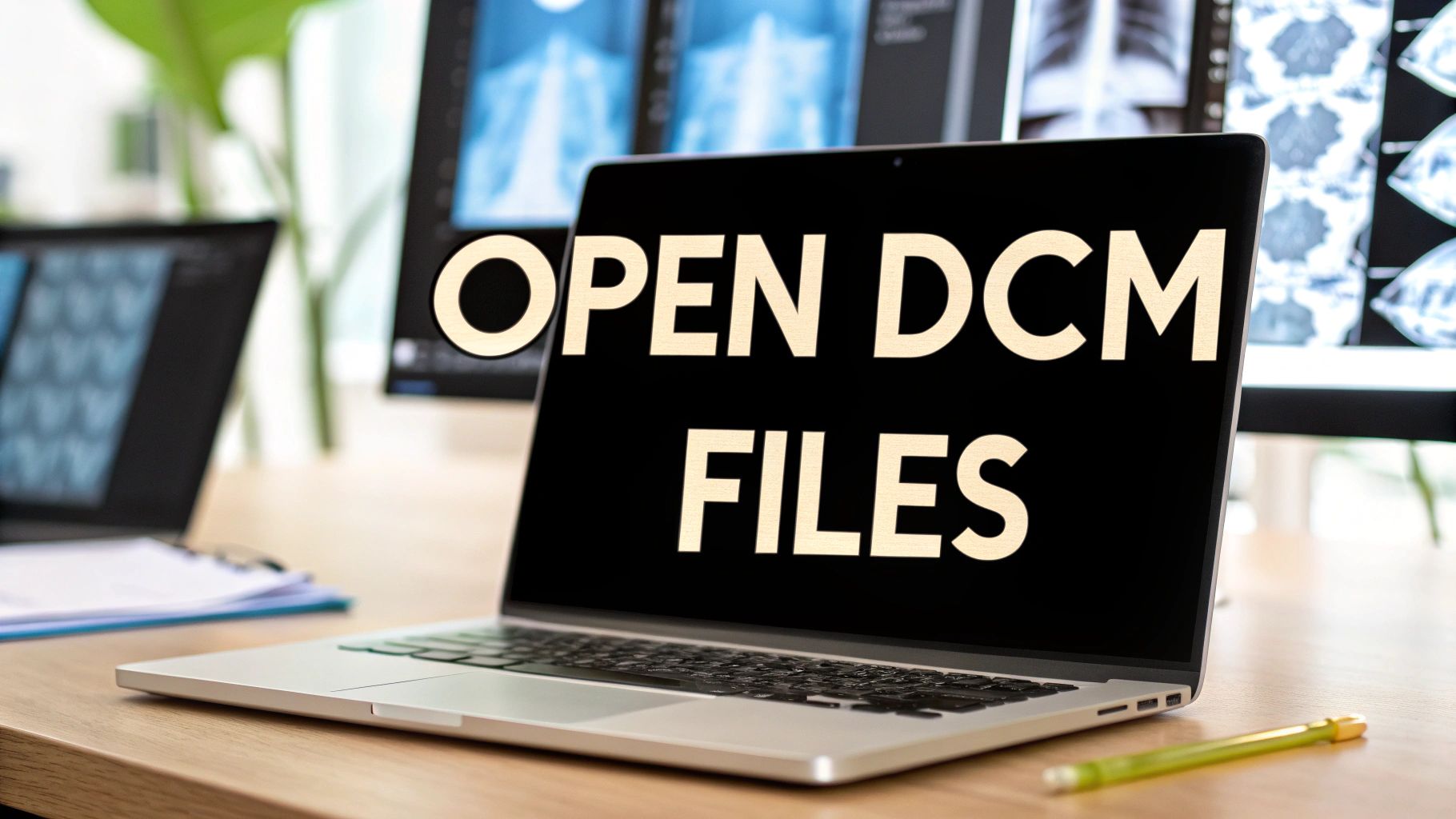
Outside the medical field, encountering a DCM file can be confusing. These files are core to medical imaging and contain more than just images. DCM is an abbreviation for DICOM, which stands for Digital Imaging and Communications in Medicine. This specialized format encapsulates images from X-rays, CT scans, MRIs, and ultrasounds, along with vital patient data, creating a complete medical record.
Opening a DCM file is akin to accessing a detailed chapter of a patient’s medical history. It provides insights into their internal structures and serves as a crucial tool for diagnosis and treatment planning. Healthcare professionals, researchers, and educators rely on reliable methods for accessing these files. Patients, too, benefit from understanding DCM files, particularly when reviewing their own medical images.
Opening DICOM files is becoming simpler due to its widespread adoption in medical imaging. As of 2023, DICOM is the industry standard used by nearly every medical equipment manufacturer. This standardization has led to the development of numerous software tools for viewing and managing these files, simplifying access and analysis for professionals. Tools like DCMTK, DCM4CHE, and PyDICOM provide robust solutions for handling DICOM data across various platforms.
Currently, approximately 95% of medical imaging data is stored in the DICOM format, highlighting its prevalence. While widely used, specialized software is still required to view DCM files, presenting a challenge for those outside the medical profession. For more information, explore this topic further here.
Why the DICOM Format Matters
The DICOM standard ensures consistent sharing and viewing of medical images across diverse devices and software platforms. This interoperability is essential for collaborative diagnoses, second opinions, and research. Specialists in different locations can readily access and review patient scans without compatibility concerns. The inclusion of patient data alongside images improves workflows and minimizes errors.
The detailed information embedded within DCM files empowers doctors to deliver more accurate diagnoses and personalized treatment plans. Metadata, often unseen by the untrained eye, offers critical context for interpreting images. This includes information about the scanning equipment, patient positioning, and any contrast agents used. Understanding DCM files and how to open them is thus crucial for everyone involved in medical imaging, from medical professionals to patients and researchers.
Your Toolkit: Free Software to Open DCM Files
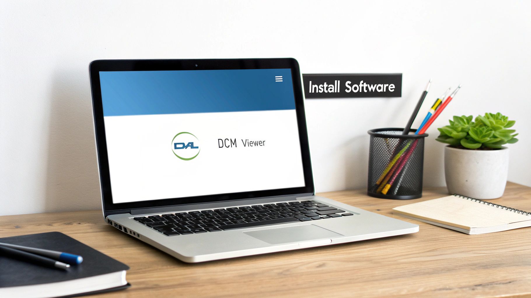
Opening and viewing DCM files doesn't require expensive software. Plenty of free and effective options are available, offering diverse features suitable for both casual users and professionals. These viewers provide convenient ways to navigate, manipulate, and analyze DICOM images without any financial burden. This section will explore some of the best free DCM file viewers available, helping you select the perfect tool for your specific requirements.
Top Free DCM File Viewers: A Detailed Look
Several free viewers are notable for their user-friendliness and comprehensive features. Let's explore a few popular choices:
-
Horos (Mac): Designed specifically for macOS, Horos presents an intuitive interface combined with powerful advanced tools. It excels in 3D visualization and image manipulation, making it a valuable resource for in-depth image analysis.
-
RadiAnt DICOM Viewer (Windows): This user-friendly option for Windows offers a straightforward installation process and an uncluttered interface. RadiAnt DICOM Viewer is known for its speed and efficiency, ideal for quickly reviewing DCM files.
-
MicroDicom (Windows): Another excellent Windows-based viewer, MicroDicom provides a lightweight yet powerful platform for viewing and manipulating DCM files. It also offers additional functionality, including burning DICOM images to discs.
-
ImageJ (Cross-Platform): A highly versatile, open-source image processing program, ImageJ is compatible across multiple operating systems. Although it needs a plugin for DICOM support, its adaptability makes it a great choice for those requiring a flexible solution.
To help you compare these options, we've compiled the following table:
Top Free DCM File Viewers Comparison: This table compares the most popular free DICOM viewers across operating systems, features, and ease of use.
| Software Name | Supported OS | Key Features | User-Friendliness (1-5) | Installation Size |
|---|---|---|---|---|
| Horos | macOS | Advanced 3D visualization, image manipulation | 4 | ~500 MB |
| RadiAnt DICOM Viewer | Windows | Fast viewing, simple interface | 5 | ~100 MB |
| MicroDicom | Windows | Lightweight, burns images to disc | 4 | ~50 MB |
| ImageJ | Cross-Platform | Extensible, requires DICOM plugin | 3 | ~30 MB |
As you can see, each viewer offers a unique set of advantages. RadiAnt excels in ease of use, while Horos provides more advanced features. MicroDicom offers a good balance of functionality and a small installation size. ImageJ shines with its cross-platform compatibility and extensibility.
Choosing the Right Viewer: A Guide to Your Needs
The best viewer for you ultimately depends on how you plan to use DCM files. For occasional viewing or quick reviews, user-friendly options like RadiAnt are excellent. Professionals requiring advanced visualization and measurement tools will likely prefer software like Horos. Understanding file compression techniques can be helpful when working with DCM files, as discussed in this article about file compression. For cross-platform compatibility and the ability to add plugins, ImageJ becomes a strong contender.
Exploring Further Functionality: Beyond the Basics
Many free viewers go beyond basic opening and viewing functionalities. Some include tools for adjusting image contrast and brightness, zooming, and even taking measurements. Several support multiple viewing layouts, enabling simultaneous comparison of different slices or orientations of the same scan. These advanced features enhance analysis and interpretation, making free viewers surprisingly powerful. They're an excellent starting point for anyone working with DCM files, providing both accessibility and valuable functionality. They represent a perfect entry into the world of DICOM without the investment required by professional tools. This allows users to familiarize themselves with opening, viewing, and utilizing basic features before considering more advanced software for specialized tasks.
The Evolution of DICOM: From Medical Revolution to Standard
The now widely used DICOM format wasn't a sudden invention. It emerged from a pressing need for a common language in the rapidly expanding world of digital medical imaging. Early on, proprietary systems were the norm, creating a disjointed environment. Imagine each manufacturer using its own unique file format for medical images. Sharing and interpreting scans would have been incredibly difficult. This lack of interoperability hampered collaboration and effective patient care.
This is where the story of DICOM begins. The necessity for a universal standard became undeniable, leading to a partnership between the American College of Radiology (ACR) and the National Electrical Manufacturers Association (NEMA). Their combined efforts in the early 1980s produced ACR-NEMA 1.0 and 2.0, the forerunners of today's DICOM standard. These initial versions laid the groundwork for a key element of modern radiology. Find more detailed statistics here.
From ACR-NEMA to DICOM: A Landmark Shift
The official launch of the DICOM standard in 1993 was a pivotal moment in medical imaging. This standardized format finally enabled seamless data exchange between different systems, transforming how medical images were stored, shared, and interpreted. Hospitals and clinics could now integrate imaging data from various sources, regardless of the manufacturer. This interoperability was, and remains, essential for collaborative diagnoses and streamlined patient care.
Ongoing Evolution: Adapting to Advancements
The story doesn't end there. Technology continues to advance, and the DICOM standard has adapted alongside it. The standard undergoes regular revisions, with updates released five times a year thanks to the diligent work of various committees. This ensures DICOM stays current and adaptable to the latest advancements in medical imaging technology.
DICOM Today: A Cornerstone of Modern Healthcare
Today, DICOM is much more than just an image format. It's a complex system for managing comprehensive patient information, encompassing everything from basic demographics to intricate 3D imaging datasets. It's integrated into a wide array of imaging modalities, supporting X-rays, MRIs, CT scans, ultrasounds, and more. The ability to open a DCM file and view a patient's CT scan along with relevant medical information is now standard medical practice.
Furthermore, DICOM's evolution has fueled innovation in related areas. More efficient data compression techniques and improved image processing capabilities have emerged, directly affecting the quality and accessibility of medical imaging. This ongoing development highlights the significance of understanding DICOM’s history. It provides context for how we interact with DCM files and emphasizes the critical role this standard plays in modern healthcare.
Professional-Grade Tools for Advanced DCM Handling

While free DCM viewers are great for basic viewing, professional-grade tools offer advanced features that significantly improve how medical professionals, researchers, and educators work with medical imaging. These premium software solutions are a valuable investment for those who need specialized features and seamless integration within a clinical or research environment. This means looking beyond simple viewers to platforms that provide a comprehensive toolkit for interacting with DCM files.
OsiriX MD: A Powerful Platform for Medical Professionals
OsiriX MD is a leading DICOM viewer specifically designed for macOS. It offers a robust suite of tools, including advanced visualization techniques, precise measurement tools, and seamless integration with hospital systems. For instance, OsiriX MD allows for complex 3D reconstructions, giving practitioners highly detailed visualizations of anatomical structures. Furthermore, its integration capabilities streamline workflows by connecting directly with Picture Archiving and Communication Systems (PACS) within a healthcare facility.
Horos Pro: Expanding the Horizons of DICOM Viewing
Horos Pro is another excellent professional-grade option, particularly for those familiar with the free Horos viewer. Horos Pro builds upon the foundation of its free counterpart, providing enhanced features for demanding clinical and research applications. This includes advanced image processing algorithms, broader support for DICOM modalities, and improved performance with large datasets. Horos Pro is ideal for specialists who need a powerful yet familiar interface for working with complex medical images.
Investing in Advanced Features: A Closer Look
Professional DICOM software like OsiriX MD and Horos Pro provides features that truly enhance diagnostic capabilities and research potential. Here are some key benefits:
- Advanced Visualization: Tools like Maximum Intensity Projection (MIP) and Volume Rendering (VR) provide more insightful image analysis.
- Precise Measurements: Accurate measurements of distances, angles, and volumes are crucial for diagnostic accuracy.
- DICOM Segmentation & 3D Printing Integration: Some tools offer segmentation of specific structures within images for more focused analysis, and this integration with 3D printing can be especially valuable for surgical planning and medical education.
- AI-Powered Diagnostics (Emerging Trend): Integration with AI algorithms is becoming increasingly common, providing features like automated lesion detection and image enhancement.
Pricing and Licensing: Navigating the Investment
Before choosing a professional-grade tool, it's important to understand the pricing models and licensing options. Many providers offer flexible plans for individual practitioners and large institutions, often with tiered pricing based on features and the number of users. Some offer perpetual licenses, while others use a subscription model. OsiriX MD, for example, has different licensing options for individual users, clinics, and hospitals. This flexibility allows users to select the best option for their needs and budget. Consider your budget and anticipated usage. How often will you use the software, and what are your primary needs for opening and manipulating DCM files? Answering these questions will help you decide if the advanced capabilities justify the cost.
Cloud Solutions: Opening DCM Files Without Installing Software
We've covered several ways to open DCM files, but a newer method is gaining popularity: cloud-based DICOM viewers. These platforms let you access medical images from any device with an internet connection, eliminating the need for local software. This offers significant convenience, but also requires careful attention to security and patient privacy.
Advantages of Cloud-Based DCM Viewers
Cloud solutions provide flexibility in how medical professionals and researchers work with DICOM data. Here are some key benefits:
- Accessibility: View images from anywhere with an internet connection. This is particularly helpful for remote consultations and on-call scenarios.
- Collaboration: Easily share studies with colleagues at other institutions, simplifying second opinions and collaborative diagnoses.
- No Installation: Avoid complex software installation and configuration, allowing faster access to critical data.
- Scalability: Cloud platforms efficiently handle large datasets, bypassing the storage limitations of personal devices.
This means healthcare providers can review images even when away from their workstations. Researchers can easily share data for collaborative projects. And patients may have easier access to their own imaging records.
Security and Privacy in the Cloud
The convenience of cloud-based viewers must be balanced with strong security. DCM files contain sensitive patient data, so compliance with regulations like HIPAA is crucial. This means using encryption, user authentication, and audit trails to protect patient information.
When choosing a cloud-based DICOM viewer, consider these factors:
- Data Encryption: Ensure the platform encrypts data both during transmission and while stored.
- Compliance: Verify the platform’s compliance with all relevant healthcare regulations, such as HIPAA in the US and GDPR in Europe.
- User Authentication: Look for robust access controls and multi-factor authentication to prevent unauthorized access.
- Data Backup and Recovery: Understand the platform's data backup and disaster recovery plans to protect against data loss.
To help you compare cloud-based and desktop viewers, we've created the table below:
Cloud vs. Desktop DCM Viewers
This table compares the advantages and disadvantages of cloud-based and desktop DICOM viewers.
| Feature | Cloud-Based Viewers | Desktop Applications |
|---|---|---|
| Accessibility | Access from anywhere with internet connection | Limited to the device with the installed software |
| Collaboration | Easy sharing and collaboration | More difficult to share files externally |
| Installation | No installation required | Installation and setup are required |
| Security | Requires careful vetting of data security and privacy practices | Local security control, but potential vulnerabilities exist |
| Updates | Automatic updates | Manual updates required |
| Cost | Typically subscription-based | Can be free or a one-time purchase |
| Storage Capacity | Scalable storage | Limited by local hard drive space |
As shown above, each type of viewer offers different benefits.
Real-World Implementations: Changing the Landscape of Medical Imaging
Platforms like Postdicom and the Open Health Imaging Foundation (OHIF) Viewer are changing how medical professionals share and view images. Postdicom provides a secure cloud PACS solution, while OHIF offers an open-source, customizable viewer deployable in various cloud environments. These tools promote collaboration across geographical boundaries, creating a more connected and efficient approach to medical imaging. Companies specializing in AI and medical imaging can help organizations leverage these cloud technologies securely and effectively, integrating them with existing systems and workflows to improve patient care and research.
Choosing the right viewer, whether desktop or cloud-based, depends on individual needs. While free viewers suit casual use, cloud-based solutions offer enhanced collaboration and accessibility, vital for today’s healthcare environment.
Troubleshooting DCM Files: Solutions That Actually Work
Working with DICOM (Digital Imaging and Communications in Medicine) images is essential for many healthcare professionals, researchers, and students. Even with the right software, opening these files can be tricky. This guide offers practical solutions to common DICOM image challenges, drawing on techniques used by PACS administrators and medical imaging specialists. With these tools, you can resolve issues quickly and efficiently, minimizing frustration and potential data loss.
Decoding Cryptic Error Messages
Often, the first sign of a problem is a cryptic error message. While these messages can seem indecipherable, they hold clues to understanding the issue. Let's look at some common examples:
-
"Invalid DICOM File": This typically points to a corrupted file header. The header contains vital file information. It's like a damaged table of contents in a book – you can't access the content easily. Specialized DICOM repair tools may help by reconstructing the header.
-
"Missing Data Elements": This indicates an incomplete dataset, similar to a puzzle with missing pieces. You might see parts of the image, but not the complete picture. Re-acquiring the image from the source is usually the best solution.
-
"Unsupported DICOM Version": This signals compatibility conflicts between different DICOM versions, like trying to play a modern video game on an old console. Older viewers might not support newer DICOM features. Updating your software or using a converter typically resolves this.
Practical Solutions for Common Issues
Now that we understand some common error messages, let's explore the corresponding solutions:
-
Corrupted Headers: Try using a DICOM repair tool for corrupted headers. These tools try to rebuild the header to allow access to the image data. If this issue happens frequently, review your data storage processes to identify and prevent corruption at the source.
-
Incomplete Datasets: If data elements are missing, contact the image source (like the imaging center) to request a complete file. If the original is unavailable, some viewers have reconstruction tools that might partially recover the image, but with possible data loss.
-
Compatibility Problems: If you have unsupported DICOM versions, make sure your viewer software is up-to-date. Many developers release regular updates to maintain compatibility with the latest DICOM standards. If older software is essential, consider using a DICOM converter to transform the file into a compatible format.
Optimizing Performance for Large Studies
Large datasets, especially those with high-resolution images or extensive studies, can be challenging. These tips can improve performance:
-
Powerful Hardware: Ensure your computer has sufficient processing power and memory (RAM) to handle large DICOM files. A faster hard drive, particularly a Solid State Drive (SSD), significantly improves loading times.
-
Optimized Viewers: Some viewers are designed for efficient handling of large datasets. They use techniques like lazy loading, which only loads data when needed, minimizing initial load times.
-
Data Caching: Many viewers use caching to store frequently used data. Clearing the cache regularly can free up resources and improve performance.
Working with Anonymized Datasets
Anonymization, the removal of identifying patient information, is critical for research and education. However, it also presents specific challenges:
-
Consistent Anonymization: Use dedicated anonymization tools to ensure complete removal of patient identifiers according to regulations.
-
Metadata Preservation: Ensure essential non-identifying metadata is preserved during anonymization for research and analysis.
-
Secure Data Sharing: Use secure methods when sharing anonymized datasets to maintain patient confidentiality.
PYCAD, a company specializing in AI-driven medical imaging solutions, understands the complexities of DCM files. They offer expertise in data handling, model training, and deployment to help optimize medical devices and improve healthcare outcomes. Learn more about how PYCAD can help you navigate the complexities of DICOM: https://pycad.co.


