Navigating the Liver's Segmental Landscape Through CT

Understanding the complexities of liver anatomy is essential for accurately interpreting CT scans of the liver segments. This detailed imaging technique allows healthcare professionals to visualize the liver's eight distinct segments, each with its own unique vascular supply and biliary drainage. This knowledge is critical for surgical planning, targeted interventions, and the accurate diagnosis of liver diseases.
Knowing the precise location of a tumor within a specific liver segment, for example, can significantly influence treatment strategies. This section will explore how we can accurately identify these segments using CT imaging.
Couinaud Classification: The Roadmap to Liver Segmentation
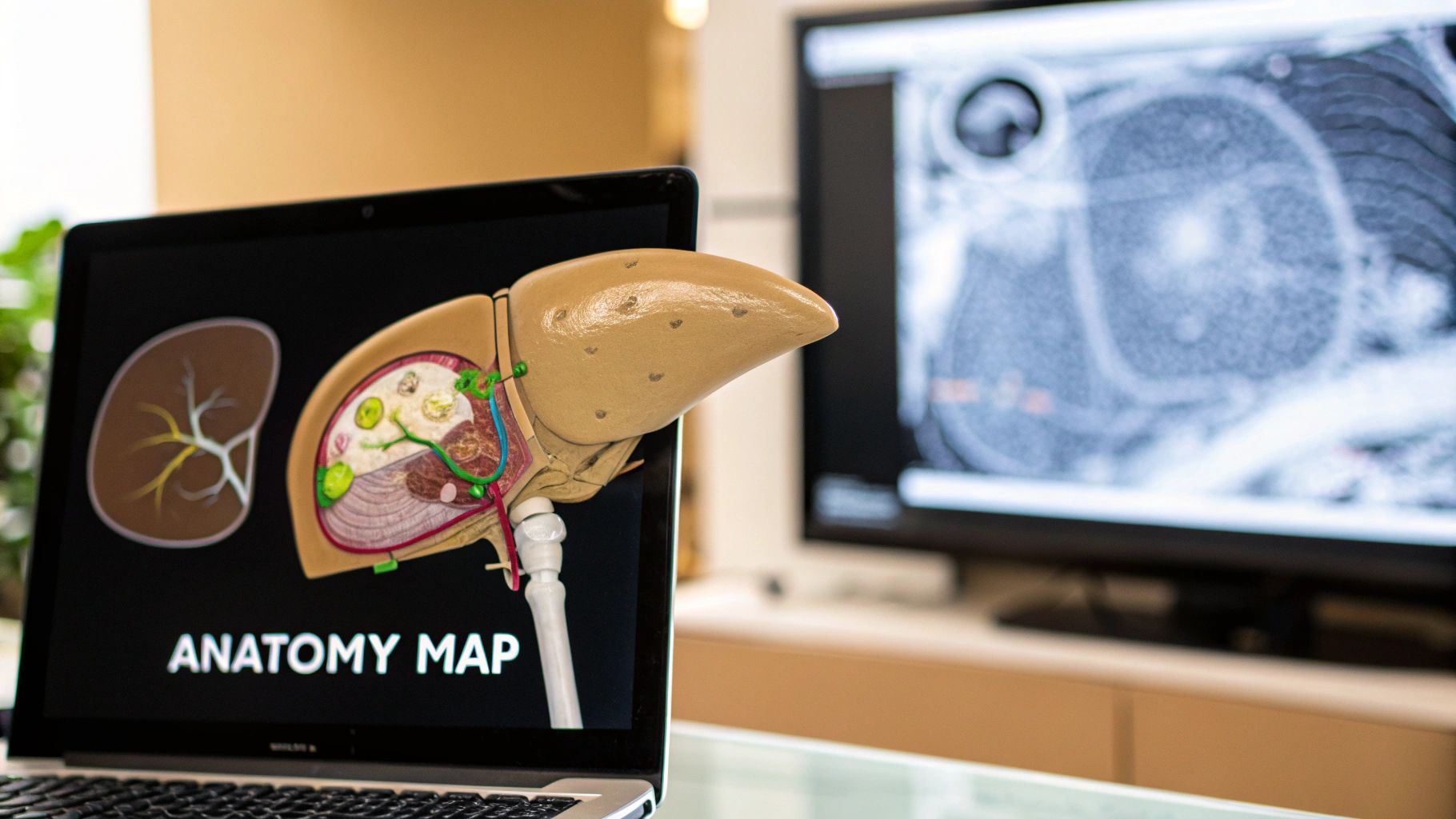
The Couinaud classification system, also known as the "French system," is the internationally recognized standard for dividing the liver into eight independent segments. This system provides a practical framework that guides surgical procedures and localized treatments. It acts as a common language for radiologists and surgeons, ensuring clear communication and facilitating the precise targeting of lesions.
This is particularly valuable in procedures like liver resection, where accurate segmentation is crucial for preserving healthy liver tissue. Understanding the Couinaud classification allows for more informed decision-making and improved patient outcomes.
To further illustrate the Couinaud system, the table below provides a detailed breakdown of each segment:
Couinaud Classification of Liver Segments
| Segment Number | Anatomical Location | Vascular Supply | Key Landmarks |
|---|---|---|---|
| 1 | Caudate Lobe | Left and Right Portal Vein | Inferior Vena Cava |
| 2 | Superior Lateral Left Lobe | Left Portal Vein | Left Hepatic Vein |
| 3 | Inferior Lateral Left Lobe | Left Portal Vein | Left Hepatic Vein |
| 4 | Medial Left Lobe | Left Portal Vein | Middle Hepatic Vein |
| 5 | Inferior Anterior Right Lobe | Right Portal Vein | Middle Hepatic Vein |
| 6 | Inferior Posterior Right Lobe | Right Portal Vein | Right Hepatic Vein |
| 7 | Superior Posterior Right Lobe | Right Portal Vein | Right Hepatic Vein |
| 8 | Superior Anterior Right Lobe | Right Portal Vein | Middle Hepatic Vein |
This table clarifies the anatomical location, vascular supply, and key landmarks for each of the eight liver segments. This detailed breakdown facilitates a more comprehensive understanding of the Couinaud classification system, which is vital for accurate interpretation of liver segment CT scans.
Liver segmentation in CT scans has become increasingly important, especially with the growing use of AI-driven diagnostics. Research on AI-based liver segmentation, particularly using deep learning models, highlights this trend.
Vascular Landmarks: Guiding the Way
Identifying liver segments on a CT scan relies heavily on recognizing key vascular landmarks. These landmarks serve as guides, marking the boundaries between segments. The portal vein, hepatic artery, and hepatic veins are the primary vascular structures used for orientation.
These vessels branch throughout the liver in a predictable manner, enabling radiologists to precisely locate each segment. The middle hepatic vein, for instance, divides the liver into right and left lobes, a fundamental distinction in understanding liver anatomy. This detailed vascular mapping provides a crucial roadmap for navigating the liver's segmental landscape.
Overcoming Interpretation Challenges
While the Couinaud system offers a valuable framework, interpreting liver segment CT scans can present challenges. Anatomical variations, the presence of disease, and image artifacts can sometimes obscure the clear delineation between segments.
However, experienced radiologists utilize specific techniques to navigate these difficulties. Advancements in CT technology, such as multiplanar reconstructions and 3D rendering, also enhance clarity, enabling more precise segment identification. This is particularly important in complex cases, where accurate segmentation is essential for effective treatment planning.
Advanced CT Techniques That Reveal Hidden Liver Segments

Advanced CT techniques offer significantly improved visualization of liver segments, going beyond the capabilities of standard imaging protocols. These techniques provide critical details essential for accurate diagnoses and effective treatment planning. Let's explore some of these advanced approaches and their practical applications in liver segment CT.
Multi-Phase Contrast Protocols: Visualizing Vascular Structures
Multi-phase contrast protocols are essential for accurately visualizing liver segments. By capturing images at different phases of contrast enhancement, radiologists can differentiate between vascular structures and the surrounding tissues. This detailed view is fundamental for accurate interpretation of liver segment CT scans.
For instance, the arterial phase highlights the hepatic artery, while the portal venous phase emphasizes the portal vein. These distinct phases help define the segmental boundaries, enabling precise localization of lesions. The delayed phase can also reveal lesions not visible in earlier phases.
Advanced Reconstruction Methods: Revealing Hidden Anatomical Details
Multiplanar reformations (MPR) and volume rendering are advanced reconstruction methods that further enhance the visualization of liver segments. MPR allows radiologists to view the liver in any plane, offering a complete understanding of its three-dimensional structure. This is especially useful for visualizing the path of vessels and their relationship to liver segments.
Volume rendering creates a 3D model of the liver, enabling detailed visualization of the segmental anatomy. This technique is particularly helpful for surgical planning, allowing surgeons to precisely map resection margins before an operation.
Overcoming Imaging Challenges in Liver Segment CT
Several factors can hinder accurate delineation of liver segments in CT scans. Motion artifacts, resulting from patient movement during the scan, can blur the image and obscure segmental boundaries. Respiratory variations can also introduce similar distortions, impacting image quality.
Modern CT scanners employ techniques like respiratory gating to address these issues. This technique synchronizes image acquisition with the patient's breathing cycle, minimizing motion artifacts and improving image clarity.
Precise contrast timing is also essential for optimal visualization. Administering the contrast too early or too late can prevent capturing the desired vascular phase, making segment identification difficult. Bolus tracking techniques are often used to precisely time the contrast injection, ensuring optimal enhancement of the vascular structures. This contributes to the accuracy and reliability of liver segment CT interpretations.
How AI Is Transforming Liver Segment CT Analysis

Medical imaging is a constantly evolving field, and liver segment CT scan analysis is no exception. Artificial intelligence (AI) is playing an increasingly vital role in improving the precision and efficiency of this critical diagnostic procedure. This empowers radiologists to utilize AI’s capabilities for interpreting these intricate images, leading to quicker and more accurate diagnoses.
Deep Learning Algorithms: Enhancing Precision and Efficiency
Deep learning algorithms, a subset of AI, are designed to mirror the human brain's learning process from large datasets. When applied to liver segment CT scans, these algorithms can be trained on thousands of scans to identify patterns and recognize liver segments with remarkable precision. This significantly reduces the time and effort needed for manual segmentation, freeing up radiologists to concentrate on more complex diagnostic tasks.
For example, AI can efficiently and accurately outline the boundaries between liver segments, even when anatomical variations or disease processes complicate visualization.
Furthermore, AI algorithms excel at detecting subtle changes in liver segment morphology that might be overlooked by the human eye. This heightened sensitivity can be especially beneficial for the early detection of liver diseases, enabling timely intervention that can significantly improve patient outcomes. AI can also quantify liver segment volumes and ratios, offering essential data for treatment planning and monitoring disease progression.
AI Architectures in Clinical Settings: Strengths and Limitations
Several distinct AI architectures are currently employed for liver segment CT analysis, each with its own advantages and disadvantages. Convolutional neural networks (CNNs) are particularly well-suited for image analysis because they effectively capture spatial relationships within image data. Another promising architecture is the U-Net, designed specifically for biomedical image segmentation. This architecture enables precise localization of liver segments, even with the presence of noise or artifacts.
However, accurately segmenting small liver structures and tumors still presents challenges. Existing AI algorithms can sometimes have difficulty differentiating between healthy and diseased tissue in these complex cases. This underscores the continuing need for research and development to further refine these algorithms and enhance their performance. Recent applications of AI in medical imaging have substantially improved the accuracy and efficiency of liver segmentation. Learn more about AI-powered liver segmentation: https://www.mdpi.com/1424-8220/24/6/1752
Integrating AI into Radiology Workflows
Leading radiology departments are actively incorporating AI tools into their workflows. Importantly, AI is not meant to replace radiologists. Instead, these tools are designed to enhance their expertise by providing valuable support in interpreting complex imaging data. This collaborative approach leverages the strengths of both human and artificial intelligence, resulting in improved diagnostic accuracy and efficiency.
For instance, AI can pre-segment liver CT scans, giving radiologists a head start on their analysis. This considerably reduces the time spent on manual segmentation, allowing them to dedicate more time to complex interpretive tasks. AI can also flag potentially concerning areas on a scan, alerting radiologists to areas needing closer scrutiny. This can be particularly helpful when subtle findings might otherwise be missed. As AI technology continues to progress, it holds immense promise for further advancements in liver segment CT analysis, ultimately benefiting both patients and healthcare professionals.
Liver Segment CT Ratios: Small Numbers, Big Clinical Impact

Beyond simply visualizing the liver's different segments, CT scans provide crucial quantitative data. These data points, expressed as ratios, offer valuable insights into liver health. These seemingly small numbers can significantly impact patient management, especially in cases of chronic liver disease. This section explores the clinical relevance of these powerful ratios.
Key Liver Segment CT Ratios: CRL-R and LSVR
Two primary ratios derived from liver segment CT scans are the caudate to right lobe ratio (CRL-R) and the liver segmental volume ratio (LSVR). The CRL-R compares the size of the caudate lobe (segment 1) to the right lobe (segments 5-8). The LSVR compares the combined volume of the left lobe (segments 2-4) and caudate lobe to the right lobe.
These ratios offer essential information about the proportional size of different liver segments, which can shift with disease progression. Understanding these changes is critical for accurate diagnosis and effective treatment planning.
For instance, in cirrhosis, the right lobe often decreases in size, while the caudate lobe remains relatively unaffected. This leads to an increased CRL-R. Similarly, the LSVR can also rise in cirrhosis, reflecting the altered balance in volume between the left and right lobes.
Calculating and Interpreting Liver Segment Ratios
Calculating these ratios requires measuring the volumes of the specific liver segments from CT scan data. While this process can be complex, it's typically automated by software and performed by trained radiologists. 3D Slicer is an example of a platform used for medical image visualization and analysis.
Interpreting these ratios necessitates understanding normal reference ranges and how values deviate in various liver diseases. A significantly elevated CRL-R, for example, could suggest advanced liver disease, warranting further investigation. The predictive value of liver segment ratios is particularly important in chronic liver disease.
To better understand the clinical significance of these ratios, let's examine the following table:
Understanding normal ranges and the diagnostic implications of deviations is crucial for effective clinical application.
| Ratio Type | Calculation Method | Normal Range | Diagnostic Significance | Clinical Applications |
|---|---|---|---|---|
| Caudate-to-Right Lobe Ratio (CRL-R) | Caudate Lobe Volume / Right Lobe Volume | 0.25 – 0.40 | Elevated in cirrhosis, hepatic venous outflow obstruction | Assessing liver disease severity, monitoring treatment response |
| Liver Segmental Volume Ratio (LSVR) | (Left Lobe Volume + Caudate Lobe Volume) / Right Lobe Volume | 0.8 – 1.2 | Elevated in cirrhosis, portal hypertension | Evaluating liver function, predicting prognosis |
This table summarizes the key characteristics and applications of CRL-R and LSVR, providing a quick reference for clinicians. The data highlights the importance of considering these ratios in the context of other clinical findings.
Find more detailed statistics here. These resources can provide a deeper understanding of the research and data behind the diagnostic significance of liver segment ratios.
This emphasizes that liver segment ratios are not merely abstract numbers; they are valuable tools for guiding clinical decision-making. Their use can significantly improve patient outcomes.
Using Ratios to Monitor Disease Progression
Liver segment CT ratios are also vital for monitoring disease progression. By tracking changes in these ratios over time, healthcare providers can evaluate treatment effectiveness and adjust as necessary. This proactive approach is crucial for managing chronic liver diseases.
Regular monitoring of CRL-R and LSVR can help identify patients at higher risk of developing complications, facilitating timely interventions. Early detection allows for prompt and appropriate management, potentially improving long-term outcomes.
Furthermore, these ratios can help predict long-term outcomes, including the need for a liver transplant. This information empowers physicians to make more informed decisions, improving the quality of life for patients with liver disease. Using data-driven insights provides a more personalized and proactive approach to patient care.
Surgical Success Stories: When Liver Segment CT Saves Lives
Liver segment CT scans are essential for surgical planning and execution, especially for complex liver conditions. The precision of these scans, interpreted by skilled radiologists, leads to improved surgical outcomes and can be life-saving.
Precise Segment Mapping: Guiding the Surgeon's Hand
Accurate liver segment mapping from high-quality CT scans gives surgeons a detailed roadmap of the liver's complex anatomy. This allows for precise surgical planning, particularly for resections, minimizing the risk to vital structures like blood vessels and bile ducts. This precise knowledge helps surgeons navigate the liver's complexities during surgery, much like a GPS guides a driver.
For example, during a liver resection to remove a tumor, accurate segment mapping is essential for defining the diseased tissue boundaries and preserving healthy liver segments. This detailed preoperative visualization from liver segment CT contributes to a more precise and safer procedure.
Collaboration: Bridging the Gap Between Imaging and Intervention
The relationship between radiologists and surgeons is critical for successful liver surgery. Effective communication and collaboration ensure that the information from liver segment CT scans translates into actionable surgical strategies. This teamwork is essential for optimal patient care.
Preoperative conferences, where radiologists and surgeons review CT scans together, are vital. This gives the surgeon a deep understanding of the patient's specific liver anatomy and potential surgical challenges. These discussions often focus on the volume of each liver segment, critical for assessing the future liver remnant (FLR). Ensuring an adequate FLR is crucial for maintaining post-surgery liver function.
Segment-Based Volumetry: Ensuring a Healthy Future Liver Remnant
Segment-based volumetry, derived from liver segment CT, is vital for determining the FLR. This involves precisely measuring the volume of liver segments that will remain after resection. This information helps determine if a resection is feasible and safe, allowing surgeons to make informed decisions. Surgeons use these measurements to calculate the FLR, ensuring enough healthy liver tissue remains after surgery.
Additionally, liver segment CT scans are crucial for identifying variations in vascular anatomy, which are common and can significantly impact surgical planning. Accurately mapping the hepatic artery, portal vein, and hepatic veins within each segment is paramount to preventing complications. This detailed vascular mapping helps surgeons avoid injuring these vital structures.
Vascular Variation Detection: Navigating Anatomical Challenges
Precise detection of vascular variations using liver segment CT allows surgeons to anticipate and address potential complications, minimizing the risk of bleeding and other adverse events. It’s like having a blueprint that shows hidden pipes and wires in a building, preventing accidental damage during renovations.
Furthermore, liver segment CT is vital in living donor liver transplantation for both the donor and recipient. Precise segmental volumetry helps determine the best liver segment for donation, ensuring both donor safety and graft viability. This accurate assessment is crucial for transplant success.
These surgical applications demonstrate how accurate interpretation of liver segment CT scans directly impacts surgical decisions, contributing to positive patient outcomes. These are real-world examples where detailed imaging has prevented complications and saved lives.
Avoiding Diagnostic Disasters: Liver Segment CT Pitfalls
Liver segment CT interpretation is a powerful diagnostic tool, but it's not without its challenges. Even seasoned radiologists can encounter difficulties. This section explores common pitfalls and offers practical strategies to improve accuracy. Understanding these challenges is vital for accurate diagnoses and effective treatment planning.
Anatomical Variations: Navigating the Unpredictable
One of the biggest challenges in liver segment CT interpretation is the prevalence of anatomical variations. These variations, such as accessory fissures and differing vascular anatomy, can complicate segment identification. An accessory fissure, for example, could be mistaken for the border between two segments, leading to inaccurate lesion localization. Variations in the portal vein branching pattern can also make defining segmental boundaries difficult.
Radiologists must be mindful of these potential anatomical variations. Careful examination of CT images, especially in multiple planes, is essential. Multiplanar reformations (MPR) can be invaluable for clarifying ambiguous anatomy. Furthermore, correlating CT findings with other imaging modalities, like MRI or angiography, can provide additional clarity for accurate segment identification.
Morphological Distortions: Seeing Through the Haze
Liver diseases, such as cirrhosis and steatosis, can significantly distort the liver's normal shape, making segmentation difficult. These distortions can obscure the usual landmarks used for segmentation, like the hepatic veins and portal vein branches. Previous interventions, such as surgery or biopsies, can also create scar tissue that further complicates interpretation.
Imagine trying to identify city blocks on a map after an earthquake. Familiar landmarks might be shifted or obscured. Similarly, morphological distortions in the liver can alter its architecture, making it difficult to apply standard segmentation rules.
Radiologists use various techniques to overcome these challenges. Careful observation of the overall liver contour and any remaining identifiable landmarks is crucial. Contrast-enhanced CT can help to highlight vascular structures and differentiate them from surrounding diseased tissue. Understanding the specific disease process and its effects on liver anatomy can also guide interpretation.
Precise Segment Localization: Ensuring Accuracy
Even without anatomical variations or morphological distortions, pinpointing lesions to specific segments can be tricky. Small lesions near segmental boundaries pose a particular challenge. Errors in segment localization can have major consequences for treatment decisions and potentially lead to suboptimal outcomes.
This is especially relevant in surgical planning, where accurate segment identification is critical for determining resection margins and preserving healthy liver tissue. Misidentifying a lesion's location could lead to inadequate resection or unnecessary removal of healthy tissue.
Radiologists employ a systematic approach for precise localization. They carefully analyze CT images in all three planes, tracing the course of the major vascular structures to define segmental boundaries. Using 3D visualization tools, they create a mental map of the liver segments and precisely locate the lesion within that framework. This methodical approach maximizes accuracy and minimizes the risk of diagnostic errors.
Distinguishing Pathology from Normal Variants: Avoiding False Positives
Another pitfall in liver segment CT is mistaking normal variants for pathology. Small areas of fatty infiltration can sometimes resemble small tumors. Variations in the shape or size of a liver segment might be misinterpreted as a disease indicator. These false-positive findings can cause unnecessary anxiety and additional investigations, placing an undue burden on patients.
To avoid these pitfalls, radiologists rely on their expertise in normal liver anatomy and its variations. They compare the CT findings with established criteria for normal variants. If there is any doubt, they may recommend follow-up imaging or other diagnostic tests to confirm or rule out disease. This careful approach helps minimize false positives and ensures appropriate patient care.


