The Evolution of Liver Segmentation CT: From Pixels to Precision
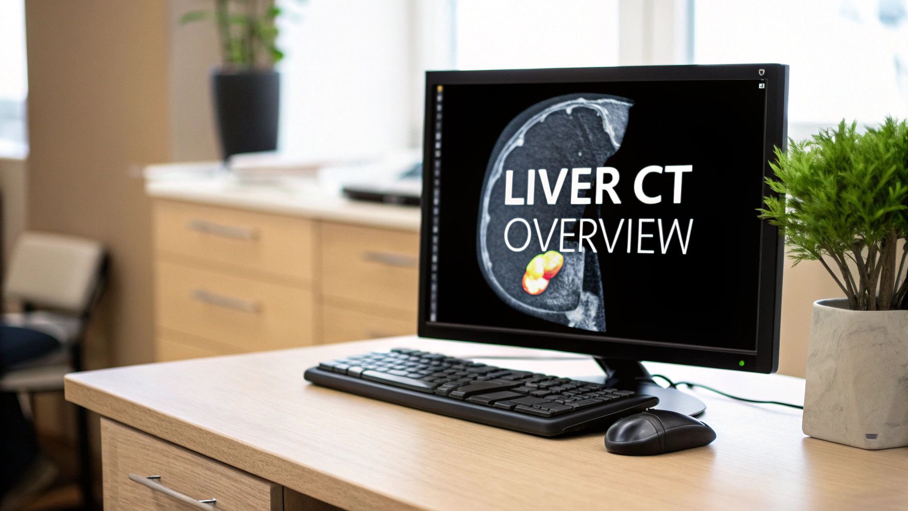
Liver segmentation CT has undergone a dramatic transformation. The journey has taken it from basic pixel evaluation to the sophisticated 3D modeling we see today. Initially, CT scans offered a grainy, two-dimensional view inside the body. While groundbreaking at the time, these early images provided limited information for accurate liver analysis.
Pinpointing the exact boundaries of the liver and identifying small lesions proved challenging with the technology then available. This lack of detail hampered diagnostic confidence and treatment planning. Accurate analysis was difficult, and medical professionals needed more information to make informed decisions.
However, the field of liver segmentation CT advanced significantly with the arrival of improved computing power and imaging techniques. The history of CT imaging itself plays a vital role in understanding the development of liver segmentation. CT History and Technology provides further insight into this historical context.
The first CT scanners, introduced in the 1970s, revolutionized medical imaging. They offered non-superimposed images of body slices, a significant leap forward. Over the decades, CT technology has evolved from first-generation pencil beam scanners to modern dual-source and photon-counting CT systems. These newer systems offer superior temporal and spatial resolutions, leading to higher quality liver imaging and more detailed segmentation and analysis. This evolution laid the foundation for more accurate and informative assessments of the liver.
Advancements in Image Quality and Resolution
The increasing sophistication of CT scanners has been a key driver in improving liver segmentation. As spatial resolution increased, the ability to distinguish fine details within the liver improved dramatically. This is analogous to switching from a standard-definition television to high-definition: details unseen before suddenly become clear. This higher resolution enabled more accurate identification of liver boundaries, blood vessels, and even small lesions.
Furthermore, improvements in image quality reduced noise and artifacts, making it easier for radiologists to interpret the scans. Clearer images provide a more accurate representation of the liver's structure and condition, improving diagnostic capabilities.
From 2D To 3D: The Impact of Volumetric Data
The transition from single-slice to multi-detector CT (MDCT) technology marked another crucial step. MDCT allows for the rapid acquisition of volumetric data, enabling the creation of detailed 3D liver models. This has revolutionized treatment planning, especially for surgical procedures.
Surgeons can now visualize the liver in three dimensions, rotate the image, and even simulate procedures before operating. This improved visualization translates to more accurate surgical planning, potentially reducing complications and improving patient outcomes.
The Rise of Automated Liver Segmentation CT
Manual liver segmentation, a time-consuming and potentially subjective process, is gradually being replaced by automated methods. Software algorithms now assist in defining liver boundaries and identifying key structures. This automation not only saves time but also improves consistency and reduces variability between different analysts.
This shift towards automated liver segmentation represents a crucial step towards more efficient and standardized liver analysis, paving the way for new possibilities in both research and clinical practice. This continued evolution promises even more precise and personalized approaches to liver care in the future.
Multi-Slice CT Revolution: Redefining Liver Segmentation
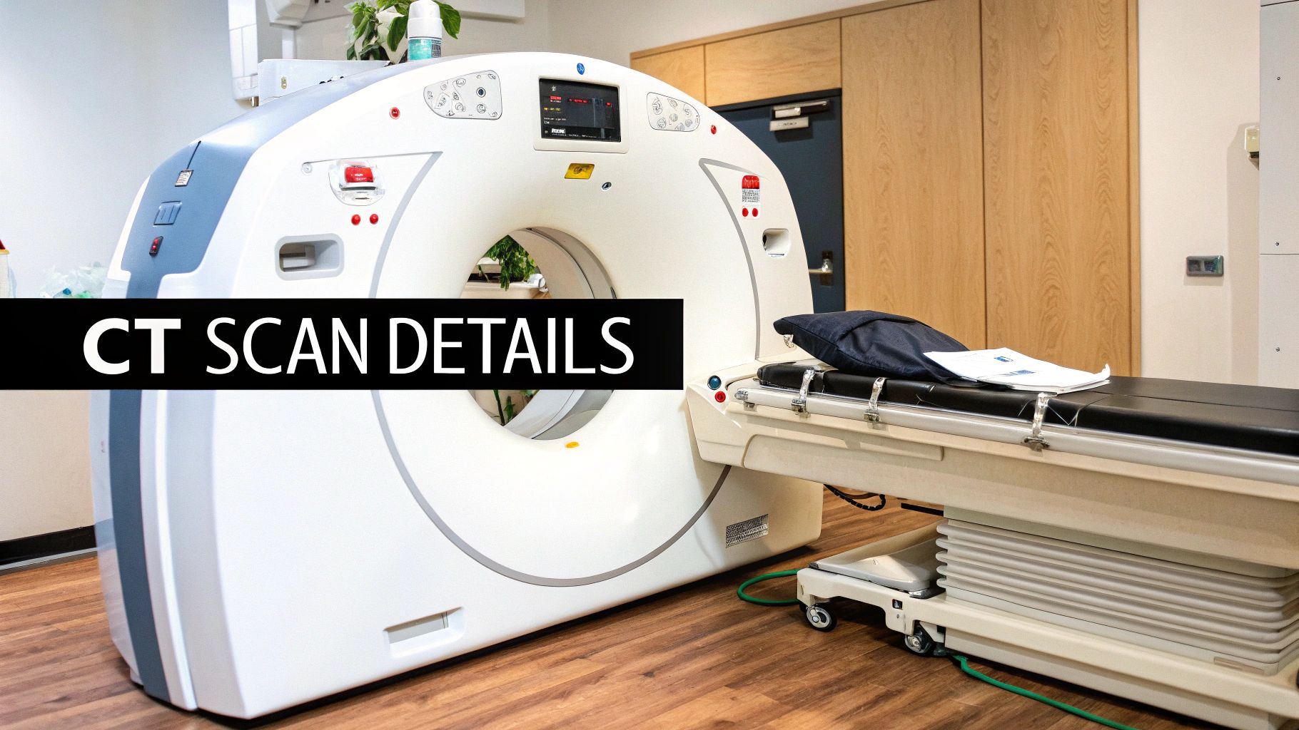
The advent of Multi-slice Computed Tomography (MDCT) has significantly changed liver segmentation in CT scans. This technology goes beyond simply enhancing image quality; it has fundamentally altered the possibilities of liver analysis. MDCT allows for the rapid acquisition of volumetric datasets, providing a wealth of information previously unavailable. Instead of capturing individual slices, MDCT acquires data as a continuous volume, enabling the creation of highly detailed 3D liver models.
Technical Advancements Driving MDCT
Several key advancements have made the power of MDCT possible. Improvements in detector technology allow for the simultaneous acquisition of multiple slices in a single rotation. This significantly increases scanning speed, reducing motion artifacts and patient discomfort. Faster rotation speeds further enhance data acquisition efficiency.
Sophisticated reconstruction algorithms process the raw data into clear, high-resolution images suitable for accurate liver segmentation. These combined advancements dramatically improve image quality and diagnostic capabilities.
The development of liver segmentation techniques in CT scans has evolved considerably with the introduction of MDCT. The introduction of MDCT in the late 1990s was a milestone. It allowed for the acquisition of larger volumes of data in a single rotation, leading to better 3D reconstructions crucial for liver segmentation.
This technology facilitated the rapid increase in detector rows, from four to sixty-four, enhancing spatial resolution and enabling more accurate liver imaging. By the early 2000s, MDCT became the standard for clinical imaging, including liver segmentations, because of its ability to obtain isotropic spatial resolution, vital for detailed 3D visualization of liver structures. Explore this topic further.
Clinical Workflow Transformation
MDCT has substantially impacted clinical workflows. Before MDCT, liver segmentation involved manually tracing liver contours on individual CT slices, a time-consuming and potentially inaccurate process. MDCT and its inherent 3D capabilities have made this process significantly faster and more precise.
Precise volume calculations, essential for surgical planning and disease monitoring, became a clinical reality. The improved resolution and 3D visualization also greatly enhanced lesion detection, allowing for earlier diagnosis and intervention. This improved workflow efficiency leads to better-informed treatment decisions and, ultimately, better patient outcomes.
Enhanced Surgical Planning and Diagnostic Accuracy
MDCT revolutionized surgical planning by allowing surgeons to visualize the liver in three dimensions. This enables accurate assessment of tumor location and size, facilitating more precise planning for complex resections. It's like having a detailed roadmap for a journey, improving navigation and minimizing unexpected issues.
This improved planning leads to more precise surgeries, potentially reducing complications and improving patient recovery. MDCT's enhanced lesion detection capabilities have also significantly improved diagnostic accuracy. Smaller lesions, previously undetectable, can now be identified and characterized, enabling earlier and more effective treatment. These improvements have significantly benefited liver care.
Breaking the Manual Barrier: Automated Liver Segmentation CT

Manual liver segmentation from CT scans has long been a time-consuming and labor-intensive task for medical professionals. The emergence of automated methods is transforming this process. Automated liver segmentation CT uses computational techniques to define the liver's boundaries and pinpoint key structures, offering substantial improvements in speed and efficiency. This automation frees up clinicians to concentrate on diagnosis and treatment planning, rather than manual contouring.
Liver segmentation is essential for accurate diagnosis of liver diseases and planning effective treatments. Automated methods aim to enhance both the speed and precision of this process. For example, a 2002 study by Saitoh et al. demonstrated an automated approach using mathematical morphology and thresholding techniques to segment the liver from abdominal CT scans. The results showed promising agreement between the automated and manual segmentations, highlighting the potential for reducing manual effort in clinical practice. Read the full research here.
Computational Techniques: A Spectrum of Approaches
A range of computational techniques underpins automated liver segmentation CT. Thresholding, a fundamental method, differentiates the liver from surrounding tissues based on variations in image intensity. Think of it like adjusting the contrast on an image to highlight a specific object. Region growing works by expanding a seed point within the liver until it reaches a boundary defined by changes in image characteristics.
More advanced algorithms utilize machine learning, especially deep learning models such as Convolutional Neural Networks (CNNs). CNNs are capable of learning intricate patterns from vast datasets of CT scans. This enables more accurate and resilient segmentations, even when dealing with image variability. This data-driven approach significantly improves the precision and reliability of automated liver segmentation.
Deep Learning: Powering Accuracy and Efficiency
Deep learning has emerged as a major catalyst for progress in automated liver segmentation CT. These models are trained on extensive datasets of annotated liver CT scans, enabling them to discern subtle features that distinguish the liver from surrounding anatomical structures. Through this training, deep learning models achieve exceptional accuracy and consistency in segmentation.
Moreover, deep learning drastically reduces processing time compared to manual segmentation. A task that might take a human expert several hours can be completed by a trained deep learning model in mere minutes, substantially streamlining clinical workflow efficiency. This speed and accuracy make automated liver segmentation CT an increasingly valuable asset for busy radiologists.
Let's take a look at how different methods compare:
To understand the landscape of automated liver segmentation from CT scans, we present a comparison of competing approaches. This table evaluates performance across various metrics relevant to clinical settings.
| Method | Accuracy (Dice Score) | Processing Time | User Intervention Required | Clinical Adoption |
|---|---|---|---|---|
| Thresholding | Moderate | Fast | Minimal | Limited |
| Region Growing | Moderate | Moderate | Moderate | Limited |
| Machine Learning (Traditional) | Good | Moderate | Moderate | Growing |
| Deep Learning (CNNs) | High | Fast | Minimal | Increasing |
As you can see, deep learning methods are emerging as a leading approach due to their high accuracy and fast processing times. However, traditional methods still hold value in specific scenarios.
Implementation Challenges and Solutions
Despite the significant potential of automated liver segmentation CT, some implementation hurdles remain. Integrating these technologies into established clinical workflows can be complicated, often necessitating adjustments to software and hardware infrastructure. Maintaining quality control of automated segmentations is also vital, as inaccuracies can have repercussions for diagnosis and treatment. Much like relying on a flawed map, inaccurate liver segmentations could result in suboptimal treatment strategies.
Leading institutions are actively addressing these challenges by developing rigorous quality control procedures and partnering with AI developers like PYCAD to streamline software integration. Such collaboration ensures that automated liver segmentation CT complements and strengthens existing clinical practices.
Realistic Performance Expectations and Clinical Integration
Automated liver segmentation CT isn't a universal solution. Performance can fluctuate depending on factors like image quality, the presence of artifacts, and the particular clinical situation. It’s crucial to maintain practical expectations regarding the technology's capabilities and acknowledge its limitations.
Even considering these variations, automated liver segmentation CT is steadily integrating into a variety of clinical workflows. Its applications range from assisting in surgical planning and radiation therapy targeting to monitoring disease progression. As the technology advances and becomes more robust, its role in liver care is poised to expand further, ultimately leading to improved patient outcomes and greater clinical confidence.
The Contrast Advantage: Enhancing Liver Segmentation CT
Building on the advancements in MDCT technology, contrast enhancement plays a vital role in liver segmentation CT. It significantly improves image quality and fundamentally changes what’s detectable within the liver. Contrast agents highlight specific tissues and structures, providing crucial functional information for a more thorough understanding of the liver’s condition. This leads to more precise and reliable segmentations.
The integration of contrast media in CT scans has greatly improved liver segmentation. This is achieved by providing functional information about liver tissues. Iodine-based contrast agents are commonly used to highlight structures like blood vessels and liver lesions, often difficult to distinguish otherwise. The use of contrast media allows for clearer delineation of liver anatomy during different phases of contrast circulation, which aids in accurate segmentation and diagnosis.
For example, the late arterial phase is particularly useful for detecting liver tumors. This is because contrast agents tend to accumulate differently in healthy versus diseased tissues.
Understanding Contrast Dynamics in the Liver
The dynamic interaction between iodine-based contrast agents and liver tissues is complex. During the arterial phase, the liver receives a rapid influx of contrast. This highlights the hepatic arteries and enhances the visibility of hypervascular lesions. This phase is critical for detecting tumors, which often have a richer blood supply than the surrounding healthy tissue.
As the contrast progresses to the venous phase, it distributes more evenly throughout the liver parenchyma. This provides a clear delineation between the liver and adjacent organs, crucial for accurate segmentation as it defines the liver's boundaries. The delayed phase, occurring several minutes later, can further enhance the visibility of certain lesions or abnormalities.
Pharmacokinetic Principles and Contrast Delivery
The pharmacokinetics of contrast agents (how the body processes them) are essential for optimizing contrast delivery. Factors like injection rate, contrast volume, and patient-specific characteristics (such as weight and kidney function) all influence the contrast enhancement pattern. Careful consideration of these factors helps ensure optimal image quality and diagnostic accuracy.
For instance, patients with impaired kidney function may require adjustments to the contrast protocol to minimize risks. Top institutions employ tailored protocols for contrast delivery, meticulously optimizing these parameters to ensure clear delineation of the liver’s intricate structures. This precision allows for reliable liver segmentation CT and helps maximize diagnostic information.
Balancing Clinical Needs and Technical Requirements
Radiologists must carefully balance clinical needs with the technical requirements of liver segmentation CT. Patient safety, particularly concerning potential adverse reactions to contrast agents, is paramount. Minimizing contrast volume while maximizing diagnostic information is a key goal, especially for patients with underlying health conditions.
Leading radiologists employ various strategies to address these challenges. One method involves adjusting the contrast injection protocol. This optimizes visualization during specific phases of contrast enhancement, capturing the most relevant information for a particular clinical question. Another approach involves using advanced image processing techniques. This reduces noise and artifacts, enhancing the quality of images obtained with lower contrast doses. These approaches strive to minimize risk while optimizing the diagnostic yield of liver segmentation CT.
Beyond Imaging: Clinical Impact of Liver Segmentation CT
Liver segmentation CT has moved beyond its role as a sophisticated imaging technique. It is now actively changing how we approach patient care across various medical specialties. This precise method allows for surgical innovations and provides objective measurements that improve clinical outcomes and enhance patient experiences.
Transforming Surgical Planning and Execution
Precise liver segmentation CT offers surgeons an exceptionally detailed view of the liver's complex anatomy. This detailed view translates to more accurate pre-operative planning, especially in complex cases like liver resections and transplantations.
Surgeons can now use 3D models, created from liver segmentation CT scans, to simulate procedures, evaluate the feasibility of different surgical approaches, and anticipate potential complications. This drastically minimizes risks and increases the likelihood of successful surgeries.
In living donor liver transplantation, for example, volumetric analysis from liver segmentation CT allows for precise measurement of liver volumes in both the donor and recipient. This ensures the optimal graft size and maximizes the chances of a successful transplant. Accurate volume measurements are essential, similar to how choosing the right engine size is critical for a car's performance.
Advancing Radiation Therapy Precision
Liver segmentation CT has also become indispensable in radiation oncology. By accurately defining the tumor's boundaries and its relationship to surrounding healthy tissue, oncologists can optimize radiation therapy planning.
This precision allows for targeted radiation delivery, maximizing tumor control while minimizing damage to healthy liver tissue and neighboring organs. This approach reduces side effects and improves treatment efficacy.
Think of it like using a focused spotlight versus a floodlight. The spotlight illuminates the target precisely, leaving the surrounding areas untouched. This targeted approach represents a significant advancement in cancer care.
Empowering Interventional Radiology Procedures
Interventional radiologists also benefit from liver segmentation CT, using it to navigate complex procedures with increased confidence. The detailed anatomical view offered by segmentation enables precise targeting during minimally invasive procedures.
Procedures such as radiofrequency ablation and transarterial chemoembolization are now more accurate, minimizing procedural risks and promoting quicker patient recovery. It's like having a detailed GPS map for navigating difficult terrain.
Personalized Treatment Approaches Through Quantitative Data
Liver segmentation CT is pushing the boundaries of personalized medicine by providing quantifiable data on liver anatomy and function. Objective measurements, such as liver volume, vascularity, and lesion characteristics, inform treatment decisions and enable tailored therapeutic strategies. This individualized approach moves away from generalized treatment plans.
For example, measuring liver stiffness with specialized CT techniques, combined with segmentation data, provides critical information about liver fibrosis and cirrhosis. This information helps clinicians make informed decisions about patient management. This personalized approach empowers healthcare professionals to deliver care tailored to each patient’s unique needs, improving treatment outcomes and overall patient experience.
To understand how this technology benefits various clinical applications, let's examine the following table:
This table highlights the key applications and benefits of liver segmentation CT across different medical specialties.
| Clinical Application | Key Benefits | Required Segmentation Accuracy | Technology Requirements |
|---|---|---|---|
| Surgical Planning (Resection/Transplantation) | Improved pre-operative planning, reduced complications | High | Advanced 3D visualization software |
| Radiation Therapy Planning | Targeted radiation delivery, minimized side effects | High | Image registration and contouring tools |
| Interventional Radiology Procedures | Precise targeting, improved treatment accuracy | High | Real-time image guidance and navigation systems |
| Disease Monitoring | Objective assessment of disease progression, treatment response | Moderate | Automated segmentation algorithms |
| Research and Clinical Trials | Standardized measurements, improved data analysis | High | Image analysis and statistical software |
As shown in the table, liver segmentation CT offers distinct advantages across multiple clinical areas. The technology’s impact on precision, planning, and ultimately patient outcomes is significant. As liver segmentation CT technology progresses, it holds immense promise for further advancements in liver care and management. The future looks bright for this technology, with ongoing innovation set to expand its clinical applications and improve patient care. AI-powered solutions, like those from PYCAD, are leading these advancements, creating innovative tools that improve the efficiency and accuracy of liver segmentation CT for even greater clinical benefit.
The Future of Liver Segmentation CT: What's Next?
Liver segmentation CT is constantly evolving. Ongoing innovations promise to redefine hepatic imaging and analysis. This progress, fueled by research and development, is pushing the boundaries of medical imaging. These advancements hold immense potential for improving the accuracy and efficiency of liver disease diagnosis and treatment.
Next-Generation AI Architectures
Researchers are developing next-generation AI architectures. These aim to increase the accuracy of liver segmentation while reducing processing demands. Advanced algorithms, especially in deep learning, are being refined for more precise CT image analysis. This allows for the identification of subtle details and variations within the liver, leading to more accurate diagnoses. Furthermore, these advancements aim to reduce the computational resources required, making these analyses more accessible.
Imagine an AI that not only delineates the liver's borders but also automatically classifies different types of lesions with speed and accuracy. This level of detail would significantly impact how radiologists interpret CT scans and make treatment decisions.
Emerging Hardware Innovations
Alongside software advancements, new hardware is reshaping liver segmentation CT. Photon-counting detectors and dual-energy CT offer more detailed tissue characterization. These detectors provide information about tissue composition and function, going beyond simply measuring tissue density. This yields a more comprehensive understanding of the liver's condition. Dual-energy CT, using two X-ray energy levels, further enhances tissue differentiation, revealing previously hidden details. These innovations offer a much more detailed view of the liver's structure and composition.
The market for CT scanners and related technologies is expanding rapidly due to the growing need for precise diagnostic tools. The incorporation of advanced algorithms for liver segmentation and other applications is a major driver of this growth. Market trends indicate significant growth in the global CT scanner market, driven by technological advancements, a rising prevalence of diseases requiring imaging, and improved healthcare infrastructure in developing nations. This, in turn, supports the development of more advanced liver segmentation methods, leading to better diagnostic accuracy and treatment outcomes globally. Find more detailed statistics here.
Multimodal Imaging Approaches
Leading institutions are pioneering multimodal imaging approaches. This involves combining CT data with information from other imaging modalities like MRI and ultrasound. Integrating these data sources provides a more complete picture of the liver. MRI, with its superior soft tissue contrast, gives detailed information about the liver's internal structure. Ultrasound offers real-time imaging, helpful for guiding biopsies and other interventions. Each imaging modality contributes to a comprehensive assessment of the liver's health, leading to better-informed diagnoses and treatment planning.
Market Forces, Regulations, and Accessibility
Several factors will shape the widespread adoption of liver segmentation CT technologies. Market forces, regulatory considerations, and accessibility will play key roles. Cost-effectiveness, regulatory approvals, and availability in different healthcare settings will influence uptake. A highly accurate but expensive technology might face adoption challenges due to cost. Conversely, a less accurate but affordable and readily available technology might see wider use. Ensuring accessibility for all patients, regardless of their location or socioeconomic status, remains a crucial challenge.
Looking Ahead: Enhanced Clinical Applications
As liver segmentation CT advances, its clinical applications will likely expand. We can expect improved surgical planning, more precise radiation therapy targeting, and better treatment response monitoring. These advancements could enable earlier diagnosis, less invasive treatments, and better patient outcomes. Imagine a non-invasive CT scan, combined with AI-powered segmentation, accurately predicting liver cancer recurrence after surgery, guiding personalized treatment.
Ready to experience the future of liver segmentation CT? PYCAD offers advanced AI-powered solutions that enhance the accuracy and efficiency of liver analysis, improving patient care and driving innovation in medical imaging. Visit PYCAD to learn more and explore how our solutions can benefit your practice or research.


