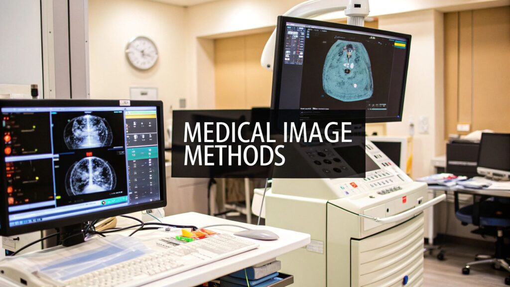
Decoding the Future of Medical Imaging
Medical imaging has fundamentally changed healthcare delivery since the early days of X-rays. Today’s advanced imaging technologies produce incredibly detailed 3D and 4D scans, but the real value comes from analyzing and interpreting these images effectively. Medical image analysis translates complex visual data into insights that guide clinical decisions. This capability is becoming essential for everyone working to advance healthcare – from device makers and researchers to hospital systems and medical startups.
Traditional medical image analysis depended heavily on radiologists carefully examining scans for abnormalities. But as imaging technologies generate increasingly complex data and healthcare demands faster diagnoses, new analytical approaches have emerged. The best methods can reliably detect subtle patterns, measure key features, and provide physicians with clear, actionable information – often for time-sensitive cases. True breakthroughs in this field reveal details invisible to human eyes while improving diagnostic precision and enabling personalized treatment plans.
This article examines eight key methods that are defining modern medical image analysis, from foundational techniques to advanced AI applications. We’ll explore each approach’s capabilities, limitations, and potential impact on patient care, medical research, and the broader healthcare system. By understanding these analytical tools, you’ll gain perspective on how imaging technology continues to evolve and enhance medical practice in meaningful ways.
1. Deep Learning-Based CNN Analysis
Convolutional Neural Networks (CNNs) have fundamentally changed medical image analysis by offering excellent accuracy in disease detection, image segmentation and other critical tasks. CNNs learn features directly from medical images, removing the need for complex manual feature extraction methods.
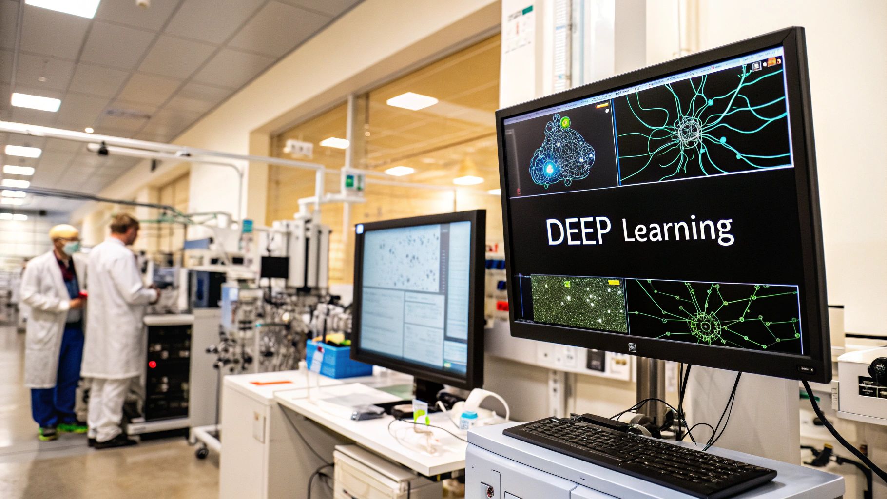
CNNs work through multiple specialized layers. The convolutional layers identify patterns like edges and textures using learnable filters. Pooling layers then compress this information while maintaining important features. This layered approach enables CNNs to understand increasingly complex image characteristics. The system learns end-to-end, going straight from raw image data to the final output, whether that’s disease classification or image segmentation.
Key Features and Benefits:
- Automatic Feature Extraction: The network learns relevant features on its own, removing the need for time-intensive manual feature engineering
- Hierarchical Learning: Complex patterns emerge as the network combines simpler features from earlier layers
- Multi-layer Processing: Enables detailed analysis and robust image interpretation
- End-to-end Learning: Creates a direct path from input images to desired results
Real-world Examples:
- ChestNet: Stanford’s system for analyzing lung X-rays matches radiologist-level performance
- DeepMind’s eye disease detection: Analyzes retinal scans to catch eye diseases early with high accuracy
- Brain tumor segmentation: Precisely maps tumor boundaries to help plan surgery and track treatment
Growth and Development:
CNNs took off in medical imaging thanks to bigger datasets, better GPUs, and groundbreaking work by researchers at places like Stanford, Google DeepMind, and pioneers like Geoffrey Hinton and Yann LeCun.
Pros and Cons:
Pros:
- Strong pattern recognition
- Handles large datasets well
- Minimal manual feature work needed
- Learns complex patterns automatically
Cons:
- Needs lots of training data
- Heavy computing requirements
- Limited insight into decisions
- Substantial hardware needs
Implementation Tips:
- Use transfer learning: Start with pre-trained models and adapt them to your medical data
- Apply data augmentation: Expand your dataset through image transformations
- Preprocess properly: Normalize and standardize image formats
- Add interpretability tools: Use techniques like saliency maps to understand the network’s choices
CNNs have become essential tools for medical device companies, healthcare tech firms, researchers and institutions. They excel at automating complex image analysis with high precision, advancing diagnostics and personalized care. Understanding both their strengths and limits helps organizations implement this powerful technology effectively.
2. Segmentation Using Active Contours
Active contours, also known as “snakes,” are powerful tools for medical image segmentation that help outline anatomical structures with precision. These dynamic models adjust their shape to match object boundaries in an image by using energy minimization principles – the contour seeks the lowest energy state that corresponds to the target boundary.
The process works through an energy minimization framework with two key components. Internal energy controls the contour’s smoothness and elasticity to prevent distortion. External energy, derived from the image data, pulls the contour toward features like edges or uniform intensity regions. Through iterations, the contour adjusts to minimize total energy and lock onto the target boundary.
A major strength of active contours is their ability to handle complex shapes and noisy images effectively. In cardiac imaging, they enable precise measurement of heart chamber volumes. For brain scans, they help map specific anatomical regions. Their capacity to detect subtle boundaries makes them essential for tumor analysis in treatment planning.
The technique gained prominence after Michael Kass, Andrew Witkin, and Demetri Terzopoulos introduced “snakes” in their landmark 1988 paper. Their mathematical framework laid the foundation for many advances, including gradient vector flow (GVF) snakes that improved initialization and convergence in difficult regions.
Features and Benefits:
- Energy Minimization Framework: Allows the contour to dynamically adjust to image features
- Deformable Curve Models: Adapts to complex object shapes
- Interactive Initialization: Provides user control over the initial contour placement
- Boundary Detection Capabilities: Accurately delineates even subtle boundaries
Pros:
- Accurate boundary detection: Achieves precise segmentation results
- Works well with noisy images: Robust to image artifacts and noise
- Can handle complex shapes: Effectively segments intricate anatomical structures
- Interactive control possible: User interaction can guide the segmentation process
Cons:
- Sensitive to initialization: Initial contour placement can influence the final result
- May get stuck in local minima: The optimization process may converge to a suboptimal solution
- Computational complexity: Can be computationally demanding for complex images
- Parameter tuning required: Optimization parameters need to be adjusted based on image characteristics
Tips for Implementation:
- Carefully initialize contour position: A good initial approximation can improve convergence and accuracy
- Adjust parameters based on image characteristics: Tailor parameters like internal and external energy weights to the specific image data
- Use a multi-resolution approach for efficiency: Start with a coarse image resolution and progressively refine the segmentation at higher resolutions
- Consider gradient vector flow (GVF) for better convergence: GVF snakes can improve performance, especially in concave regions
Active contours remain a key medical image analysis method due to their accuracy in segmenting complex anatomical structures, even with noisy or artifact-filled images. Their flexibility, adaptability and interactive control capabilities make them invaluable for diagnosis, treatment planning and research applications.
3. Atlas-Based Registration
Atlas-based registration helps doctors analyze medical images by matching a patient’s scan with a pre-made anatomical map called an atlas. This approach allows doctors to label body parts, find abnormalities, and measure anatomical differences by aligning the patient’s image with the standardized atlas. It has become essential for many medical applications.
How it Works
The method finds the best way to map the patient’s image onto the atlas through movements like shifting, rotating, and scaling. Special measurements check how well the images line up, while matching algorithms look for similar features between them to improve alignment accuracy.
Features and Benefits:
- Template Matching: Accurately identifies anatomical structures even with noisy or varied image quality
- Spatial Alignment: Precisely aligns patient images with atlas through position and size adjustments
- Multi-Modal Support: Works across different scan types like MRI, CT, and PET
- Standardized Framework: Provides consistent way to label and analyze anatomy across patients
Real-World Applications:
Atlas registration helps doctors in several ways:
- Brain MRI Studies: Measures brain region volumes for research, like tracking changes in Alzheimer’s disease
- Neurological Diagnosis: Helps find and assess brain lesions from strokes, tumors, and multiple sclerosis
- Surgery Planning: Creates detailed 3D models of patient anatomy to guide complex procedures
Development Over Time
Better computers and digital imaging pushed atlas registration forward. Early methods used single atlases, which had limitations. Now, multiple atlases can be combined for each patient, greatly improving accuracy. Along with faster computers and better algorithms, this has made atlas registration a key medical imaging tool.
Advantages:
- Reliable labeling: Accurately identifies anatomical structures
- Works with multiple scan types: Can analyze different kinds of medical images together
- Consistent approach: Enables comparable analysis between studies
- Automation capable: Can be automated to improve workflow efficiency
Limitations:
- Atlas quality matters: Results depend on having good reference atlases
- Unusual anatomy challenges: May struggle with significant anatomical differences
- Processing demands: Needs substantial computing power
- Error effects: Registration mistakes can impact later analysis steps
Implementation Tips:
- Use multiple atlases: Combine several atlases for better results
- Check quality regularly: Monitor registration accuracy
- Choose specific atlases: Use reference images matched to patient groups
- Verify results: Compare with expert review to ensure accuracy
This method helps medical device companies, researchers, and healthcare providers make better decisions through standardized anatomical analysis. It offers valuable insights for both research and clinical applications.
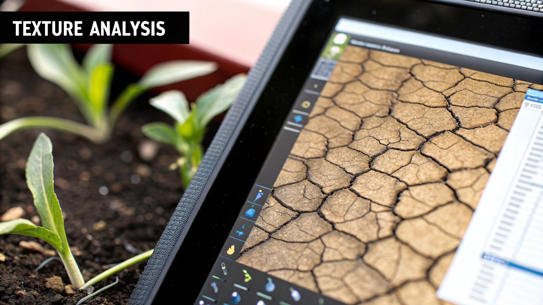
4. Texture Analysis
Texture analysis is a key method in medical image analysis that helps quantify and characterize subtle patterns within medical images. These patterns, which are often impossible to see with the naked eye, provide important clues about tissue properties and disease progression. This method has become increasingly important because it offers objective ways to assess tissue characteristics, leading to more accurate diagnoses.

The method combines statistical and structural approaches to extract meaningful data from medical images. Statistical approaches focus on measuring features like smoothness and regularity, while structural methods examine how image elements are organized spatially. These measurements help identify abnormalities and understand tissue characteristics.
Key Features of Texture Analysis:
- Statistical feature extraction: Uses measurements like gray-level co-occurrence matrix (GLCM) and run-length matrix (RLM) to quantify texture properties
- Pattern recognition: Uses machine learning to identify and classify specific texture patterns linked to different tissues or diseases
- Multi-scale analysis: Studies texture features at various scales to capture details at different levels
- Quantitative measurements: Provides measurable data for precise and repeatable tissue assessments
Advantages of Texture Analysis:
- Objective assessment: Gives unbiased measurements of tissue properties, reducing variation between observers
- Works across imaging types: Can be used with CT, MRI, ultrasound, and microscope images
- Detects small changes: Can find tiny variations in tissue texture that doctors might miss visually
- Non-invasive: Evaluates tissue properties without needing biopsies or other procedures
Challenges and Limitations:
- Image quality matters: Noise and artifacts can affect accuracy
- Feature selection is difficult: Choosing the most useful texture features requires careful consideration
- May need normalization: Images often need adjusting to account for differences in intensity and contrast
- Hard to interpret: Understanding texture features requires special knowledge
Real-world Examples:
- Liver health: Helps measure liver scarring using ultrasound and MRI images
- Cancer analysis: Helps tell benign from cancerous tissues and predict treatment outcomes
- Bone studies: Assesses bone structure and estimates fracture risk
Practical Tips:
- Keep imaging consistent: Use standard protocols to reduce variation
- Use multiple features: Combine different texture measurements for better results
- Consider image resolution: Remember resolution affects texture features
- Check against clinical data: Compare findings with patient records and tissue samples
Texture analysis has grown more powerful as computing and image processing have advanced. Its importance comes from its ability to reveal tissue properties and disease signs that help doctors make better diagnoses and treatment plans. This makes it an essential tool in modern medical imaging and research.
5. Machine Learning Classification
Medical image analysis has made significant progress through machine learning classification methods. These techniques help doctors and researchers categorize medical images into specific groups by analyzing key visual patterns and features. The process is fast, reliable, and provides clear insights that medical professionals can understand and trust.
The classification process works in two main steps. First, the system identifies and measures specific aspects of the image like shapes, textures, and intensity levels. Then, established algorithms like Support Vector Machines (SVM), Random Forests, and Decision Trees analyze these measurements to sort images into categories. The system also provides a confidence score to show how certain it is about each classification.
Key Features and Benefits:
- Targeted Analysis: Focuses on specific, measurable image characteristics that matter most for diagnosis
- Algorithm Flexibility: Different classification methods available to match specific medical needs
- Learning from Examples: Uses labeled training data to achieve high diagnostic accuracy
- Confidence Metrics: Provides reliability scores to support clinical decisions
- Clear Results: Medical professionals can understand why the system made specific classifications
Advantages:
- Clear Decision Process: Medical staff can follow how classifications are made
- Efficient with Limited Data: Works well even with smaller image collections
- Quick Processing: Provides fast results for both training and analysis
- Proven Methods: Based on well-tested scientific principles
Limitations:
- Complex Setup: Requires expert knowledge to identify and extract relevant image features
- Pattern Recognition Limits: May miss subtle image details that humans can spot
- Feature Dependencies: Results depend heavily on choosing the right image characteristics
- Scope Constraints: Can only analyze features that were specifically programmed
Real-World Applications:
- Mammogram Analysis: Helps identify potential breast cancer markers
- Skin Cancer Screening: Evaluates skin lesions using standard medical criteria
- Tissue Sample Review: Classifies microscope images for disease diagnosis
Historical Development:
Machine learning classification gained momentum in medical imaging when researchers Vladimir Vapnik developed SVM algorithms and Leo Breiman created Random Forest methods. The release of open-source tools like scikit-learn made these techniques widely available to medical researchers.
Implementation Guidelines:
- Choose Features Carefully: Select image characteristics that align with medical diagnostic criteria
- Validate Thoroughly: Use comprehensive testing to ensure reliable performance
- Balance Training Data: Include equal representation of different medical conditions
- Consider Combined Methods: Use multiple algorithms together for better accuracy
Machine learning classification continues to serve as a valuable tool in medical image analysis. While it has some limitations in handling complex patterns, it offers clear benefits through its speed, reliability, and understandable results. As deep learning methods advance, they complement these traditional techniques to provide even better medical imaging solutions.
6. Morphological Processing
Morphological processing helps analyze medical images by examining the shapes, sizes and structures within them. It relies on mathematical principles to process images in ways that extract key information and features. The technique is particularly good at tasks like finding objects, separating overlapping structures, cleaning up noise, and bringing out important details. It works by using a small “structuring element” shape that probes the image and changes pixel values based on their surroundings.
The core operations in morphological processing are:
- Erosion: Makes objects smaller by removing pixels from their edges. This helps eliminate small bits of noise and separate objects that are touching.
- Dilation: Makes objects larger by adding pixels to their edges. Great for filling holes and connecting broken parts.
- Opening: Combines erosion then dilation to smooth object edges and remove small items while keeping larger ones intact.
- Closing: Combines dilation then erosion to fill holes and gaps while maintaining overall object size.
Key Benefits:
- Shape Analysis: Directly examines object shapes – perfect for medical tasks like tumor detection and bone analysis
- Versatile Processing: Works on both black/white and grayscale images for broader applications
- Customizable Results: Different structuring element shapes produce different effects
- Clean Output: Very effective at removing noise while keeping important features
- Fast Performance: Operations are quick to compute, even on large images
Advantages and Limitations:
| Advantages | Limitations |
|---|---|
| Keeps shape details intact | May alter fine features |
| Removes noise effectively | Results depend on structure element |
| Fast processing speed | Works best for specific uses |
| Good for pre-processing | Can create unwanted artifacts |
Medical Applications:
- Bone Analysis: Measuring bone density and structure in osteoporosis studies
- Cell Counting: Separating and counting cells in microscope images of blood or tissue samples
- Blood Vessel Enhancement: Making vessels more visible in angiograms and retinal scans
Implementation Tips:
- Choose Structure Elements Carefully: Test different shapes/sizes to find what works best
- Combine Operations: Using multiple steps often gives better results than single operations
- Check Results Visually: Always inspect output to catch unwanted effects
- Use with Other Methods: Works well alongside segmentation and classification techniques
Historical Context:
Mathematical morphology emerged in the 1960s and grew popular as computers became more powerful. Its ability to handle complex shapes reliably made it essential for medical imaging. Today, morphological processing remains a key part of many medical image analysis systems, helping advance diagnosis, treatment planning, and research.
7. Multi-Scale Analysis
Multi-scale analysis is a key method in medical image analysis that examines images at different resolutions, capturing both precise details and broader contextual features. It efficiently analyzes important information that exists across multiple scales. For example, in mammograms, the method can detect tiny microcalcifications while also analyzing the broader tissue patterns and density variations that provide vital medical context.
The process works by using mathematical tools like wavelet transforms or image pyramids. These tools break down images into different frequency parts that correspond to different scales or create layered image representations through systematic downsampling.
Key Features:
- Multi-resolution capabilities: Examines features from fine details to large structures
- Scale representation: Provides complete image analysis across all scales
- Step-by-step processing: Analyzes coarse features first before moving to finer details
- Information retention: Keeps important features intact across scales
Advantages:
- Detects features at all scales: Can find both subtle changes and major abnormalities
- Handles noise well: Multiple scale analysis helps filter out image artifacts
- Processing efficiency: Hierarchical approach reduces overall computation needs
- Better feature detection: Excels at finding varying sizes and shapes of features
Limitations:
- Complex setup: Requires expertise in wavelet transforms and pyramids
- Parameter selection: Must carefully choose decomposition levels and functions
- Storage needs: Multi-resolution data requires more space
- Processing demands: Can still need significant computing power
Real-World Applications:
- Mammography: Detects both tiny calcifications and broader tissue patterns that may indicate cancer
- Eye imaging: Maps blood vessels of varying sizes to diagnose retinal conditions
- Brain scans: Identifies brain structures from small folds to major regions for neurological analysis
Implementation Tips:
- Select scale levels wisely: Match scales to the features you need to detect
- Manage resources: Balance detail level with available computing power
- Check all scales: Verify analysis quality across each resolution level
- Use proper reconstruction: Choose methods that minimize data loss when rebuilding images
Why It Matters:
Multi-scale analysis effectively bridges a key gap in medical imaging – the need to analyze both small details and large structures simultaneously. Its ability to examine images comprehensively across different scales makes it vital for accurate diagnosis, treatment planning, and research. The method’s systematic approach to handling multiple resolutions has made it a fundamental tool in modern medical image analysis.
8. Graph-Based Analysis
Graph-based analysis is a powerful technique that converts medical images into graph structures. The analysis represents regions of interest, anatomical elements, or individual pixels as nodes, while the connections between them become edges based on factors like proximity, intensity, or texture similarities. This approach provides valuable insights into how different parts of medical images relate to each other.
Understanding the Process
Graph theory principles form the foundation of this analysis method. Converting images into graphs allows us to study complex anatomical structures and spot subtle patterns that standard image processing might miss. For example, we can model connections between brain regions or analyze blood vessel networks to assess vascular health and detect abnormalities.
Recent Growth and Development
Graph-based analysis has gained significant traction as computing power has increased and graph algorithms have improved. While early applications used basic graph structures, today’s systems can process large, complex graphs from high-resolution 3D medical images. This advancement has created new opportunities in areas like brain mapping, disease detection, and treatment planning.
Key Features and Benefits:
- Structural Focus: Concentrates on relationships between elements rather than pixel data, making it resistant to image noise and variations
- Clear Connections: Shows how different image regions interact and relate to each other
- Proven Methods: Uses established graph theory algorithms for tasks like segmentation and pattern detection
- Pattern Discovery: Identifies complex patterns that may indicate medical conditions
Advantages:
- Accurate Spatial Analysis: Effectively shows how anatomical structures connect and relate
- Adaptable Format: Works well with different types of medical images and structures
- Handles Complexity: Well-suited for analyzing detailed networks and hierarchies
- Manages Variation: Maintains accuracy despite irregular shapes and sizes
Limitations:
- Resource Intensive: Creating graph representations requires significant processing power
- Processing Demands: Complex graph algorithms need substantial computing resources
- Memory Usage: Large graphs require considerable memory storage
- Setting Sensitivity: Results depend heavily on chosen parameters
Real-World Applications:
- Brain Studies: Using graphs from brain scans to understand neural connections and identify disorders
- Blood Vessel Analysis: Examining vascular structures to assess health and spot issues like aneurysms
- Tissue Research: Studying tissue arrangement in microscope images to improve cancer diagnosis
Implementation Guidelines:
- Optimize Construction: Choose node and edge definitions carefully to balance speed and accuracy
- Use Sparse Data: Reduce memory needs by using efficient data structures
- Select Smart Tools: Pick algorithms that match your needs and scale well
- Verify Results: Check that graphs accurately represent image data and analysis methods suit your goals
Graph-based analysis stands out for its ability to reveal complex relationships in medical images. By examining images from a structural perspective rather than just pixel values, it enables deeper understanding of anatomy and disease processes, leading to better diagnosis and treatment planning.
8-Point Comparison: Medical Image Analysis Methods
| Method | Complexity (🔄) | Resources (⚡) | Outcomes (📊) | Advantages (⭐) | Insights (💡) |
|---|---|---|---|---|---|
| Deep Learning-Based CNN Analysis | High – multi-layer, end-to-end training | High – large datasets & advanced hardware | High accuracy with automatic feature extraction | Automatic learning & end-to-end capability | Use transfer learning and data augmentation |
| Segmentation Using Active Contours | Moderate – sensitive to initialization | Moderate – needs iterative tuning | Accurate boundary detection | Robust with noisy images and complex shapes | Initialize carefully & tune parameters |
| Atlas-Based Registration | Moderate to High – spatial transformations | High – computationally intensive | Robust anatomical labeling | Standardized, cross-modality analysis | Use multi-atlas approaches & quality control measures |
| Texture Analysis | Low to Moderate – statistical methods | Low to Moderate – less intensive but quality sensitive | Quantitative tissue characterization | Detects subtle changes with non-invasive assessment | Standardize image acquisition & use multiple features |
| Machine Learning Classification | Low – relies on feature engineering | Low – works with smaller datasets | Interpretable classification outcomes | Fast training, clear results, and established methods | Emphasize careful feature selection and cross-validation |
| Morphological Processing | Low – based on simple shape operations | Very efficient – minimal computational cost | Effective noise removal and shape preservation | Excellent preprocessing and speed | Choose appropriate structural elements and sequences |
| Multi-Scale Analysis | High – complex multi-resolution processing | High – significant computational overhead | Captures features at various scales | Robust feature detection across scales | Optimize decomposition levels and manage resources |
| Graph-Based Analysis | High – complex graph construction | High – memory intensive and computationally heavy | Detailed spatial and relational modeling | Excellent for handling irregular and complex structures | Optimize graph sparsity and use efficient algorithms |
The Expanding Horizon of Medical Image Analysis
Medical imaging is a vital field with significant advances in technology and analysis methods. Deep learning with convolutional neural networks (CNNs), graph-based analysis, and other advanced techniques enable medical professionals to extract meaningful insights from medical images. Healthcare providers can now select from multiple proven methods – from CNNs that automate feature extraction to active contours for precise segmentation and atlas-based registration that standardizes anatomical images.
Success in medical image analysis requires matching the right techniques to your specific needs. Key factors include the characteristics of your imaging data, your desired outcomes, and available computing resources. It’s essential to carefully evaluate these elements when implementing any analysis solution.
The field continues advancing rapidly with new developments in:
- Federated learning for privacy-protected collaborative model training
- Radiomics techniques for quantifying image features
- Explainable AI approaches that enhance result interpretation
Combining multiple analysis methods often leads to better results. For example, integrating multi-scale analysis with deep learning can help uncover subtle patterns and improve diagnostic accuracy.
Key Takeaways:
- Medical image analysis tools significantly improve diagnostics, treatment planning, and patient care
- Method selection should align with specific applications and data types
- Ongoing learning is essential to stay current with new developments
- Combined analysis approaches often provide more complete insights
The future looks promising for medical image analysis, with opportunities for more precise diagnostics and improved patient outcomes. To maximize the potential of your medical imaging data, consider working with experienced partners. PYCAD provides comprehensive AI solutions for medical imaging – from data processing through deployment. Our expertise in deep learning, computer vision, and regulatory compliance enables robust and scalable solutions customized for your needs.