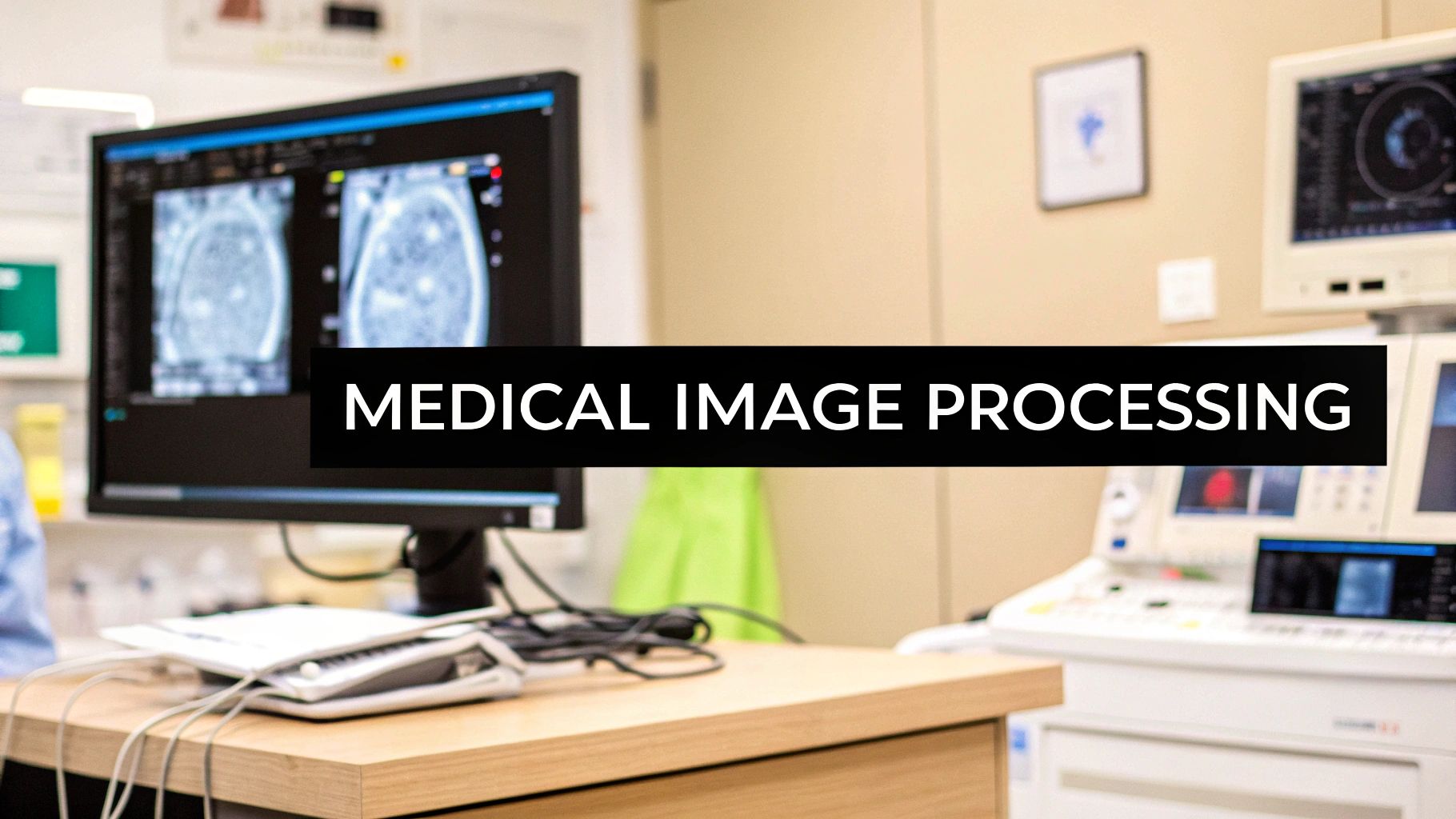
The Evolution and Impact of Medical Image Processing
Medical image processing has fundamentally changed how doctors diagnose and treat patients. What started with basic X-rays has grown into an advanced field that combines AI capabilities with sophisticated analysis techniques. Medical image processing tutorials play a key role in helping healthcare professionals stay current with these rapid advances.
From X-rays to AI: A Historical Overview
The early days of medical imaging focused on converting physical X-rays into digital formats and managing those files effectively. By the 1980s, the field expanded to include tools for enhancing image quality and speeding up analysis. This allowed doctors to see clearer images and review them more efficiently.
The 1990s brought major steps forward in image analysis capabilities. Key developments included image registration for aligning multiple scans, segmentation to isolate specific structures, and classification to automatically categorize images. Since the first X-ray in 1895, the technology has made incredible progress. Learn more about this journey here: Explore the evolution of medical image processing. Understanding this history helps put modern advances in context.
The Impact on Modern Healthcare
Today’s medical professionals rely heavily on image processing in their daily work. These tools support everything from initial diagnosis to surgical planning and real-time guidance during procedures. For example, surgeons now routinely use detailed 3D models created from CT scans when preparing for complex operations.
Future Trends and Best Practices
The field continues to advance rapidly, especially with new AI applications that enable automated diagnosis and personalized treatment approaches. This makes understanding core image processing concepts more important than ever for healthcare workers. Current best practices emphasize ongoing education to keep pace with new developments.
High-quality tutorials and training resources are essential for helping medical professionals effectively use these powerful tools. As the technology grows more advanced, creating clear standards will be crucial for smoothly integrating new capabilities into clinical practice. This will lead to better coordination between different parts of the healthcare system and improved patient outcomes through enhanced medical imaging.
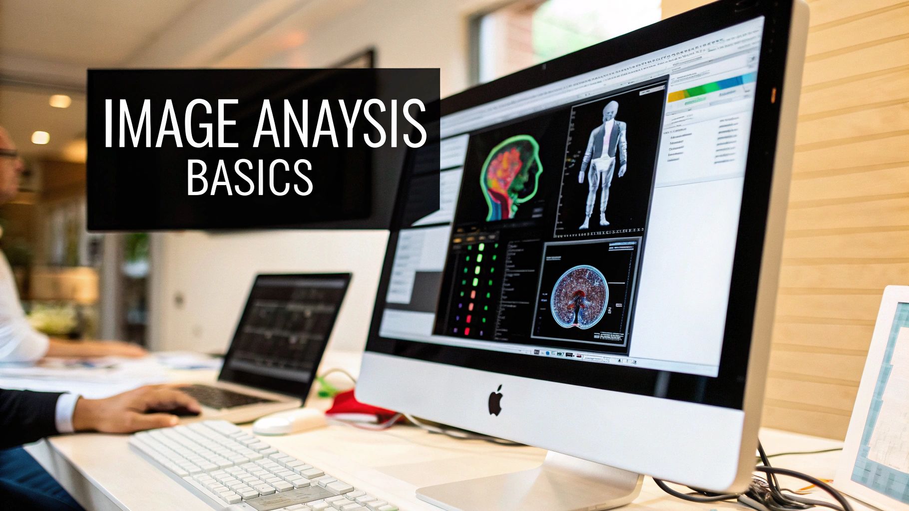
Selecting and Mastering Essential Processing Tools

Choosing the right medical image processing tools requires careful evaluation and planning. Key platforms like 3D Slicer and ITK-SNAP offer different capabilities that need to be assessed based on your specific needs. Making informed decisions about software involves weighing factors such as costs, user-friendliness, and how well tools work together.
Evaluating Software Options: Open-Source vs. Enterprise
Open-source platforms provide an accessible starting point for medical image processing tutorials, particularly in research and education. These tools come with active user communities that offer support and learning resources. For instance, 3D Slicer shines in 3D visualization and analysis, while ITK-SNAP focuses on segmentation. However, open-source tools may need more technical knowledge and could lack some advanced features.
Enterprise solutions pack more features, including direct support and smooth integration with hospital systems. They often include AI analysis tools and automated reporting capabilities, making them ideal for busy clinical settings. The main consideration here is cost, which can be substantial.
Combining Tools for Efficient Workflows
Many healthcare facilities blend different tools to create smooth workflows. They might start with open-source software for basic processing before using enterprise systems for complex analysis. Cloud-based options are becoming more popular, making it easier to share data and work together. This mixed approach helps organizations get the best from each tool while managing resources wisely. The industry numbers reflect this trend – the medical image analysis software market grew from USD 2.43 billion in 2016 and is expected to reach USD 4.26 billion by 2025. Learn more at Medical imaging software market statistics. All-in-one solutions are gaining ground while standalone software use decreases.
Practical Tips for Tool Selection
When choosing tools, match your clinical needs with your budget. Start by listing exactly what tasks you need to complete. Then check different options based on their features, ease of use, and how well they connect with other systems. Consider how much technical skill your team needs to use and maintain the software. Testing tools in real situations before making final choices provides valuable insights. This careful approach ensures your selected tools fit your goals and help improve medical image processing.
Mastering Medical Image Dataset Management
Managing medical image datasets requires careful attention to organization, security, and processing methods. Proper handling of these valuable assets ensures both research success and patient confidentiality. Let’s explore the key aspects of medical image dataset management.
Organizing and Preprocessing Medical Image Data
The foundation of medical image management starts with the DICOM standard (Digital Imaging and Communications in Medicine). This standard enables consistency across different imaging platforms and systems. Good organization requires clear file naming rules and an organized storage structure that makes finding and analyzing images simple.
Key preprocessing steps prepare images for analysis:
- Noise reduction to remove unwanted artifacts
- Image enhancement to improve visibility
- Normalization to ensure consistent data ranges
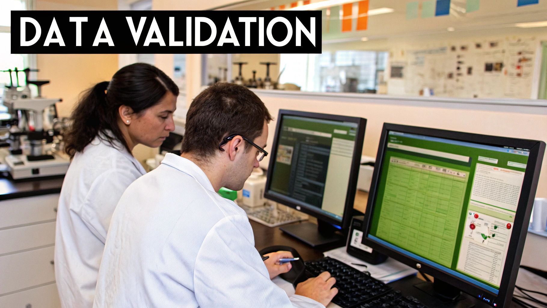
Maintaining Data Integrity and Patient Privacy
Data integrity is essential in medical imaging. Regular quality checks and backup systems help prevent data loss or corruption. Validation procedures confirm that images remain accurate and reliable for clinical or research use.
Patient privacy protection requires strict data security measures. This includes removing identifying details from images and metadata. Organizations must follow privacy regulations like HIPAA when handling medical data.
Accessing and Utilizing Public Datasets
Public medical image collections offer valuable resources for research and algorithm development. Major repositories include:
- The National Institutes of Health (NIH) chest X-ray dataset with 100,000+ images from 30,000+ patients
- The Cancer Imaging Archive (TCIA) with extensive oncology imaging collections
These datasets help advance AI development in healthcare applications from diagnosis to treatment planning. Learn more: Medical imaging dataset resources guide.
Building Robust Data Pipelines
Effective data pipelines support both clinical work and research by automating image handling workflows. A well-designed pipeline:
- Converts images to DICOM format automatically
- Performs necessary preprocessing
- Stores data securely
- Handles multiple imaging types (CT, MRI, etc.)
- Integrates different data formats
This automation allows medical professionals to focus on analysis and patient care rather than manual data management tasks.
Implementing Deep Learning for Medical Image Analysis
Deep learning has become a vital tool for analyzing medical images, helping doctors make more accurate diagnoses and treatment plans. This section explores how healthcare organizations are putting these techniques to work in their daily operations.
Choosing the Right Deep Learning Architecture
The success of medical image analysis depends heavily on selecting appropriate deep learning models. Convolutional Neural Networks (CNNs) work well for tasks like spotting tumors or abnormalities in scans. For tracking disease progression through image sequences over time, Recurrent Neural Networks (RNNs) often provide better results.
Fine-tuning Models for Specific Applications
While starting with pre-trained models is helpful, customizing them for specific medical uses is essential. For example, a model meant to detect lung cancer needs training on many lung CT scans. This focused training helps the system learn exactly what to look for, improving its accuracy for that particular use.
Medical imaging generates massive amounts of data – in fact, images make up 90% of all medical data according to IBM research. This provides plenty of material to train deep learning systems effectively. Learn more at Deep Learning Applications in Medical Imaging.
Balancing Automation With Clinical Expertise
While deep learning can speed up image analysis, it works best as a tool to support doctors rather than replace them. These systems help highlight potential issues, but the final decisions about diagnosis and treatment should always come from qualified medical professionals.
Validating AI Results in Medical Contexts
Thorough testing is crucial when using deep learning in healthcare. The stakes are high, as mistakes could harm patients. Testing must happen with separate image sets that weren’t used in training. Key metrics like sensitivity, specificity, and precision need careful evaluation to ensure the system performs reliably in real clinical settings.
Real-world Implementation Strategies and Performance Optimization
Successful deep learning projects need clear goals, quality data, and the right technical resources. Regular monitoring helps maintain accuracy over time. As medical knowledge grows and new data comes in, models often need updates and retraining. This ongoing refinement helps get the most benefit from deep learning in medical imaging.
Building Effective Clinical Applications
Medical image processing applications require thoughtful planning and implementation to enhance patient care and fit seamlessly into medical environments. The key is creating tools that both improve clinical outcomes and work smoothly for medical staff.
Integrating New Tools into Clinical Workflows
The best medical imaging tools complement existing clinical processes rather than disrupting them. For example, an AI diagnostic system should integrate directly into a radiologist’s current viewing system (PACS) and provide insights within their normal workflow. This approach minimizes disruption and helps busy medical professionals readily adopt new technology.
Ensuring Regulatory Compliance and Maintaining Performance
Clinical applications must meet strict medical standards like HIPAA in the US. Patient privacy and data security are essential considerations when implementing any new system. Applications also need consistent performance under real clinical conditions. This requires ongoing testing and quality checks to maintain accuracy and reliability in actual medical settings.
Strategies for Training Staff and Managing Change
Getting staff comfortable with new technology is crucial for successful implementation. Clear training programs help medical personnel learn to use tools effectively. It’s important to address concerns about changes to workflows. Showing concrete benefits through case examples, providing continuous support, and involving staff in implementation decisions helps build acceptance.
Measuring Clinical Impact and Quality Assurance
The real value of clinical applications comes from improving patient care outcomes. Key metrics might include:
- Increased diagnostic accuracy
- Faster result turnaround
- Reduced need for invasive procedures
A strong quality assurance program is essential. Regular performance reviews and validation testing ensure applications maintain high standards over time. This helps translate the potential of medical image processing into measurable clinical benefits.
Preparing for the Future of Medical Imaging

Medical image processing continues to advance at a rapid pace. Healthcare professionals and organizations must stay informed about new developments and adapt their practices to provide the best patient care. Understanding how to effectively manage these changes is essential for success in this field.
Emerging Trends and Their Impact
Several key developments are reshaping medical imaging practices. Artificial intelligence is becoming essential for automating image analysis and supporting diagnoses. For instance, NVIDIA’s MAISI platform now generates synthetic 3D CT images with segmentation masks. Cloud solutions are also gaining prominence, enabling better collaboration and access to imaging data.
These advances are improving diagnostic accuracy in meaningful ways. AI tools help spot subtle abnormalities that humans might miss. As noted in Ramsoft’s analysis, cloud-based systems provide remote access capabilities and improved data sharing. However, organizations need careful planning to successfully implement these new technologies.
Strategies for Technological Adaptation
A systematic approach is crucial when integrating new imaging tools. Organizations should:
- Evaluate their current systems and infrastructure
- Identify where new technologies can add the most value
- Test compatibility with existing PACS and RIS platforms
- Run pilot programs before full implementation
As discussed in Ramsoft’s integration guide, ensuring smooth integration between systems is essential. Change management and staff training are just as important as the technical aspects of implementation.
Investing in Future Capabilities
Success in medical imaging requires ongoing investment in both equipment and people. Organizations should:
- Provide regular training on new techniques and technologies
- Support continuing education for staff
- Create an environment that welcomes innovation
- Stay current with emerging trends and best practices
Continuous learning and development help teams master new approaches while maintaining high standards of patient care.
Empowering the Future of Medical Imaging with PYCAD
PYCAD offers AI solutions designed specifically for medical imaging workflows. Their services include:
- Data management and processing
- AI model development and training
- Implementation support
- Ongoing technical assistance
These tools help healthcare organizations make the most of advances in medical imaging technology while maintaining focus on patient care.


