Think about the last time you saw a medical drama on TV.Remember those scenes where doctors pull up incredibly detailed, glowing images of the human body on a big screen? That's medical imaging software in action. It's the essential digital engine that takes the raw, unprocessed data from machines like MRI, CT, and X-ray scanners and turns it into the detailed pictures doctors use to see what's happening inside us.
This software is more than just a picture viewer; it's a powerful diagnostic hub and a secure digital archive all rolled into one, completely changing how clinicians work with patient scans.
The Digital Darkroom and an Intelligent Filing Cabinet
A great way to understand this software is to see it as two things at once: a high-tech digital darkroom and a super-smart filing cabinet. Not so long ago, radiologists would physically hold up large films to a lightbox. It was a slow, clunky process that feels worlds away from what's possible now. Today's software is the digital darkroom, giving doctors the power to tweak and enhance images with incredible detail.
At the same time, it acts as that intelligent filing cabinet. Every single scan, whether it's a basic X-ray or a complex 3D reconstruction from a CT, is carefully stored, tagged, and ready to be pulled up in seconds. This two-part role is absolutely vital in modern medicine, where every moment counts and accuracy is everything.
From Raw Data to Actionable Insights
When a patient gets a scan, the imaging machine captures a huge amount of raw data. By itself, this data is just a jumble of numbers and signals. Medical imaging software is what makes sense of it all, piecing it together into a clear visual map of the body. But its real power lies in what it lets medical professionals do with that map.
This software comes loaded with tools that allow doctors to:
- Manipulate Images: They can adjust brightness and contrast to make subtle abnormalities pop, or zoom in to examine a tiny area of concern.
- Reconstruct Views: They can build interactive 3D models from a series of 2D image "slices," giving them a 360-degree view of an organ or bone structure. This is a game-changer for planning surgeries.
- Perform Measurements: The software allows for precise measurement of tumors, blockages, or other structures, which is essential for monitoring how a disease is progressing or if a treatment is working.
The core purpose of medical imaging software is to bridge the gap between data acquisition and clinical decision-making. It translates complex scanner outputs into a language that doctors can use to diagnose conditions, plan treatments, and ultimately improve patient outcomes.
To give you a clearer picture, here’s a quick breakdown of what this software really does behind the scenes.
Core Functions of Medical Imaging Software at a Glance
This table breaks down the essential roles of medical imaging software, offering a quick summary of its impact across the clinical workflow.
| Core Function | Primary Purpose | Impact on Patient Care |
|---|---|---|
| Image Visualization & Manipulation | To render raw data into 2D/3D images and allow for adjustments (zoom, pan, contrast). | Enables clinicians to spot subtle issues that might be missed on a static image, leading to earlier and more accurate diagnoses. |
| Advanced Analysis & Measurement | To provide tools for measuring anatomical structures, quantifying changes, and performing complex calculations. | Allows for objective tracking of disease progression or treatment effectiveness, moving beyond subjective visual checks. |
| Secure Archiving & Retrieval (PACS) | To store vast amounts of imaging data securely and make it instantly accessible to authorized personnel. | Ensures a patient's complete imaging history is available on-demand, improving continuity of care and preventing repeat scans. |
| Workflow Integration & Collaboration | To connect with other hospital systems (like EMRs) and allow for easy sharing among specialists. | Creates a more efficient diagnostic process, enabling faster consultations and more coordinated treatment planning. |
Ultimately, these functions work together to turn a simple scan into a powerful diagnostic tool that informs nearly every step of a patient's journey.
The Foundation of Modern Diagnostics
Without this software, even the most advanced imaging machines would be like fancy cameras with no way to see the pictures. Just consider this: in the United States alone, healthcare providers perform over 70 million CT exams every year. Each one produces a massive dataset that would be impossible to interpret without specialized software to process and display it.
This technology isn't just a nice-to-have extra; it's the bedrock of the entire diagnostic process. It ensures every scan is stored safely, can be found easily, and comes with the analytical tools needed for a rock-solid evaluation. From finding a tiny fracture in a bone to mapping out a delicate brain surgery, medical imaging software is the indispensable control room for modern medicine.
Breaking Down the Different Types of Imaging Software
When you hear "medical imaging software," it's easy to picture a single, all-encompassing program. The reality is much more interesting. It’s actually a connected ecosystem of specialized tools, each playing a critical role in a patient's diagnostic journey. To really get a handle on how modern healthcare facilities work, you need to understand the two systems that act as the central nervous system: PACS and RIS.
Think of them as a perfectly coordinated team. One is the master librarian, managing a massive archive of visual data. The other is the logistical mastermind, handling all the administrative tasks that keep the department running. Together, they create a smooth workflow, from the moment a patient books an appointment to the second a radiologist signs off on a report.
PACS: The Digital Image Library
The Picture Archiving and Communication System (PACS) is the heart and soul of image management. It's essentially a high-security digital vault built to store, retrieve, and display massive medical imaging files. Every single CT scan, MRI, and X-ray captured in a hospital or clinic finds its home here.
Its main purpose is to give authorized clinicians instant access to a patient's entire imaging history from any secure workstation. This simple function completely changes the game. It gets rid of the cumbersome process of handling physical film, helps avoid unnecessary repeat scans, and gives doctors a complete visual timeline for better long-term patient care.
This infographic breaks down some of the biggest benefits that facilities see when they implement modern imaging systems like PACS.
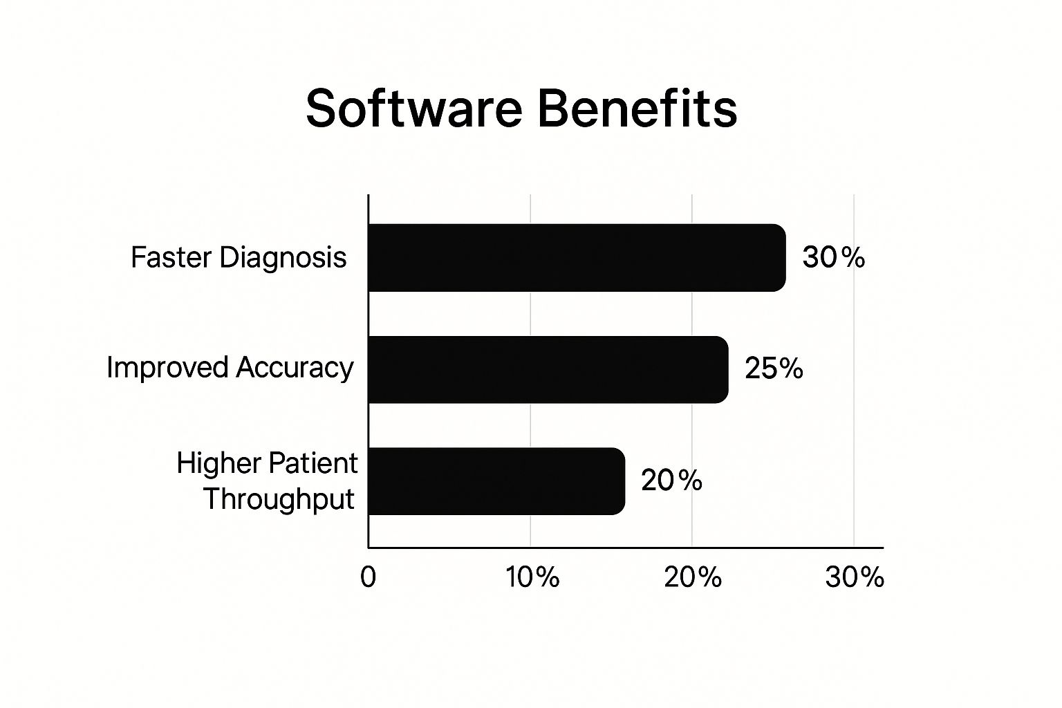
As you can see, the data points to huge wins in efficiency, leading to faster diagnoses and the ability to see more patients—major advantages in any healthcare setting.
RIS: The Operational Brain
If PACS is the library, then the Radiology Information System (RIS) is the clinic's brain—the meticulous operations manager. This software platform doesn't deal with the images directly. Instead, it manages all the crucial information and workflows surrounding them.
The RIS has a lot on its plate. It's responsible for a wide range of essential jobs, including:
- Patient Scheduling: Juggling appointments for all the different imaging procedures.
- Billing and Reporting: Handling the financial side of things and generating administrative reports.
- Workflow Management: Tracking a patient’s entire journey through the radiology department, from the moment they check in to the final report being sent out.
So, while a radiologist is using the PACS to examine a scan, the RIS is humming along in the background, making sure the correct patient data is attached, the right billing codes are applied, and the final report gets to the referring physician without a hitch.
Advanced Visualization Platforms
Beyond the foundational duo of PACS and RIS, a third type of software has become absolutely essential: advanced visualization platforms. These are the high-octane engines that give clinicians powerful tools for 3D and 4D image rendering, Multiplanar Reconstruction (MPR), and other complex analytical tasks.
While a standard PACS viewer is great for routine reviews, these advanced platforms are where the magic happens for complex cases. For instance, a surgeon preparing for a delicate operation can use this software to build an interactive 3D model of a patient's anatomy. This lets them map out the entire procedure with incredible precision before they even step into the operating room.
These platforms do more than just create pretty pictures. They turn flat, 2D image slices into a navigable, measurable, and interactive diagnostic environment. It's this functional depth that helps clinicians spot critical details that might otherwise be missed.
The market for this kind of software is growing fast, fueled by constant innovation in scanner technology and a rising demand for more precise diagnostic tools. In fact, the global medical image analysis software market is on track to hit roughly $4.26 billion by 2025. This growth is also reflected in technology shifts, with modalities like CT and MRI expected to grow their market share from 15% to 16% and 12% to 14%, respectively, over that period. You can dig deeper into market dynamics and technology trends to see the full picture.
When you bring it all together, PACS, RIS, and advanced visualization platforms form a powerful trio. Their integration ensures the entire imaging lifecycle—from scheduling and scanning to deep analysis and reporting—is handled efficiently, accurately, and securely. This is the bedrock of modern patient care.
Must-Have Features of Modern Imaging Platforms
So, what really separates a simple image viewer from a truly powerful, modern diagnostic platform? It boils down to a handful of critical features that help clinicians work smarter, make workflows feel effortless, and ultimately lead to better patient outcomes. These are the capabilities that turn medical imaging software from a digital lightbox into an essential analytical partner.
A top-tier platform needs to be a central hub, one that can pull in different types of data without a hitch. This ensures a radiologist or surgeon gets the complete, cohesive picture of a patient's health, all from a single, intuitive screen.
Seamless Multi-Modality Support
In any busy hospital, patients get all kinds of scans. One person might have an MRI on Monday and a CT scan on Wednesday. Multi-modality support is the magic that lets a clinician view and compare images from different sources—like CT, MRI, PET, and X-ray—all in one place.
Think about a doctor trying to compare a new PET scan (which shows metabolic activity) with an older MRI (which details anatomy). Without this feature, they’d be stuck toggling between two separate programs, trying to line up the images by eye. With it, they can instantly overlay the scans, fuse them together, and get a much richer, more complete story about the disease. This unified view is absolutely critical for complex cases, especially in fields like oncology and neurology.
Advanced Visualization and Reconstruction Tools
Flat, two-dimensional images only tell you so much. The best medical imaging software brings this data to life with advanced tools, allowing clinicians to see inside the body in ways that were once pure science fiction.
These tools aren't just for show; they offer real clinical advantages, particularly for surgical planning and tough diagnostic challenges.
- 3D Rendering: This feature takes a stack of 2D image "slices" and builds a detailed, interactive 3D model. A surgeon can spin this model around, look at it from any angle, and even digitally peel back layers of tissue to see what lies beneath. It’s a game-changer for planning delicate procedures like tumor removals or reconstructive surgeries.
- Multiplanar Reconstruction (MPR): Scanners capture images in a set orientation, like axial slices from top to bottom. MPR lets a clinician instantly rebuild and view that data from any plane—coronal (front to back), sagittal (side to side), or even a custom oblique angle. This makes it easy to trace a tricky blood vessel or get the perfect view of a spinal fracture.
These visualization tools shift diagnosis from a passive act of just looking at images to an active process of exploring them. Clinicians can interrogate the data, test their theories, and build a solid mental model of the patient’s condition before making a life-altering decision.
Interoperability and Communication Standards
For all the different pieces of medical technology to play nicely together, they have to speak the same language. In medical imaging, the two most important languages are DICOM and HL7. Any worthwhile software must be fluent in both to ensure data flows smoothly across the entire healthcare system.
Think of DICOM (Digital Imaging and Communications in Medicine) as the universal file format for the images themselves. It's the standard that guarantees a CT scan from a GE machine can be opened and read perfectly by software from a totally different company.
HL7 (Health Level Seven), on the other hand, handles the exchange of clinical and administrative data. It's how the Radiology Information System (RIS) sends patient details to the scanner and how the final report gets sent back to the patient's Electronic Health Record (EHR).
Solid support for these standards isn't optional. It’s what prevents data from getting trapped in silos, makes sure information moves freely and accurately, and creates a genuinely connected clinical environment.
Rock-Solid Security and Compliance
Given how sensitive patient data is, security isn't just another feature—it's the foundation of the entire platform. Great medical imaging software is built from the ground up with security and regulations like HIPAA (Health Insurance Portability and Accountability Act) in mind.
Essential security elements include:
- Strong Encryption: Protecting data whether it’s just sitting on a server (at rest) or being sent across the network (in transit).
- Strict Access Controls: Making sure only authorized people can view or edit patient information, often based on their specific job role.
- Comprehensive Audit Trails: Keeping a detailed log of every single action taken within the software, so there's always a clear record of who did what, and when.
These measures are vital for protecting patient privacy, keeping data accurate, and shielding the organization from the fallout of data breaches and legal trouble. Beyond these industry-specific needs, it's also smart to think about the broader key features to look for in a custom software app when you're evaluating your options.
How AI Is Transforming Medical Imaging
Artificial intelligence in medical imaging isn't about replacing the seasoned expertise of radiologists. Instead, think of AI as a highly-trained co-pilot, sitting alongside the human expert to make their job faster, more accurate, and more impactful. This technology gives clinicians a set of digital superpowers, allowing them to see more and work smarter.
AI algorithms are designed to analyze vast quantities of imaging data at incredible speeds, spotting subtle patterns and abnormalities that might be nearly invisible to the human eye. The core idea is to automate tedious, time-consuming tasks while simultaneously flagging areas of interest that deserve a closer look from a medical professional. This frees up clinicians to focus on what they do best: complex decision-making and patient care.
Enhancing Diagnostic Accuracy and Speed
One of the most powerful applications of AI is its ability to learn from millions of past scans. By training on enormous datasets of anonymized images, these systems develop a deep understanding of what both normal and abnormal anatomy look like. This training enables medical imaging software to perform tasks with remarkable precision.
For example, AI-powered image segmentation can outline organs or highlight potential tumors in seconds—a task that could take a radiologist many minutes to do manually. A study by Stanford University found that one AI model could detect pneumonia from chest X-rays more accurately than human radiologists. This level of performance shows how AI can act as a reliable second opinion, catching issues that might otherwise be missed and reducing diagnostic errors.
Artificial intelligence serves as a powerful augmentation tool. It enhances the radiologist's perception by pointing out potential areas of concern, ensuring that even the smallest, most subtle anomalies are brought to their attention for review.
This assistance directly translates into better patient outcomes. When diseases are caught earlier and with greater certainty, treatment can begin sooner, significantly improving a patient's prognosis.
Automating Workflows and Predicting Outcomes
Beyond just analyzing images, AI is also reshaping clinical workflows. In busy radiology departments, AI can help manage and prioritize caseloads. It can automatically flag scans that show signs of critical conditions, like an intracranial hemorrhage, pushing them to the top of the queue for immediate review.
This automation extends to other practical applications:
- Lesion Classification: AI can help classify breast lesions in mammograms, potentially reducing false positives by as much as 30% in some systems, which means fewer unnecessary follow-up procedures and less patient anxiety.
- Predictive Modeling: Advanced AI models can go a step further by forecasting disease progression. By analyzing a current CT scan and comparing it to historical data, these systems can help predict how a condition might develop over time.
This predictive capability is a key step towards more proactive and personalized medicine. Instead of just reacting to what an image shows today, clinicians can start to anticipate future health events, allowing for earlier, more effective interventions.
The Technology Powering the Change
The engine behind this shift is machine learning, specifically a technique called deep learning. Models like Convolutional Neural Networks (CNNs) are particularly well-suited for image analysis. They function much like the human visual cortex, processing images in layers to recognize increasingly complex features, from simple edges and textures to entire anatomical structures.
Google's CT Foundation tool is a prime example of this technology in action. It processes entire 3D CT volumes and creates a compact numerical summary—an "embedding"—that captures the most important information. This allows researchers to rapidly train new AI models for specific tasks, like lung cancer detection, with far less data and computing power than was previously required. This democratization of AI development is accelerating innovation across the field, making advanced diagnostic tools more accessible than ever.
How to Choose the Right Medical Imaging Software

Picking the right medical imaging software is one of the most important decisions a practice can make. It’s about much more than just ticking off features on a list; you're essentially choosing a long-term partner for your entire clinical operation. The best platform won't just solve today's problems—it will be flexible enough to grow and change right along with you.
This decision is a direct investment in your facility's efficiency, diagnostic accuracy, and ultimately, patient care. The wrong choice can create frustrating workflow bottlenecks and trap critical data in silos. But the right one? It can open up a new world of diagnostic insight and operational smoothness. To get there, you need a smart, methodical approach to make sure you land on a solution that provides real value for years to come.
Start with a Deep Needs Analysis
Before you even think about watching a vendor demo, your first move should be to look inward. You need to map out every single one of your clinical and operational needs. This internal audit is the most important part of the whole process. Get everyone involved—from the radiologists and techs who will use it every day to the IT staff and administrators who manage it.
Start by asking the big questions about your day-to-day. What imaging modalities do you rely on most? Where do things get bogged down in your current diagnostic workflow? If you have an existing system, what are the team's biggest headaches? Answering these questions gives you a clear blueprint of the problems your new software absolutely must solve.
Choosing software isn't about finding the platform with the most features; it’s about finding the one with the right features for your unique clinical environment. A system overloaded with tools you'll never use can be just as inefficient as one that's missing a critical function.
This analysis becomes your personal scorecard for evaluating every potential vendor. It keeps your decision-making grounded in real-world needs, not just flashy marketing promises.
Key Evaluation Criteria for Vendors
With a solid grasp of your requirements, you can start looking at what's out there. Your evaluation should focus on a few core pillars that will determine if the software can actually work for you in the long run. This is a major investment, and it’s happening in a rapidly growing market.
Just look at North America—in 2024, the medical imaging software market was valued at USD 2.61 billion. It’s expected to climb to USD 2.86 billion by 2025 and keep growing at a rate of about 8.79% a year, potentially hitting USD 4.36 billion by 2030. This boom shows just how critical these advanced diagnostic tools are becoming. You can find more details on this market growth to get a sense of the landscape.
Here are the essential areas to dig into with any vendor:
- Integration and Interoperability: How well does the new software play with your existing systems? You absolutely need to know how it will connect to your Electronic Health Record (EHR) and Radiology Information System (RIS). Confirm that it's fluent in DICOM and HL7 standards to avoid creating isolated data islands.
- Scalability and Future-Proofing: Can this system grow with your practice? Talk to the vendor about their architecture. Is it cloud-based, on-premise, or a hybrid? A truly scalable solution can handle more data and more users down the road without slowing to a crawl.
- Security and HIPAA Compliance: This is a deal-breaker. Period. Ask for their detailed security documentation. You need to know about their data encryption methods (for data at rest and in transit), access controls, and their history with HIPAA compliance audits.
- Training and Support: A powerful tool is worthless if your team can't use it. What does the vendor's training program look like? Is it a one-and-done session, or is it ongoing? And what about tech support? When something goes wrong—and it will—you need to know you can get prompt, knowledgeable help.
What's Next for Medical Imaging Technology?
The world of medical imaging isn't just evolving; it's accelerating at an incredible pace. As we look to the future, a few powerful trends are set to completely reshape how we diagnose and treat patients, creating a more connected, intelligent, and personalized healthcare experience.
A fundamental part of this shift is happening in the background with the system's infrastructure. We're seeing a massive move toward cloud hosting in healthcare, which gives facilities the freedom to scale up their storage and computing resources as needed, without the headache and expense of buying and maintaining bulky on-site hardware.
The Rise of Enterprise Imaging
Think about how medical images have traditionally been handled. A CT scan lives in the radiology department's system, while an echocardiogram is stored in cardiology's. They're stuck in silos. Enterprise imaging is the movement to tear down those walls.
The goal is to create one central, unified hub for every single piece of visual data a health system generates—from MRIs and digital pathology slides to photos taken on a clinician's phone. This gives any provider with the right permissions a full, longitudinal visual record for their patient. It’s about building a seamless data environment where everyone is on the same page, leading to better collaboration and smarter clinical decisions.
Enterprise imaging turns a collection of scattered images into a coherent patient story. It means a surgeon in the OR can pull up a critical X-ray from the ER just as easily as the radiologist who originally read it, creating true continuity of care.
Radiogenomics and Hyper-Personalized Care
This is where things get really exciting. A groundbreaking field called radiogenomics is starting to merge medical imaging data with a patient's unique genetic and molecular profile. The core idea is simple but profound: the physical traits of a disease we see on a scan (the phenotype) are a direct result of its underlying genetic code (the genotype).
By using AI to analyze these connections, advanced imaging software can help predict things like:
- How aggressive a specific tumor is likely to be.
- Which targeted therapies will work best for an individual patient.
- The statistical chance of a disease coming back after treatment.
This convergence of imaging and genomics is pushing medicine beyond one-size-fits-all protocols and into a new era of truly personalized care. It’s also a major reason the market is growing so quickly. The global medical imaging software market is projected to hit an estimated USD 13.25 billion by 2030, growing at a steady 7.25% CAGR between 2024 and 2030. This growth is largely driven by the increasing need for precise imaging to manage chronic diseases. You can explore the full medical imaging market forecast to dig deeper into the numbers.
Ultimately, these forward-thinking developments are cementing medical imaging software as an indispensable tool. They're helping to build a healthcare ecosystem that's more collaborative, more intelligent, and laser-focused on the individual needs of every single patient.
Frequently Asked Questions
Diving into the world of medical imaging software can feel a bit like learning a new language. There's a lot of specific terminology and rapidly changing technology. Let's break down some of the most common questions to clear up the confusion and give you a solid understanding of the essentials.
What's the Real Difference Between a PACS and a VNA?
This is a classic point of confusion, but a simple analogy helps.
Think of a PACS (Picture Archiving and Communication System) as a specialized departmental library—say, the radiology department's private collection. It's purpose-built for their specific workflow, optimized for how radiologists view and report on images. It does its job incredibly well, but it's designed for that one department.
A VNA (Vendor Neutral Archive), on the other hand, is like the main, central library for an entire hospital system. It's designed from the ground up to be an enterprise-wide solution. It can pull in and store images from radiology, cardiology, pathology, and more, regardless of the original machine or software vendor. The key here is that it uses a standard, non-proprietary format. This breaks down data silos between departments and makes sure all that critical imaging data is accessible for the long haul.
Is Cloud-Based Imaging Software Actually Secure?
Absolutely. In fact, modern, reputable cloud-based medical imaging platforms are often more secure than traditional on-premise servers. They are built from the ground up with robust, multi-layered security to be fully HIPAA-compliant.
It's a common misconception that "on-premise" automatically means more secure. The reality is that top-tier cloud providers offer end-to-end encryption for data, sophisticated user access controls, and constant, 24/7 security monitoring. This level of dedicated management and auditing is something many local server setups simply can't match.
Can You Explain DICOM in Simple Terms?
DICOM stands for Digital Imaging and Communications in Medicine, and it's the single most important standard in the medical imaging world.
Put simply, it's the universal language for all medical images. DICOM is the international protocol that dictates how medical images and their related information are stored, viewed, and shared.
Without it, healthcare would be a mess. An MRI from a GE machine couldn't be read on a Philips workstation. A CT scan from a hospital in New York would be unreadable by a specialist in London. DICOM guarantees this interoperability, ensuring that data flows seamlessly and accurately between different devices, software, and institutions. It's the bedrock that makes modern, collaborative healthcare possible.
At PYCAD, we focus on weaving advanced AI into medical imaging platforms to unlock new capabilities. We specialize in everything from complex data handling to training and deploying sophisticated models that boost diagnostic accuracy and streamline operations. See how we're shaping the future of medical imaging at https://pycad.co.
Keep Reading
Related Articles
Explore the full DICOM HubAll services, tools, and guides on this topic — in one place.
Visit Hub →