A standard MRI gives us an incredibly detailed, but completely still, picture of our internal anatomy. It’s like looking at a single, high-quality photograph of a car engine. You can see all the parts clearly, but you have no idea how they actually work together.
An MRI with cine, on the other hand, is like watching a video of that same engine running. It captures the body in motion, allowing doctors to see dynamic processes—like a heart beating or blood flowing—as they happen.
Seeing Your Body in Motion
This ability to see movement is what makes cine MRI so powerful. The "cine" part of its name comes from cinema, and that's a perfect way to think about it. The technology creates a short, looping video that tells the functional story of your organs.
The Flipbook Analogy
The easiest way to grasp how it works is to picture a classic flipbook. You have a stack of pages, and each one has a drawing that’s just slightly different from the one before it. When you thumb through the pages quickly, those static images blend together to create the illusion of animation.
A cine MRI operates on a very similar principle:
- The scanner rapidly snaps a series of images of a specific part of the body.
- Each image is timed perfectly to capture a different point in a repeating cycle, like one phase of a heartbeat.
- These individual frames are then digitally stitched together in order.
When a radiologist plays this sequence, it becomes a smooth, continuous video loop, revealing exactly how an organ is functioning or how fluid is moving through a vessel. It’s a level of insight that a static image could never provide.
Key Takeaway: Think of it this way: a traditional MRI shows anatomy (the structure), while a cine MRI shows physiology (the function). It closes the gap between knowing what something looks like and truly understanding what it does.
Why This Matters in Cardiology
Nowhere is this more critical than in cardiology. The heart is defined by its constant, powerful motion, and being able to see that motion is fundamental to diagnosing problems. This is why cardiac cine MRI is often considered the gold standard for evaluating heart function.
With cardiovascular diseases (CVDs) causing an estimated 17.9 million deaths around the world each year, the need for precise diagnostics is immense. In fact, diagnostic models that analyze cine MRI images have demonstrated incredible accuracy in identifying cardiac conditions, reaching scores as high as 0.98 AUC. You can find the full details of this advanced diagnostic research on Nature.
How a Cine MRI Captures Motion
So, how do we get a movie of a beating heart or flowing spinal fluid? It’s not like hitting "record" on a camcorder. Instead, cine MRI uses a clever bit of technical timing to build a movie frame by frame, piecing it together over several cycles of motion. The whole process hinges on a technique called gating.
Think of it like creating a stop-motion animation of a spinning top. To get a smooth video, you'd need to snap a photo at the exact same point in each rotation. Gating does something similar for the body. When scanning the heart, for instance, an electrocardiogram (ECG) is used to track the patient's heartbeat, giving the MRI scanner a perfect timing signal.
Synchronizing the Scan with the Heartbeat
The ECG tells the MRI machine exactly where the heart is in its pump-and-relax cycle (systole and diastole). The scanner then grabs a single image—one "frame"—at a specific point in the first heartbeat. For the next heartbeat, it takes another frame just a little further along in the cycle. This continues, heartbeat after heartbeat.
This approach ensures that even though the images are gathered from dozens of different heartbeats, each frame represents a very specific and consistent moment. When these individual static shots are stitched together in order, they create a seamless, dynamic video loop.
Creating High-Contrast Images
Just timing the shots isn't enough. We need to clearly see the difference between the moving blood and the heart muscle itself. This is where specific imaging sequences come into play, with one of the most common being Steady-State Free Precession (SSFP).
SSFP is a go-to for cine imaging because it makes fluid—like blood—show up as incredibly bright, while the surrounding muscle tissue appears much darker. This high contrast gives us a sharp, clear picture, making it easy to watch the blood flow through the heart's chambers and valves. For a radiologist trying to measure how well the heart is working, that visual distinction is absolutely critical.
This graphic breaks down the journey from raw scan data to a final, actionable diagnosis.
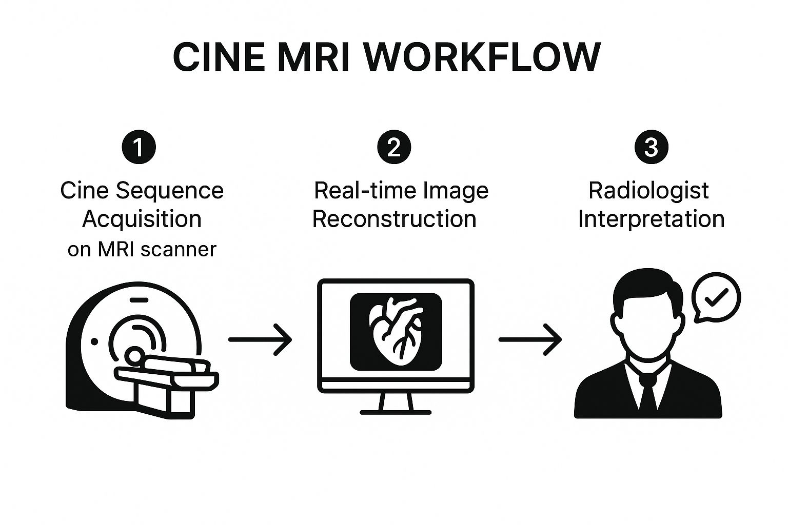
As you can see, the workflow transforms the initial data from the scanner into a diagnostic video that a radiologist can interpret to truly understand a patient's condition.
2. Setting Up a Cine MRI: The Art of Technical Finesse
Getting a crystal-clear, useful cine MRI isn't as simple as just hitting "start." It’s a genuine art form, a delicate dance of physics and clinical need. Technologists and radiologists have to carefully tweak a whole host of settings to get images that can answer a very specific question about a patient.
Think of it like a professional photographer adjusting their camera. They don't just point and shoot; they manipulate shutter speed, aperture, and ISO to capture the perfect image. In the world of MRI, our "dials" have names like Repetition Time (TR) and Echo Time (TE). TR dictates how often we send a pulse sequence into the body, while TE determines the precise moment we listen for the signal "echoing" back. Getting these two just right is what creates the contrast and clarity in the final video loop.
The Constant Tug-of-War: Speed vs. Detail
One of the most important ideas to grasp in cine MRI is temporal resolution. This is essentially the "frame rate" of our medical movie. A high temporal resolution gives us more frames per second, which is absolutely critical when we need to see something moving very quickly, like the delicate leaflets of a heart valve snapping open and shut.
But there's a catch. Ramping up the frame rate often means we have to give something up. In this case, it’s usually image detail, a property we call spatial resolution. This creates a fundamental trade-off that experienced imaging specialists navigate with every single scan.
The core challenge boils down to this: Do we need a super-smooth, high-speed video to catch rapid movement, or do we need an ultra-sharp, high-definition still frame to inspect tiny anatomical structures? The answer is always dictated by what we're trying to find.
For instance, if we're measuring how effectively a heart chamber is squeezing blood out (its ejection fraction), high temporal resolution is king. But if we’re hunting for a tiny, non-moving growth on that same heart valve, we might sacrifice some of that speed to boost the spatial resolution for a clearer picture. It’s this kind of expert judgment that makes the scan so powerful.
Key Cine MRI Sequence Parameters and Their Impact
To really appreciate the skill involved, it helps to understand what the main control parameters do. The table below breaks down the core settings a technologist adjusts and shows how each choice influences the final images.
| Parameter | What It Controls | Impact on Image |
|---|---|---|
| Temporal Resolution | The "frame rate" of the cine loop. | Higher: Better for fast motion (e.g., valve function). Lower: May miss rapid events. |
| Spatial Resolution | The level of detail or sharpness in each frame. | Higher: Clearer view of small structures. Lower: Images may appear less sharp or pixelated. |
| Repetition Time (TR) | The time between successive pulse sequences. | Shorter TR: Faster scanning, higher temporal resolution. Can reduce the signal-to-noise ratio. |
| Echo Time (TE) | The time at which the signal is captured. | Shorter TE: Minimizes motion artifacts. Affects tissue contrast. |
This constant balancing act is precisely why a cine MRI is never a cookie-cutter procedure. Each and every scan is meticulously planned and fine-tuned for the patient on the table and the specific medical puzzle that needs solving. It’s what turns a routine imaging appointment into a highly focused diagnostic investigation.
How Cine MRI Transforms Patient Care
While the physics behind a cine MRI is impressive, its real power comes to life in the clinic. This is where it stops being a collection of images and starts providing life-changing answers for patients. Dynamic imaging goes far beyond showing static anatomy; it reveals function, offering insights that a still picture could never capture. Nowhere is this more true than in cardiac care, where seeing the heart in motion is everything.
Cardiologists depend on cine MRI to get hard, quantifiable data on heart performance. One of the most critical metrics is the ejection fraction (EF), which tells us exactly what percentage of blood is pumped out of a ventricle with each heartbeat. A low EF is a tell-tale sign of heart failure, and cine imaging gives doctors the precise measurements they need to diagnose the condition and track how well a treatment is working.
It’s also an incredible tool for spotting valvular heart disease. By creating a video loop of blood flow, radiologists can directly see problems like regurgitation (leaky valves) or stenosis (narrowed valves). They can watch jets of blood shooting backward through a valve that isn't sealing properly—a finding that is both unmistakable and essential for planning a patient's next steps.
Assessing Damage and Planning Procedures
When a patient has a heart attack, a cine MRI can map the extent of the damage to the heart muscle with remarkable precision. It clearly shows which areas of the heart wall have stopped contracting (akinesis) or are moving abnormally (dyskinesis). This information is fundamental to predicting a patient's long-term outlook and building a recovery plan that's right for them.
The applications reach into some of medicine’s most delicate areas. Think about its impact in pediatrics:
- Congenital Heart Defects: For a baby born with a complex heart issue, surgeons can use cine MRI to understand the unique blood flow and functional problems before they even make an incision. This allows for incredibly detailed surgical planning that can radically improve a child's chances.
- Cerebrospinal Fluid (CSF) Flow: Neurologists also use cine MRI to watch the movement of CSF around the brain and spinal cord. This is critical for diagnosing conditions like Chiari malformation, where blockages can disrupt this vital flow and cause serious symptoms.
The real magic of a cine MRI is its ability to answer the question, "How is it working?" instead of just, "What does it look like?" This functional data leads directly to more confident diagnoses and treatments built around the individual patient.
Of course, technology has played a huge role in making cine MRI the gold standard for evaluating cardiac function. Since the goal is to capture moving images beat-by-beat, speed is non-negotiable. Advanced reconstruction methods, like undersampled and parallel imaging algorithms, have made faster scans possible without compromising image quality. You can dive deeper into these advanced imaging techniques on Insights into Imaging.
Beyond the Heart
But this technology isn't just for cardiologists. Orthopedic specialists can use cine MRI to watch joints in motion, catching subtle instabilities or ligament problems that would be completely invisible on a static scan. Seeing the joint mechanics in real-time helps athletes recover more effectively and guides surgeons performing complex reconstructions.
From the heart to the brain and our joints, cine imaging adds a layer of diagnostic certainty that truly makes a difference in people's lives.
How AI Is Sharpening Cine MRI Analysis
Traditionally, analyzing a cine MRI has been a painstakingly manual job for radiologists. It involves carefully tracing the borders of the heart's chambers on image after image to calculate volume and function. While this method gets the job done, it's slow, tedious, and prone to slight variations between different doctors—a real bottleneck in a packed clinic. Now, artificial intelligence is arriving on the scene to completely overhaul this process.
AI, and more specifically deep learning, can tackle these complex analytical tasks with remarkable speed and consistency. Picture an expert assistant who can outline the ventricles on hundreds of frames in mere seconds, a task that would take a human specialist a considerable amount of time. The real win here isn't just about saving time; it's about achieving a standard of precision that's difficult to maintain from one person to the next.
These AI models are trained on vast libraries containing thousands of cine MRI scans. Through this training, they learn to recognize the subtle visual cues of heart muscle and blood flow, allowing them to perform segmentation—the process of automatically identifying and outlining specific parts of the heart.
Automating Key Cardiac Measurements
The most immediate and practical benefit of AI is how it automates the calculation of cardiac function. Once an AI model has segmented the heart's chambers across the entire cardiac cycle, it can instantly spit out the crucial metrics needed for a diagnosis.
Here are a few key calculations that AI can automate:
- Ejection Fraction (EF): This is the main number we look at to gauge the heart's pumping power, and AI calculates it with high reproducibility.
- Stroke Volume: This tells us the exact amount of blood pushed out of a ventricle with every single beat.
- Myocardial Mass: Calculating the weight of the heart muscle itself is critical for spotting conditions like hypertrophy.
This automation frees up radiologists from the monotonous task of drawing lines, letting them zero in on the tougher interpretive aspects of the diagnosis.
By taking over the detailed grunt work, AI essentially acts as a force multiplier. It enhances a radiologist's ability to produce faster, more consistent, and data-rich reports, ensuring every measurement is performed the exact same way, every time.
The rise of AI foundation models is another major step forward. Unlike older AI tools built for just one specific job, these powerful models are trained on massive, diverse datasets. This broad training allows them to analyze a whole spectrum of cardiac diseases. One groundbreaking model, CineMA, has shown it can segment the heart more accurately and produce more consistent ejection fraction measurements than older methods. This improvement in both sensitivity and specificity helps cut down on misdiagnoses. Better yet, the creators have made the code public to help clinics around the world adopt it faster. You can dive into the research behind this AI-augmented cine MRI analysis on arXiv.
Balancing the Benefits with Real-World Challenges
As exciting as AI's potential is, rolling it out into everyday clinical practice comes with its own set of challenges. A huge area of ongoing research is making sure these models are robust enough to work accurately across different patient groups, MRI machine brands, and hospital systems.
Of course, there are also ethical questions and regulatory hurdles to clear before these tools can be deployed responsibly. The end goal isn't to replace the radiologist but to arm them with a smarter, more efficient set of tools. As the technology continues to mature, it points toward a future where the incredible diagnostic detail of an MRI with cine is more accessible and trustworthy than ever before.
Common Questions About Cine MRI
It's completely normal to have questions before any medical procedure, and a cine MRI is no different. Knowing what to expect can go a long way in making you feel more comfortable and prepared. Let's walk through some of the most common things patients ask about the experience.
So, what makes a cine MRI different from a regular one? From your perspective, not much. The machine looks and feels exactly the same. The key difference is the addition of small, sticky ECG leads on your chest. These monitor your heart's rhythm, allowing the scanner to time its pictures perfectly with your heartbeat. It's this precise timing that creates the "movie" of your internal structures in action.
The process itself will feel familiar if you've had a standard MRI. You'll lie on a movable table that slides into the center of the large, tube-shaped scanner. The most important thing you can do is lie as still as possible—this is what ensures the final images are crisp and clear.
How Should I Prepare for the Scan?
Thankfully, preparing for a cine MRI is pretty straightforward. You'll be asked to change into a hospital gown and remove anything metallic, like jewelry, glasses, or hearing aids. The MRI machine uses powerful magnets, so this is a critical safety step.
Before you head in, be sure to let the technologist know about a few things:
- Any metal inside your body, such as pacemakers, artificial joints, or surgical clips.
- If you are pregnant or suspect you might be.
- If you struggle with claustrophobia or feel anxious in tight spaces.
Don't worry if enclosed spaces make you nervous. Your medical team can help make you more comfortable, sometimes with a mild sedative. Many places also offer special headphones so you can listen to music, which can be a great distraction.
What Happens During and After the MRI?
Once the scan starts, you'll hear a lot of loud thumping and buzzing noises. It might be a bit startling, but it's just the normal sound of the machine's powerful internal parts doing their job. The technologist will be in an adjacent room, but they can see and communicate with you the entire time.
They will guide you through the process, often asking you to hold your breath for brief moments. This simple action helps get perfectly still images. A typical cardiac cine MRI usually takes about 30 to 60 minutes.
After it’s all over, you can get back to your day right away—no recovery time needed. The images are then sent to a radiologist, a doctor who specializes in interpreting medical scans.
What Are the Radiologists Looking For? The radiologist isn't just looking at a picture; they're watching a movie. For a heart scan, they measure how well the heart is pumping blood and check for leaky valves. For a brain scan, they might watch the flow of cerebrospinal fluid to spot any blockages.
This detailed, dynamic analysis gives your doctor functional insights that a static image just can't provide. Seeing how your body is actually working helps them arrive at a precise diagnosis and develop the right treatment plan for you.
At PYCAD, we're focused on bringing AI into medical imaging to help radiologists analyze complex scans like cine MRI faster and more accurately. Learn how our AI-powered tools are helping medical device companies improve diagnostic outcomes by visiting our PYCAD homepage.


