The Hidden Power of Pulmonary Segmentation
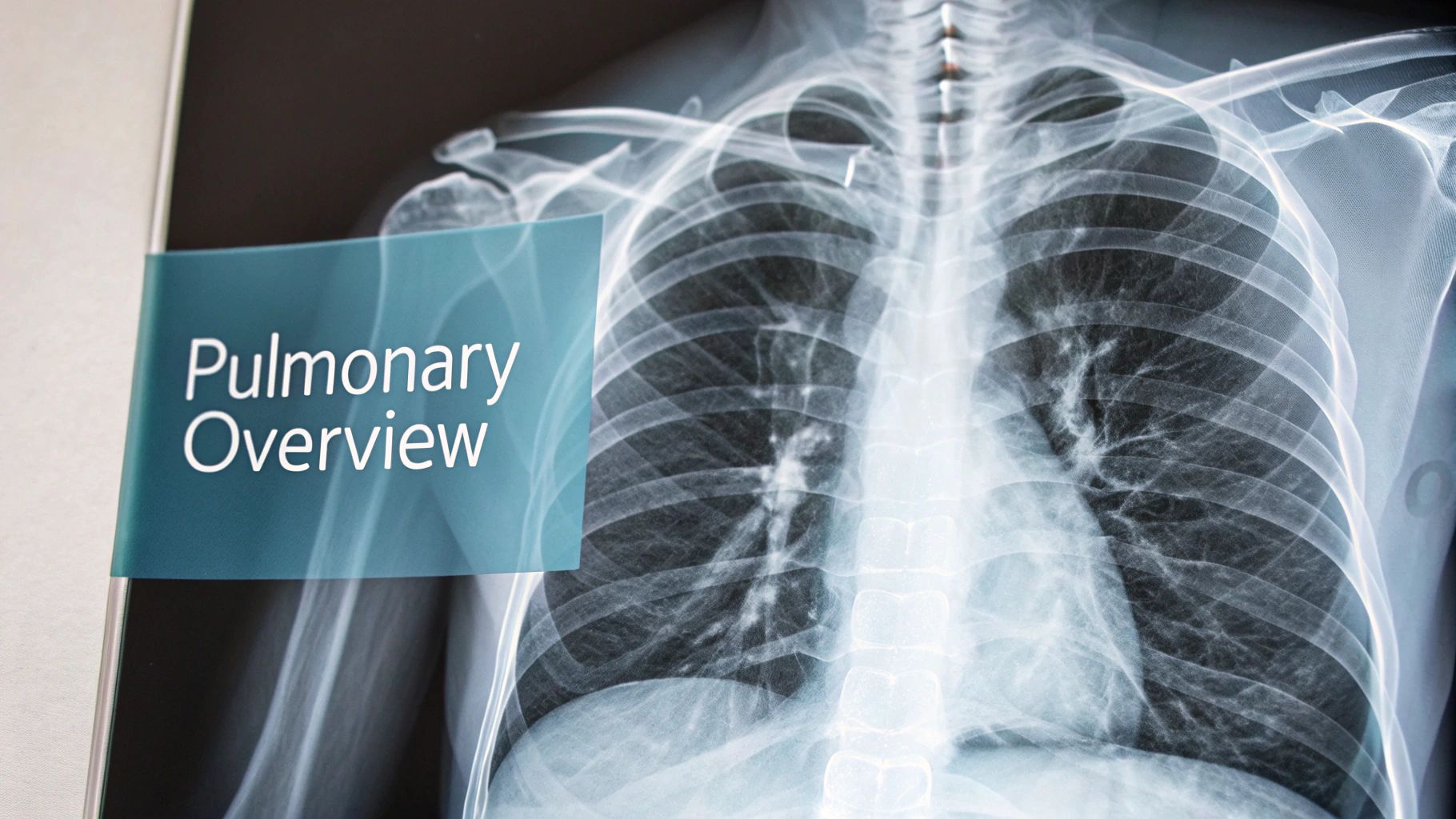
Pulmonary segmentation, the process of identifying individual bronchopulmonary segments within the lungs, plays a vital role in modern respiratory medicine. This detailed anatomical understanding is critical for targeted interventions and improved patient outcomes. Think of the lungs as an intricate tree. Each bronchopulmonary segment acts as a separate "branch" with its own air supply and blood vessels.
This complex structure enables localized treatment, minimizing the impact on healthy lung tissue.
Why Pulmonary Segmentation Matters
The ability to isolate and treat specific lung segments allows for minimally invasive procedures. This precision enables surgeons to focus on diseased areas while preserving healthy lung function. For instance, if a small, localized tumor is present, a segmentectomy (removal of a single segment) might be sufficient instead of a more extensive lobectomy (removal of an entire lobe).
Preserving lung tissue is essential for long-term respiratory health, especially for patients with pre-existing conditions. Furthermore, understanding the unique characteristics of these segments helps clinicians accurately assess disease spread and tailor treatment strategies accordingly. This is particularly important for lung cancer staging and surgical planning.
The human lungs are divided into these bronchopulmonary segments, each supplied by specific sections of the bronchial tree and its own artery. The right lung typically has ten segments, while the left lung usually has eight to nine due to the fusion of some segments. This segmentation is essential for clinical interventions, allowing for targeted treatment of specific lung areas.
The development of these segments is a complex process that begins during the embryonic stage. Learn more about bronchopulmonary segments here: Bronchopulmonary Segments. This detailed anatomical knowledge allows for a more nuanced understanding of lung disease and facilitates personalized treatment approaches.
The Impact of Segmentation on Clinical Practice
Pulmonary segmentation has significantly improved several key areas of respiratory care:
- Targeted drug delivery: By precisely identifying affected segments, clinicians can deliver medications directly to the diseased area, minimizing systemic side effects.
- Improved surgical planning: Detailed 3D models created from segmented images provide surgeons with a roadmap for complex procedures, improving precision and reducing operative time.
- Accurate disease monitoring: Pulmonary segmentation allows for precise tracking of disease progression and treatment response over time, enabling more informed clinical decisions.
- Personalized medicine: The detailed anatomical information provided by pulmonary segmentation supports the development of personalized treatment plans based on individual patient characteristics.
This approach represents a shift from traditional, generalized treatments to highly specific interventions. This precision leads to better patient outcomes, minimizing complications and maximizing the preservation of healthy lung function. The future of pulmonary care relies on this ability to visualize, understand, and interact with the lung at the segmental level.
From Confusion to Clarity: The Evolution of Lung Mapping
The human lung, with its intricate network of airways and blood vessels, has always posed a challenge for medical professionals. Understanding pulmonary segmentation, the process of dividing the lungs into distinct bronchopulmonary segments, is crucial for accurate diagnosis and effective treatment. However, this understanding wasn't always as straightforward as it is today.
Early Challenges in Lung Anatomy
Historically, the terminology used to describe lung anatomy was a major source of confusion. Different medical communities employed varying terms, leading to inconsistencies and communication breakdowns. Imagine a construction crew where each member has a different name for the same tools. Progress would be slow and riddled with errors. Similarly, the lack of standardized terminology hampered the advancement of thoracic medicine for decades.
This lack of clarity made it difficult to share knowledge and compare findings. This inconsistency also presented significant challenges in surgical planning and execution, where precise anatomical understanding is paramount.
The Rise of Standardization
This anatomical confusion began to clear in the mid-20th century. The concept of pulmonary segmentation was formalized in 1950 by R.C. Brock, following the work of The Thoracic Society of Great Britain to standardize bronchial nomenclature and anatomy. This important step led to an international agreement on bronchial naming conventions, further solidified by the first Nomina Anatomica in 1955.
This standardization wasn't just about creating a universal language. It signified the maturation of pulmonary surgery as a distinct and sophisticated field. Learn more about this critical evolution: Evolution of Pulmonary Segmentation Terminology.
The Impact of Standardized Terminology
The adoption of standardized terminology significantly impacted pulmonary segmentation. It established a common language, enabling clearer communication and understanding between medical professionals across the globe. This shared understanding fostered collaboration and accelerated advancements in both surgical techniques and patient care. Surgeons could now confidently discuss specific lung segments, ensuring everyone was on the same page.
To illustrate the evolution of terminology, let's look at the following table:
Evolution of Pulmonary Segmentation Terminology
This table shows the historical development of pulmonary segmentation terminology and standardization efforts over time.
| Time Period | Key Contributors | Major Developments | Clinical Impact |
|---|---|---|---|
| Early 20th Century | Various Anatomists | Inconsistent nomenclature across different medical communities | Difficulty in communication, limited collaboration |
| Mid-20th Century | R.C. Brock, The Thoracic Society of Great Britain | Formalization of pulmonary segmentation concept, standardized bronchial nomenclature, Nomina Anatomica (1955) | Improved communication, enhanced surgical planning, foundation for advanced imaging techniques |
| Late 20th Century – Present | International anatomical societies, Radiological and surgical communities | Refinement of terminology based on imaging advancements (CT, MRI) | More precise diagnoses, development of minimally invasive surgical procedures, personalized treatment strategies |
The standardization of lung segment terminology allowed for accurate communication, which has significantly improved patient care and surgical outcomes. This shared language enabled precise identification of affected segments, leading to better surgical planning, less invasive procedures, and more targeted treatments.
From 2D to 3D: Imaging Advancements
Standardized terminology further paved the way for advancements in imaging technology. Initially, imaging techniques provided basic 2D representations of lung anatomy. However, the arrival of Computed Tomography (CT) and Magnetic Resonance Imaging (MRI) revolutionized the field.
These technologies allowed for the creation of detailed 3D visualizations of lung segments. Surgeons could now visualize the intricate details of each segment, improving surgical planning, minimizing invasiveness, and increasing precision during complex procedures. This shift from 2D to 3D represents a giant leap in our ability to understand and interact with the complex structures of the lungs. This visualization continues to drive innovation in pulmonary medicine, contributing to improved patient outcomes.
Seeing What Was Once Invisible: Imaging Breakthroughs

Visualizing the bronchopulmonary segments within a living, breathing patient has always been a significant hurdle for medical professionals. However, advancements in imaging technology have dramatically changed this, allowing us to move from simple outlines to intricate 3D models. These models are now crucial for surgical procedures, revolutionizing how we understand and interact with the complex structures of the lungs.
The Power of Modern Imaging Modalities
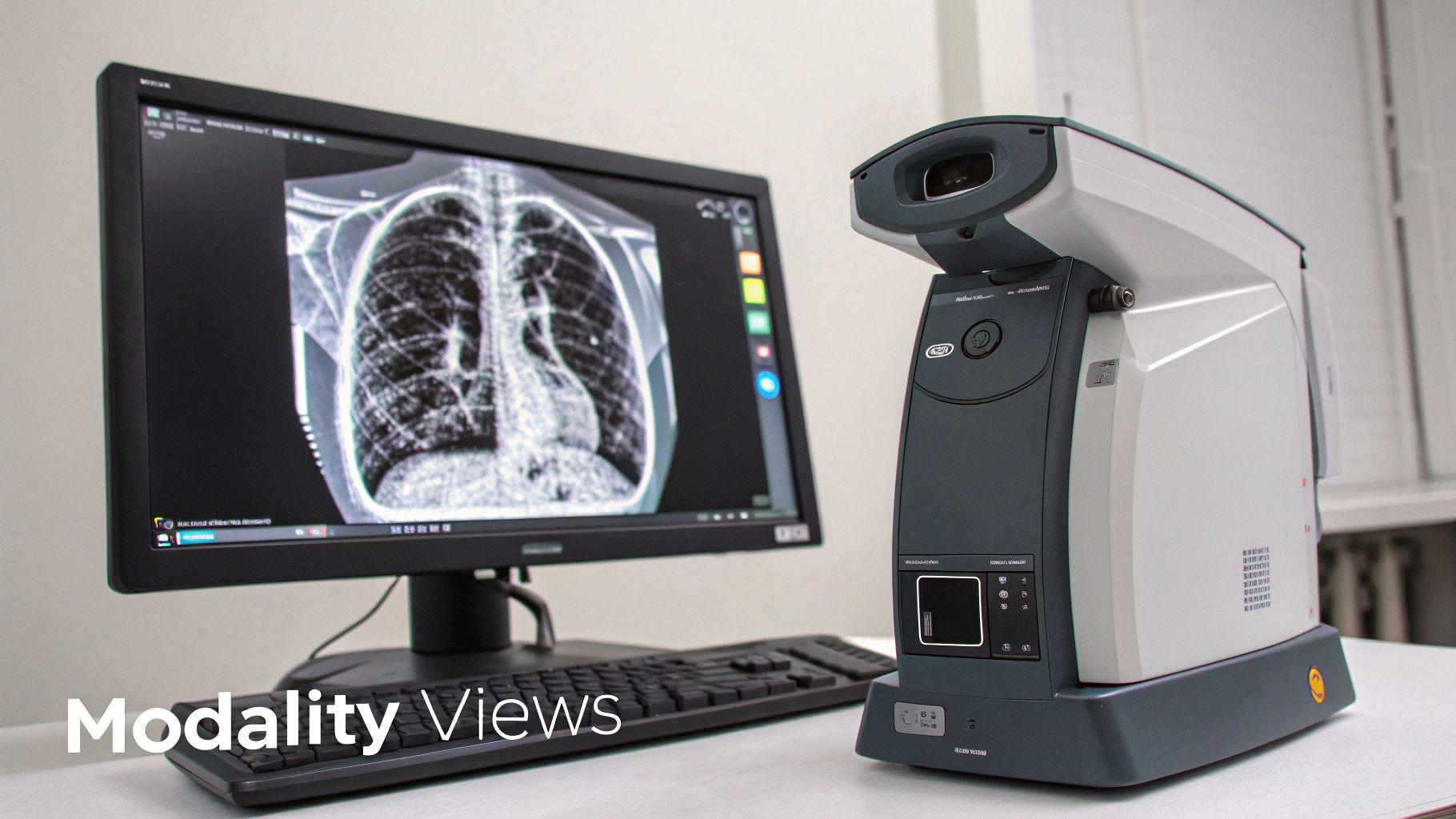
Selecting the appropriate imaging modality is paramount for visualizing these segments. Computed Tomography (CT) scans are frequently employed for pulmonary segmentation due to their ability to create detailed cross-sectional images of the lung. CT scans provide crucial information regarding lung tissue density and anatomical structures. Magnetic Resonance Imaging (MRI), while used less often for this purpose, excels at visualizing soft tissue, providing invaluable insights in specific clinical situations. The decision between CT and MRI often hinges on the specific clinical question and the patient's individual circumstances.
Optimizing imaging protocols is equally vital. Adjusting parameters such as slice thickness and reconstruction algorithms can substantially improve the clarity of segmental boundaries, especially in more complicated cases. For instance, thinner slices in a CT scan can expose subtle anatomical details that thicker slices might miss. This precision is fundamental for accurate diagnosis and treatment planning.
Optimizing CT and MRI Protocols
Careful adjustments to CT and MRI protocols can significantly elevate the quality of pulmonary segmentation. However, this requires a delicate balance. High-resolution images are desirable, but minimizing radiation exposure in CT scans is absolutely essential. Experienced radiologists utilize techniques to achieve the best possible visualization while adhering to the As Low As Reasonably Achievable (ALARA) principle for radiation dosage. This involves tailoring scan parameters to each patient and employing iterative reconstruction techniques to reduce image noise while preserving crucial details.
The selection of contrast agents, particularly in CT scans, also significantly influences image quality. Different contrast protocols can highlight particular anatomical structures, further refining the delineation of segmental boundaries. The expertise of a radiologist is invaluable here, as they can adapt the contrast protocol to the individual patient and their specific clinical needs. Pulmonary segmentation, especially in CT scans, involves identifying the major airways and aligning them longitudinally, which is crucial for diagnostic and therapeutic accuracy. Furthermore, methods used for abdominal imaging can inform the approach to pulmonary segmentation. Using attenuation-based metrics for organ dosage estimation in CT scans underscores the importance of precise segmentation for minimizing radiation exposure and ensuring accurate diagnosis. For more detailed statistics on abdominal CT exams, please see: Abdominal CT Exams.
3D Visualization and Surgical Planning
Modern imaging now enables the creation of highly detailed 3D reconstructions of the lungs. This has transformed pulmonary segmentation from 2D representations to complete 3D models. These models are indispensable for pre-operative planning, enabling surgeons to virtually "navigate" the lungs and examine the anatomy before surgery. This allows for precise mapping of the target segment, reducing the risk to surrounding healthy tissue.
Moreover, 3D visualizations enhance communication among healthcare professionals. Radiologists can effectively share critical information with surgeons and other clinical team members, fostering a collaborative approach to patient care. This enhanced communication is essential for informed decision-making and optimal surgical outcomes. The ability to visualize segments in three dimensions has significantly improved the precision and effectiveness of lung interventions, ultimately leading to better outcomes for patients with complex pulmonary conditions.
When AI Meets Lung Architecture: The Automation Revolution

The field of pulmonary segmentation is rapidly evolving, moving away from traditional manual methods. Artificial intelligence (AI) is the driving force behind this shift, offering automated solutions for greater speed and accuracy. This advancement has the potential to reshape how medical professionals analyze and interpret lung imaging data.
The Rise of Deep Learning in Pulmonary Segmentation
Deep learning algorithms, a subset of AI, are leading the way in this automation. These algorithms can learn intricate patterns from massive medical image datasets. This ability enables them to pinpoint and outline bronchopulmonary segments with remarkable precision.
Convolutional neural networks (CNNs), a specific type of deep learning architecture, are particularly effective in image analysis, including pulmonary segmentation. CNNs offer a substantial improvement over manual methods, which are often susceptible to human error and time-intensive.
However, the effectiveness of AI algorithms varies. Some struggle with the natural anatomical differences between individuals. The complexity of the human lung presents challenges for some AI models, as even slight variations in lung architecture can affect performance.
Ongoing research is dedicated to creating more robust AI solutions. These solutions must be able to handle anatomical variations and maintain high accuracy across diverse patient populations. Training AI models on extensive and diverse datasets is crucial for their ability to generalize to real-world clinical scenarios.
Implementing AI in Clinical Practice
AI-powered pulmonary segmentation tools are increasingly being integrated into clinical workflows. Hospitals and research institutions are adopting these technologies to streamline image analysis. This automation saves radiologists valuable time, allowing them to concentrate on more demanding diagnostic tasks.
AI can also boost diagnostic confidence by providing consistent and objective segmentations. This reduces the impact of inter-observer variability, which is especially beneficial in complex cases where segment boundaries are unclear or distorted by disease.
Pulmonary segmentation also plays a role in biomedical engineering. Automated techniques are being developed for various medical imaging modalities. For pulmonary imaging, this involves distinguishing the lungs and their subdivisions from other thoracic structures. Advances in segmentation algorithms have led to greater precision, improving both diagnosis and treatment planning. Learn more: Latin American Congress on Biomedical Engineering.
Evaluating AI-Powered Solutions
Selecting the right AI solution for pulmonary segmentation requires careful evaluation. There are both commercial and open-source options available, each with its own advantages and disadvantages. The following table provides a comparison:
Comparison of Pulmonary Segmentation Methods
This table compares different pulmonary segmentation approaches based on accuracy, processing time, and clinical applicability.
| Segmentation Method | Accuracy (%) | Processing Time | Required Expertise | Best Applications |
|---|---|---|---|---|
| Manual Segmentation | Variable (dependent on user skill) | High (time-consuming) | High (specialized training) | Complex cases, quality control |
| Traditional Image Processing | Moderate | Moderate | Moderate (image processing knowledge) | Research, basic segmentation tasks |
| Deep Learning (Commercial) | High (potential for >95%) | Low (fast processing) | Low (user-friendly interfaces) | Clinical routine, high-throughput analysis |
| Deep Learning (Open-Source) | High (potential for >95%) | Low (fast processing) | High (programming skills) | Research, algorithm development |
Key factors to consider include accuracy, processing speed, required expertise, and cost. The optimal choice depends on the specific requirements and resources of the institution. Choosing the right AI solution is vital for realizing the full potential of automated pulmonary segmentation. This will lead to more precise diagnoses, better treatment plans, and improved patient care in respiratory medicine.
Beyond Theory: Pulmonary Segmentation in Clinical Practice
Pulmonary segmentation has moved beyond a theoretical anatomical concept. It is now actively transforming how we approach patient care. This level of precision allows for medical interventions previously thought impossible, offering hope for individuals facing complex lung conditions. This section explores the practical applications of pulmonary segmentation and its impact on thoracic medicine.
Precision in Radiation Oncology
Radiation oncologists now leverage pulmonary segmentation to deliver radiation with remarkable accuracy. By precisely defining the tumor boundaries and the surrounding healthy tissue, they can maximize the dose to the tumor while minimizing the impact on vital structures. This targeted approach allows for higher radiation doses, potentially increasing treatment effectiveness and reducing side effects. This translates to more effective treatments and a better quality of life for patients.
Navigating the Lungs with Unprecedented Accuracy
Pulmonary segmentation also benefits interventional pulmonologists. Reaching small and difficult-to-access peripheral lung nodules has always posed a challenge. Segmentation provides a detailed map, guiding instruments with millimeter-level precision. This accuracy is crucial for gathering tissue samples for diagnostic purposes and performing minimally invasive procedures. Accurate targeting improves the diagnostic yield and facilitates earlier intervention, leading to better patient outcomes.
Essential for Transplant Surgery
In transplant surgery, pulmonary segmentation plays a vital role in successful donor-recipient matching. The precise assessment of lung segment volumes and anatomical compatibility is essential for optimizing graft function and minimizing complications after transplant. Accurate matching increases the likelihood of successful transplants, extending and improving the lives of recipients.
Quantifiable Improvements in Surgical Outcomes
Pulmonary segmentation demonstrably improves surgical outcomes. Studies show reduced blood loss, shorter hospital stays, and better preservation of functional lung capacity in patients undergoing segmentation-guided procedures. These measurable benefits highlight the technology's potential to improve patient well-being and lower healthcare costs. In segmentectomies, for instance, removing only the affected segments, rather than an entire lobe, significantly impacts long-term lung function. This precision is especially valuable for patients with pre-existing conditions or those requiring future interventions. The rise of AI has significantly impacted pulmonary segmentation; you can explore how an AI agency can help develop solutions.
The Future of Pulmonary Segmentation
The future of pulmonary segmentation is promising, with continued advancements expanding its potential. Real-time segmentation is set to transform interventional procedures by offering surgeons dynamic, intraoperative guidance. This technology allows for real-time adjustments during surgery, enhancing precision and reducing unforeseen complications.
Augmented reality overlays are also evolving, giving surgeons enhanced "X-ray vision" during minimally invasive procedures. This improved visualization boosts accuracy and minimizes invasiveness. As these technologies advance, we can anticipate even more substantial improvements in patient care and surgical outcomes. Research is ongoing to address the limitations of existing algorithms, especially in segmenting severely diseased lungs. Successfully addressing these complex cases will expand the applications of pulmonary segmentation even further.
Breaking Barriers: The Future of Pulmonary Segmentation
Pulmonary segmentation is no longer a static process limited to pre-operative planning. Exciting advancements are pushing the boundaries, offering dynamic tools and real-time insights that are transforming interventional procedures.
Real-Time Segmentation: A Game Changer in the Operating Room
Imagine a surgeon with enhanced vision, able to see the intricate details of the lungs during a minimally invasive procedure. Real-time pulmonary segmentation makes this possible. As surgeons operate, updated images are segmented instantaneously, providing a dynamic visualization of the target area and surrounding structures. This allows for adjustments during the procedure, improving precision and reducing the risk of complications. This represents a significant improvement over static pre-operative images, where intraoperative changes would rely solely on the surgeon's judgment and tactile feedback.
Augmented Reality: Enhancing Surgical Precision
Augmented reality (AR) takes real-time segmentation a step further. By overlaying segmented images onto the surgeon's view of the patient, AR provides a comprehensive roadmap during minimally invasive procedures. This technology enhances precision, minimizes invasiveness, and can lead to faster recovery times. This real-time data integration allows surgeons to navigate complex lung anatomy with greater confidence. Think of it as a GPS for the lungs, guiding surgical instruments with exceptional accuracy. AR can highlight critical structures like blood vessels or nerves, allowing for more precise maneuvering and tissue preservation.
Overcoming Technological Hurdles
While the potential of real-time segmentation and AR is significant, challenges remain. One hurdle is the computational power needed for instant processing. High-resolution images demand significant computing resources for quick segmentation and AR overlays. Another challenge lies in the robustness of the algorithms. Variations in lung anatomy, particularly in diseased lungs, can be problematic for current algorithms. Researchers are actively addressing these limitations, developing more robust algorithms and exploring high-performance computing solutions to ensure accuracy and reliability. Leading institutions are spearheading these efforts, pushing technological boundaries and developing innovative solutions to benefit more patients.
Segmenting Diseased Lungs: A Continuing Challenge
Severely diseased lungs, with their distorted anatomy and irregular structures, present a unique challenge for segmentation algorithms. Current methods often struggle to accurately define segments in these complex cases. This limits the application of segmentation in advanced lung disease. However, ongoing research focusing on advanced image processing and deep learning algorithms shows promise in overcoming this obstacle. Novel approaches incorporating multi-modal imaging are also being developed.
Multi-Modal Integration: A More Complete Picture
The future of pulmonary segmentation lies in combining information from multiple imaging modalities. Integrating data from CT, MRI, and even functional imaging like PET scans can provide a more comprehensive understanding of lung structure and function. This multi-modal approach offers a deeper insight into the complex relationship between anatomy and physiology, especially in diseased lungs. By combining structural information from CT with functional data from PET scans, clinicians can better identify areas of active tumor growth within specific segments, thus improving treatment targeting and evaluation.
Implementing Advanced Segmentation in Resource-Limited Settings
Cost and resource availability remain a significant concern for many institutions. However, strategic implementation can extend these benefits to a broader range of facilities. Collaborations with research institutions and technology providers can offer access to advanced technology without substantial capital investment. Cloud-based solutions can further reduce infrastructure costs, making advanced pulmonary segmentation tools accessible even in resource-limited environments.
Want to learn more about how AI can improve your approach to medical imaging? PYCAD offers advanced solutions for pulmonary segmentation and other medical imaging challenges. Visit PYCAD to discover how we can help you optimize your medical devices, enhance diagnostic accuracy, and improve patient care.
