Picture this: you're trying to read the fine print on a blurry photograph. Now, imagine that same photo is perfectly sharp and crystal clear. That's the difference high-resolution makes, and it's the core idea behind the resolution of a CT scan. It's all about the machine's ability to capture incredibly fine details inside the body, transforming a potential medical mystery into a clear, actionable diagnostic image.
Simply put, the higher the resolution, the sharper and more detailed the final picture.
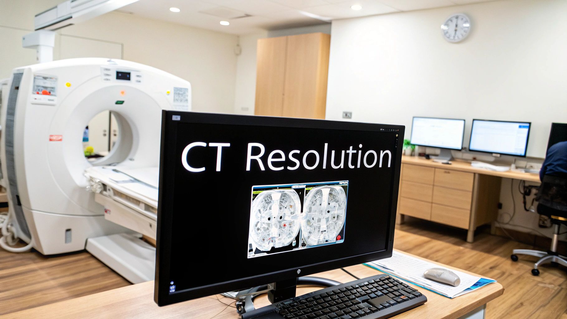
What Is CT Scan Resolution, Really?
It's best not to think of CT scan resolution as a single setting you can dial up or down. Instead, see it as the overall clarity and precision of the final image. This quality determines how well a radiologist can distinguish between two tiny, adjacent structures or tell the difference between healthy tissue and diseased tissue.
For example, a low-resolution scan might just show a vague, indistinct shadow in the lungs. But a high-resolution scan can reveal precisely what that shadow is—perhaps a harmless scar or, more critically, a small, developing nodule. This level of detail is the foundation of modern medical imaging, giving doctors the confidence they need to make the right call. It’s absolutely essential for everything from spotting minuscule bone fractures to mapping out the plan for a complex surgery.
The Three Pillars of Diagnostic Clarity
The concept of CT resolution is actually built on three different, but deeply connected, components. Each one adds a unique layer of detail to the final image. Getting the balance between them just right is the key to producing a truly useful diagnostic scan.
These are the three pillars:
- Spatial Resolution: This is about seeing small objects clearly. Think of it as the "sharpness" of the image.
- Contrast Resolution: This is the ability to tell different types of tissue apart, even when they have very similar densities.
- Temporal Resolution: This refers to the speed of the scan. A high temporal resolution can "freeze" motion, which is vital for imaging a beating heart or breathing lungs.
This drive for better resolution isn't anything new; it's been the engine of medical imaging innovation for decades. The number of CT scans performed globally has skyrocketed, jumping from about 3 million by 1980 to over 68 million annually by 2005.
This explosion in use was powered by relentless improvements in scanner technology. Scans that used to take nearly 30 minutes can now be done in just a few seconds, a direct result of the ongoing quest for higher resolution. You can learn more about this journey by reading about the evolution of computed tomography on ISCT.org.
Grasping these foundational elements is the first step to truly appreciating how CT technology gives us such powerful insights into the human body.
The Three Pillars of CT Scan Clarity
When we talk about the quality of a CT scan, it’s easy to think of it as a single score. But in reality, true diagnostic power comes from three distinct types of resolution working in harmony: spatial, contrast, and temporal. Each one offers a unique piece of the diagnostic puzzle.
Getting a handle on these three pillars is key to understanding why modern CT imaging is such an incredibly powerful tool in medicine.
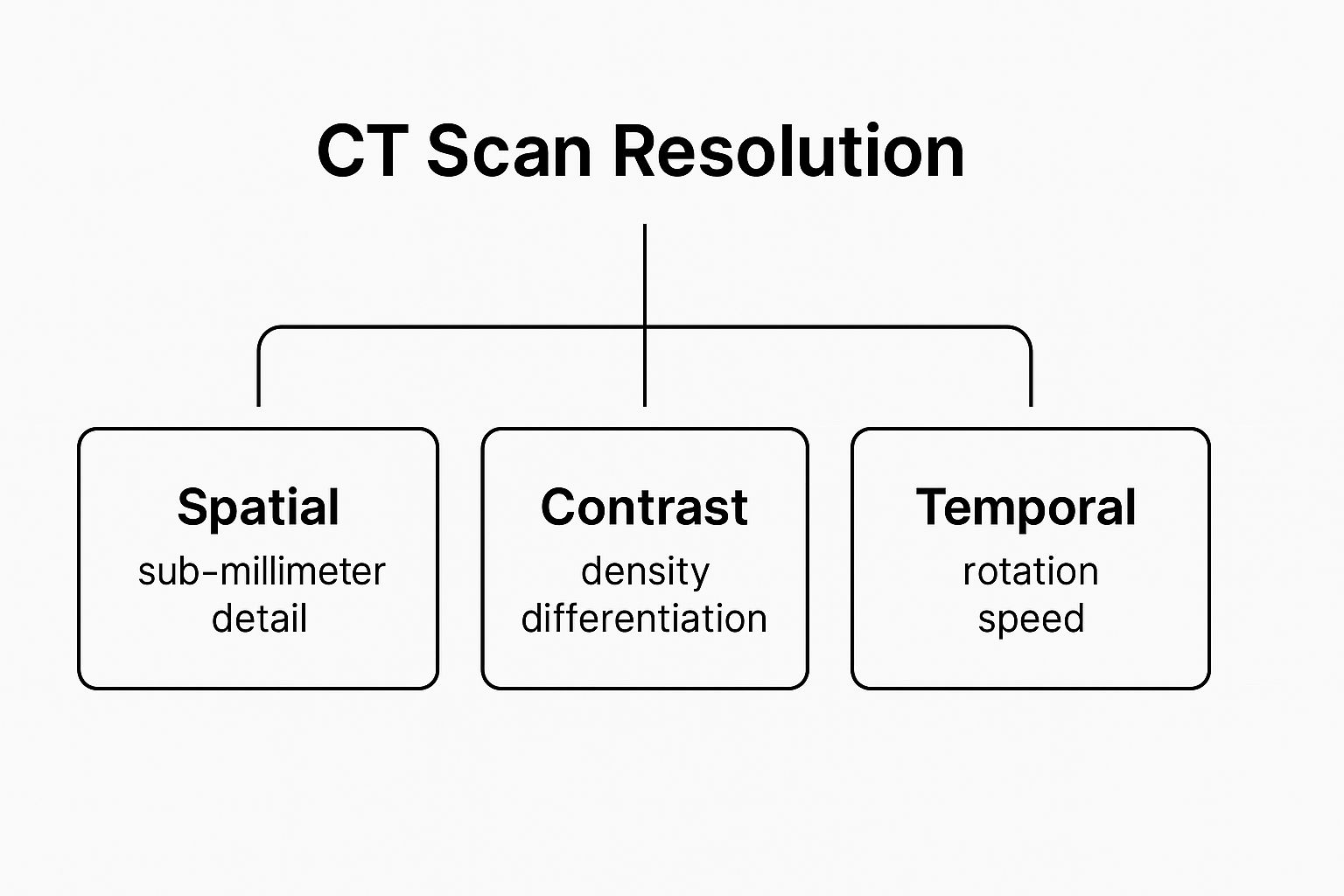
This visual breaks down how we get a complete and detailed picture—it's not just about sharpness, but also about differentiating tissues and capturing clear images of things in motion.
To make sense of this, let's look at each component individually. We'll explore what they measure, how they work, and why they are so vital for accurate diagnosis in the real world.
Key Components of CT Scan Resolution
| Resolution Type | What It Measures | Clinical Importance |
|---|---|---|
| Spatial | The ability to distinguish between two very small objects placed closely together. Essentially, it's about image sharpness and detail. | Critical for spotting tiny structures like hairline fractures, small lung nodules, or the intricate bones of the inner ear. |
| Contrast | The ability to tell apart tissues with very similar densities, like distinguishing between different shades of gray. | Essential for soft tissue imaging, such as identifying a tumor or lesion that has a slightly different density than surrounding healthy tissue. |
| Temporal | The speed at which a complete image is captured. It measures how well the scanner can "freeze" motion. | Absolutely vital for imaging moving organs, especially the heart. It prevents motion blur and allows for clear views of coronary arteries. |
Each of these resolution types plays a specific role. A great scan isn't just sharp; it's also clear and fast, giving doctors the confidence they need to make the right call.
Spatial Resolution: Seeing the Smallest Details
Think of spatial resolution like the pixel density on a 4K television screen. The more pixels packed into the display, the sharper the image and the finer the details you can make out. In a CT scan, high spatial resolution is what allows radiologists to see incredibly small anatomical structures with pinpoint accuracy.
The big question it answers is: how small of an object can we actually see?
This is typically measured in line pairs per centimeter (lp/cm) or micrometers (μm). For context, many modern CT scanners can achieve a spatial resolution of less than 0.5 millimeters—a level of detail that is absolutely necessary for a huge range of diagnoses.
A perfect example is finding a hairline fracture in a bone. A scan with poor spatial resolution might miss it completely. But with a high-resolution image, that tiny crack becomes clearly visible, leading to the right treatment and preventing a more serious injury down the road. It's just as crucial for examining the delicate structures of the inner ear or detecting small nodules in the lungs.
A scan with high spatial resolution provides a clear, sharp map of the body's structures. It's the difference between seeing a blurry shape and a distinct, defined object. This clarity is fundamental for making confident diagnoses.
Contrast Resolution: Differentiating Similar Tissues
While spatial resolution is all about sharpness, contrast resolution is about telling the difference between things with very similar physical densities. Imagine trying to sort a dozen shades of gray on a black-and-white photo—high contrast resolution makes each shade distinct and easy to identify.
This is where CT technology truly shines. It’s absolutely essential for imaging soft tissues, giving doctors the ability to clearly separate organs, fluid, fat, and muscle from one another. More importantly, it helps them distinguish between healthy tissue and a tumor, which might only have a subtle difference in density.
Radiologists often use intravenous contrast agents—special dyes injected into a patient's bloodstream—to give contrast resolution a major boost. These agents make blood vessels and certain organs appear much brighter on the scan, effectively highlighting abnormalities that would otherwise be invisible. For instance, in a scan of the abdomen, contrast can make a lesion on the liver stand out against the surrounding healthy tissue, making it impossible to miss.
Temporal Resolution: Freezing Motion in Time
Finally, we have temporal resolution, which is all about speed. It refers to the time it takes for the scanner to acquire all the data needed to create a single image. The best analogy is a high-speed camera that can capture a hummingbird's wings in perfect, frozen detail instead of a blurry mess.
This capability is non-negotiable when you’re imaging organs that are in constant motion, and the heart is the prime example.
A beating heart is a massive challenge for any imaging system. A slow scan would result in a smeared, completely useless image. But modern scanners with high temporal resolution can acquire a full picture in a tiny fraction of a second. The most advanced systems today boast temporal resolutions as fast as 65 milliseconds, quick enough to "freeze" the heart's motion between beats. This incredible speed is what makes non-invasive procedures like coronary CT angiography (CTA) possible, giving doctors a crystal-clear view of the coronary arteries to spot dangerous blockages without ever making an incision.
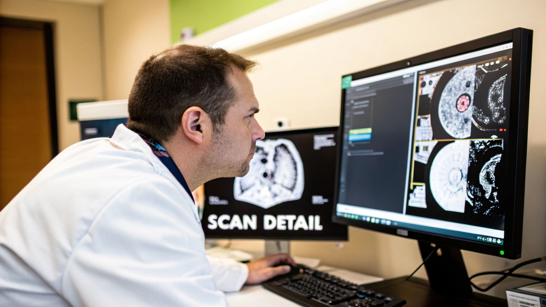
How Scanner Hardware Defines Image Quality
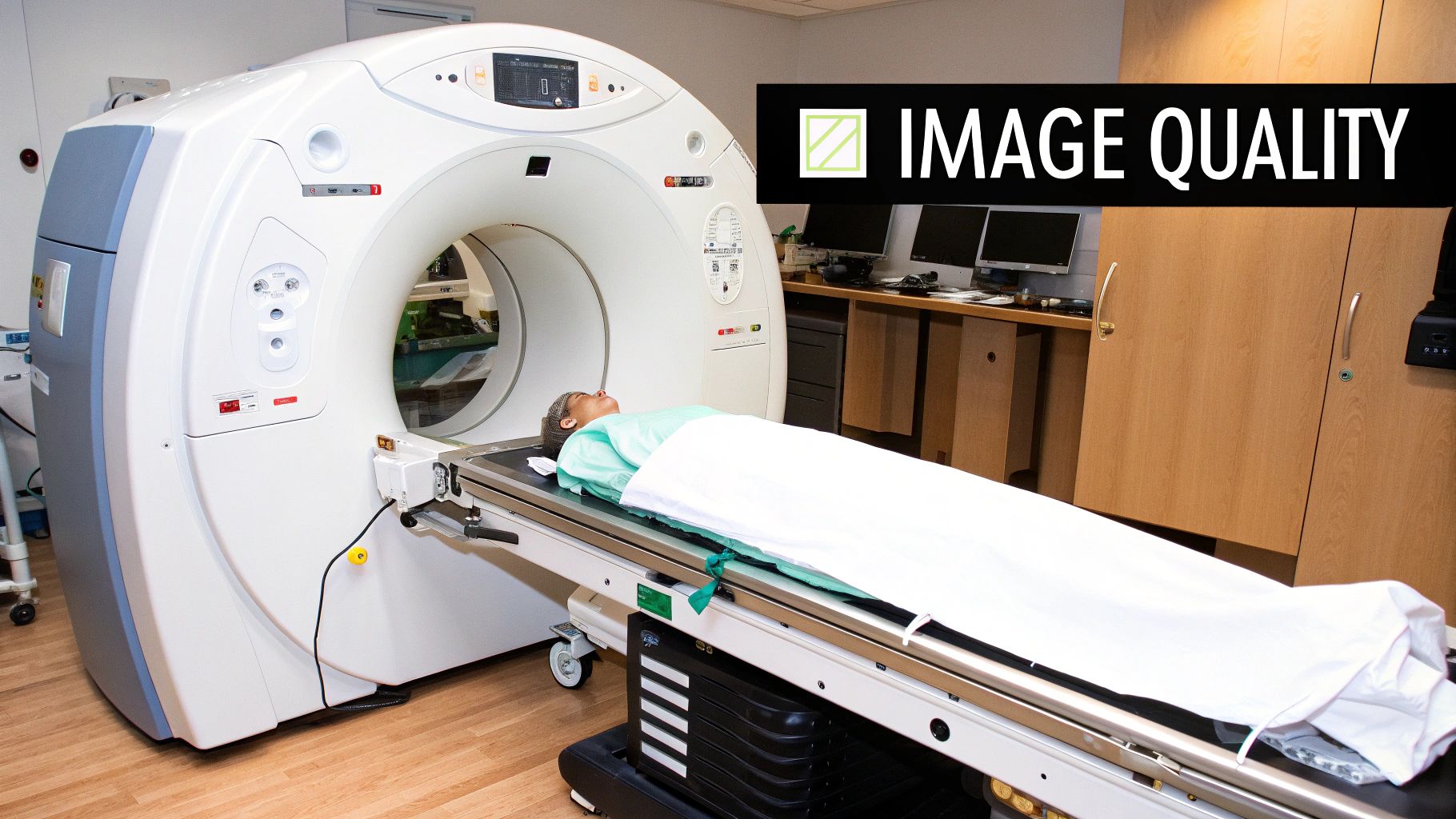
The quality of any CT image is fundamentally tied to the machine that created it. You can think of a CT scanner as a highly specialized camera—its internal components directly dictate the clarity and detail of the final picture. When we talk about what hardware truly matters for the resolution of a CT scan, two pieces stand out: the X-ray tube and the detector array.
The X-ray tube is the source, generating the energy that passes through the body. On the other side, the detectors are what "see" and measure that energy. The precision of these two elements working together is what separates a fuzzy, hard-to-read scan from a crystal-clear, diagnostic one.
Detectors and X-ray Tubes
The detectors in a modern scanner are incredibly sophisticated, made up of thousands of tiny, individual elements. The smaller and more tightly packed these elements are, the better the scanner's spatial resolution. It’s the same principle as a digital camera's megapixel count. More elements capturing information in the same space means you can resolve much finer details, letting clinicians see anatomical structures that would otherwise be invisible.
At the same time, the size of the focal spot in the X-ray tube is just as important. A smaller, more concentrated focal spot creates a sharper X-ray beam. This reduces what's known as geometric unsharpness, resulting in crisper image outlines. It's this powerful combination of tiny detector elements and a focused X-ray beam that forms the foundation of a high-quality scan.
The core idea is simple: better hardware captures better raw data. It's not just about sharpness. Superior detectors are also more sensitive to slight variations in tissue density, which directly boosts contrast resolution. This is what allows a radiologist to confidently distinguish between healthy tissue and a potential abnormality.
From Single-Slice to Multi-Detector Systems
Perhaps the single greatest leap in CT hardware was the shift from single-slice scanners to multi-detector CT (MDCT) systems. The original scanners had just a single row of detectors, meaning they could only capture one slice of the body with each full rotation of the gantry. As you can imagine, this was a painstakingly slow and limiting process.
MDCT scanners changed everything by incorporating multiple, parallel rows of detectors. Suddenly, a scanner could acquire data for dozens—or even hundreds—of slices in a single, quick rotation. This breakthrough had a massive impact:
- It led to dramatically faster scan times, which helps minimize blur from patient movement.
- Clinicians could now scan large sections of anatomy, like the entire chest or abdomen, in just a few seconds.
- It enabled the creation of isotropic voxels (perfect cubes of data), which are essential for generating pristine 3D reconstructions.
This wasn't an overnight revolution, but a steady evolution. The groundwork was laid over decades, particularly with third-generation systems in the late 1970s that introduced rotating tubes and wide fan beams, slashing rotation times to under a second and significantly improving spatial resolution. For those interested in the engineering journey, you can see a great overview of the historical progression of these scanner designs on PMC. Today’s advanced MDCT technology stands on the shoulders of these pioneers, delivering the speed and detail that are now indispensable in modern medicine.
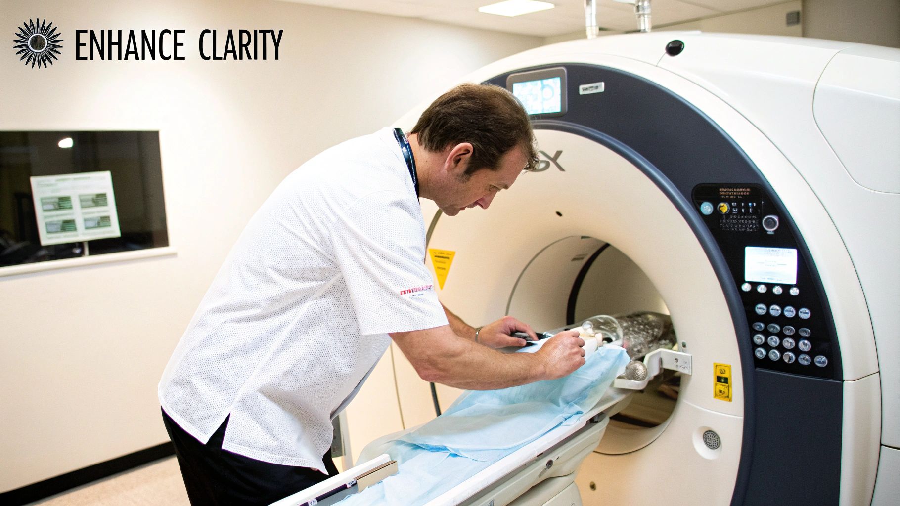
The Human Element in Achieving a Perfect Scan
While the specs on a scanner tell you what’s possible, they don't guarantee a perfect image. The truth is, technology is only half the equation. The final resolution of a CT scan hinges on a crucial collaboration between the patient and the technologist running the machine. Even the most powerful scanner can produce a fuzzy, inconclusive image if these human factors aren't managed well.
A patient's own body, for example, has a direct impact. Larger patients naturally absorb more of the X-ray beam, which can introduce "noise" into the image, sometimes washing out the subtle differences in tissue. A skilled technologist knows how to adjust the scan parameters on the fly, tailoring the dose and technique to get a clear picture regardless of body size.
But the single most important thing a patient can do to help? Hold perfectly still.
The Impact of Patient Cooperation
Any movement during a scan—even something as simple as breathing—can create motion artifacts. These show up as streaks and blurring across the image, obscuring tiny details and potentially hiding the very thing the doctor is looking for. It's a huge issue for scans of the chest and abdomen, where organs are always in subtle motion.
To get around this, technologists give clear, simple instructions. The most common one is asking you to hold your breath for just a few seconds. Following that one direction is probably the most powerful way a patient can contribute to their own diagnostic quality. That moment of stillness gives the scanner the clean, crisp "snapshot" it needs.
A perfect scan is a team effort. When a patient holds their breath precisely as instructed, they are actively helping to eliminate motion blur. This simple act of cooperation is vital for diagnostic confidence because it directly improves both spatial and temporal resolution.
This is a game-changer for pediatric scans. The incredible speed of modern CT machines, combined with a child's cooperation, can often mean the difference between a quick, easy scan and one that requires sedation.
Enhancing Clarity with Contrast Agents
Beyond stillness, the clinical team has other tools to make anatomy pop. The most common is the use of contrast agents, usually an iodine-based liquid given through an IV. These agents are designed to temporarily make certain tissues and blood vessels denser from the X-ray's perspective.
As the contrast flows through your system, it makes specific structures light up on the scan. Think of it like taking a highlighter to a page of dense text—it makes the most important information stand out immediately.
This technique is a massive boost to contrast resolution, giving radiologists the power to:
- Visualize Blood Vessels: Arteries and veins that would otherwise be invisible suddenly glow, revealing blockages, aneurysms, or other problems with stunning clarity.
- Identify Organ Lesions: A tumor in the liver or kidney, which might otherwise blend in with the surrounding healthy tissue, becomes sharply defined and easy to measure.
- Assess Organ Function: Watching how quickly an organ absorbs the contrast agent gives doctors valuable clues about its blood flow and overall health.
By combining powerful hardware with skilled technologists, cooperative patients, and smart procedural choices, the medical team can capture the high-resolution images needed for a fast and accurate diagnosis.
How High-Resolution CT Changed Heart Imaging for Good
If you want to see the real-world power of CT scan resolution, look no further than the heart. For a long time, getting a sharp image of a constantly beating organ was one of the biggest hurdles in medical imaging. The early scanners were just too slow, resulting in blurry, useless pictures that gave doctors very little to work with.
This is a classic example of why temporal resolution is so critical. The heart’s constant motion demanded a technology that could essentially "freeze time," turning a fuzzy blob into a clear, three-dimensional diagnostic map. The journey from those first blurry scans to today's incredibly detailed images shows exactly how engineering innovations directly lead to better patient care.
Conquering the Motion Problem
The main enemy was motion blur. To see the coronary arteries—those tiny, vital vessels wrapped around the heart—the scanner needed to be quicker than a single heartbeat. This sparked a race to drastically improve the temporal resolution of CT scans, with engineers pushing to build machines that could grab an entire image in a tiny fraction of a second.
The answer wasn't just software tweaks; it required brand-new hardware. The development of dual-source CT systems was the real game-changer. These machines use two X-ray sources and two detector arrays at once, basically doubling the speed at which they can gather data.
This leap forward slashed acquisition times, allowing modern scanners to capture an image in as little as 65 milliseconds. That’s fast enough to get a crystal-clear picture in the quiet moment between heartbeats, making coronary CT angiography (CTA) a go-to diagnostic tool for chest pain. You can dive deeper into these cardiac CT advancements on PMC.
A New Chapter in Non-Invasive Diagnosis
This incredible boost in speed has completely reshaped how we investigate heart disease. Before high-resolution CTA, the only reliable way to check the coronary arteries was with cardiac catheterization—an invasive procedure where a catheter is physically threaded through your blood vessels all the way to your heart.
Now, with the fantastic temporal and spatial resolution of modern CT, we have a powerful, non-invasive option. A coronary CTA lets doctors:
- See Blockages Clearly: Clinicians can spot and measure plaque buildup in the arteries without ever making an incision.
- Check Heart Function: The sharp, 3D images give a clear view of how effectively the heart is pumping.
- Plan Surgeries: Surgeons can use these scans as a detailed road map for planning complex procedures like bypass surgery with pinpoint accuracy.
This isn't just a simple tech upgrade. It means less risk and more comfort for patients, all while delivering faster, more accurate diagnoses. The evolution of cardiac CT is the perfect illustration that improving the resolution of a CT scan doesn't just create prettier pictures—it truly saves lives.
The Future of CT Scans: It’s All About Smart Software
The next leap forward in CT scan resolution isn't just about building bigger, more powerful machines. The real revolution is happening in the software. Artificial intelligence is stepping in, completely rewriting the old rules that forced a trade-off between image quality and patient safety.
We're seeing this play out with advanced algorithms, particularly iterative reconstruction and its even smarter cousin, deep learning reconstruction. Think of these AI models as master photo editors, but working on a microscopic level right inside the scanner. They take the raw, noisy data from a scan and intelligently refine it, removing the statistical "fuzz" and bringing out details that would otherwise be lost.
This is a true game-changer. For decades, getting the sharpest images meant accepting a higher radiation dose. Now, AI can deliver brilliantly clear, high-resolution images from scans that use significantly less radiation.
Smarter Algorithms Mean Sharper, Safer Images
Instead of just passively collecting data, modern AI systems actively reconstruct it. They’ve been trained to pinpoint and eliminate image noise—that grainy static that can hide tiny, critical details—with incredible accuracy. A recent AI-equipped scanner installation at UC Davis Health, for instance, was highlighted for its ability to greatly reduce the radiation dose needed for a scan, all thanks to its AI reconstruction engine.
This leap in technology brings some huge wins for doctors and patients alike:
- Serious Noise Reduction: AI algorithms are trained on massive datasets of medical images, so they know the difference between a real anatomical feature and random signal noise.
- Enhanced Detail: The AI can sharpen the edges of tissues and bones, making subtle fractures or small tumors much easier for a radiologist to spot.
- Lower-Dose Scans: By creating high-quality, diagnostic-level images from less raw data, AI directly tackles the long-standing concern of radiation exposure.
The real takeaway here is a new benchmark for diagnostic confidence. AI doesn't just make the pictures look prettier; it uncovers information that was once invisible, giving clinicians a clearer, more certain path to a diagnosis.
As we watch AI reshape the world of medical imaging, it helps to understand the broader context. Building these sophisticated algorithms is a specialized field, often relying on expert AI services to manage the complex journey from data processing to model deployment.
This blend of medical science and intelligent software points toward a future where CT scans aren't just faster and more powerful—they're fundamentally safer, too.
Answering Your Questions About CT Scan Resolution
When you're trying to understand the ins and outs of medical imaging, it's natural to have questions. Getting clear answers about the resolution of a CT scan is crucial for understanding what the final images truly represent. Let's tackle some of the most common questions to clear things up.
Does Higher Resolution Always Mean a Better Scan?
It's a common assumption, but surprisingly, the answer is no. The "best" resolution isn't about getting the highest number possible; it’s about striking the right balance for the specific medical question at hand.
Think of it this way: if a doctor is looking for a hairline fracture in a bone, incredibly high spatial resolution is exactly what's needed. But achieving that detail might mean a higher radiation dose. On the other hand, if the goal is to spot subtle differences between two types of soft tissue, contrast resolution becomes the hero of the story.
The guiding principle here is to match the scan’s capabilities to the diagnostic need. It's all about getting the clearest possible answer while adhering to the ALARA principle—keeping the radiation dose As Low As Reasonably Achievable.
Is There a Trade-Off with Radiation Dose?
Yes, historically, there has always been a direct trade-off between image resolution and radiation dose. It was a fundamental compromise in CT imaging: if you wanted a sharper, more detailed picture, you had to accept a higher radiation exposure for the patient.
But this is where modern technology is rewriting the rules. The introduction of AI-powered reconstruction algorithms has been a game-changer. These intelligent systems are now capable of creating remarkably clear and detailed images from scans performed at a much lower dose, which is a massive leap forward for patient safety.
A patient’s role is surprisingly important. Following your technologist's instructions is the best way to help. Simply staying still and holding your breath when asked can dramatically reduce motion artifacts, making a huge difference in the final image quality and clarity.
This simple act of cooperation allows the technology to capture the best possible images.
At PYCAD, we develop the very AI models that make these safer, clearer scans a reality. We work with medical device companies to build intelligent imaging solutions that improve diagnostic confidence and streamline workflows. Find out more about our AI development services.


