
Why Segment Lung Knowledge Is Crucial For Clinical Practice
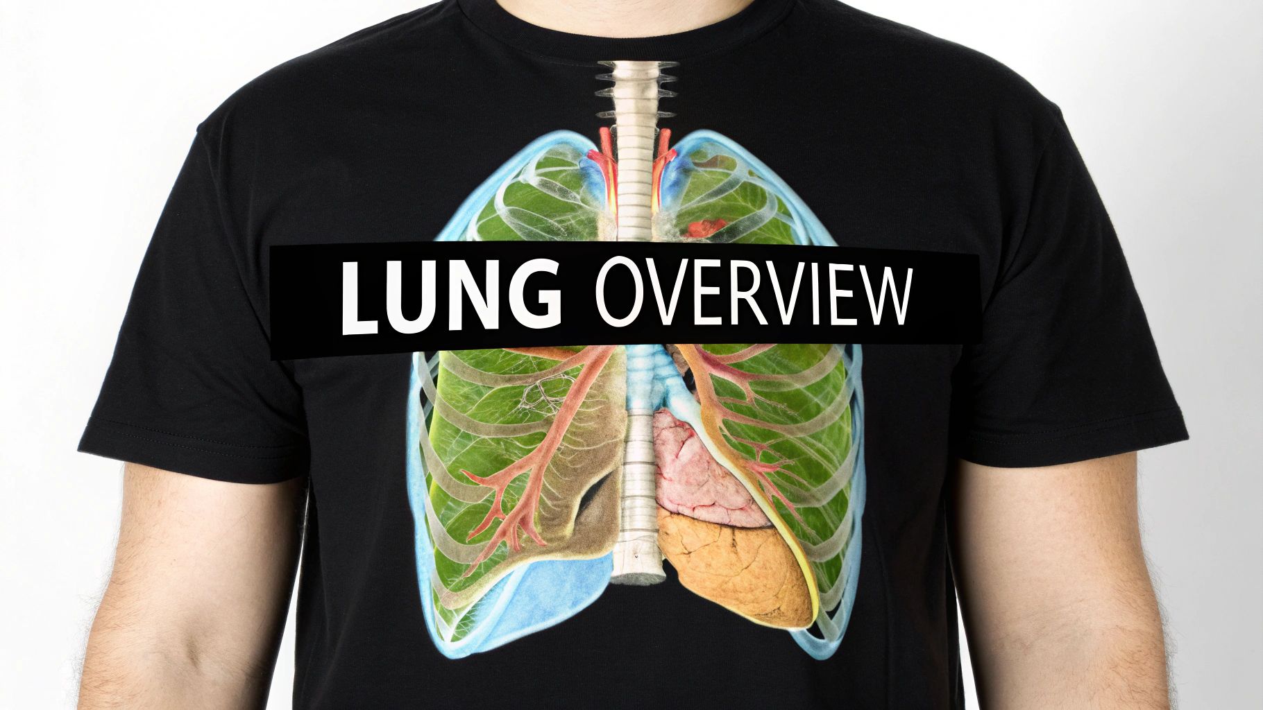
The lung segment, a self-contained anatomical unit within each lung, plays a vital role in modern respiratory care. Understanding this intricate architecture is essential for precision medicine, enabling targeted treatments and improved patient outcomes. Thoracic surgeons rely on this knowledge for informed surgical decisions.
This detailed anatomical understanding significantly improves surgical results, particularly in procedures like segmentectomy. This procedure allows for the removal of diseased lung segments while preserving healthy tissue.
In cases of early-stage lung cancer, for instance, a segmentectomy targets cancerous tissue while sparing the surrounding healthy lung. This minimizes lung function loss and reduces complications compared to more extensive procedures like a lobectomy, leading to a better quality of life for the patient.
History of Broncho-Pulmonary Segments
The evolution of our understanding of lung segments highlights advancements in thoracic medicine. The concept of broncho-pulmonary segments was first introduced in 1932.
Later, in 1943, Jackson and Huber proposed a standardized division based on bronchial distribution. They classified the right lung into three lobes with 10 segments and the left lung into two lobes with 8-10 segments. Each segment is named after the tertiary bronchus that supplies it. This standardization is critical for surgical procedures, especially segmentectomies.
However, anatomical variations and naming inconsistencies still exist, making precise segment identification a challenge. The left lung, for example, can have variations where the medial and anterior basal segments combine into an anteromedial segment. Understanding these variations is crucial for accurate diagnoses and treatment planning. Explore this topic further
The Importance of Segmental Anatomy in Surgical Planning
Understanding segmental anatomy is fundamental to improved patient care. This detailed knowledge allows surgeons to plan procedures with greater precision, reducing unnecessary tissue removal and preserving lung function. It also minimizes post-surgical complications like air leaks and other respiratory issues.
Preserving Lung Function With Segmental Resection
Imagine a patient with a localized lung tumor contained within a single segment. By precisely identifying and removing only that affected segment, the surgeon preserves the maximum amount of healthy lung tissue.
This preservation is paramount for maintaining the patient's respiratory function and quality of life. It also allows for future treatment options if another lung issue develops.
The focus on lung segments represents a high standard in modern respiratory care. It showcases how a thorough understanding of anatomy empowers clinicians to deliver more effective and less invasive treatments, improving patient outcomes. Ongoing research and development continue to refine surgical techniques, promising further advancements in respiratory health.
Navigating the Hidden Challenges of Lung Segment Identification
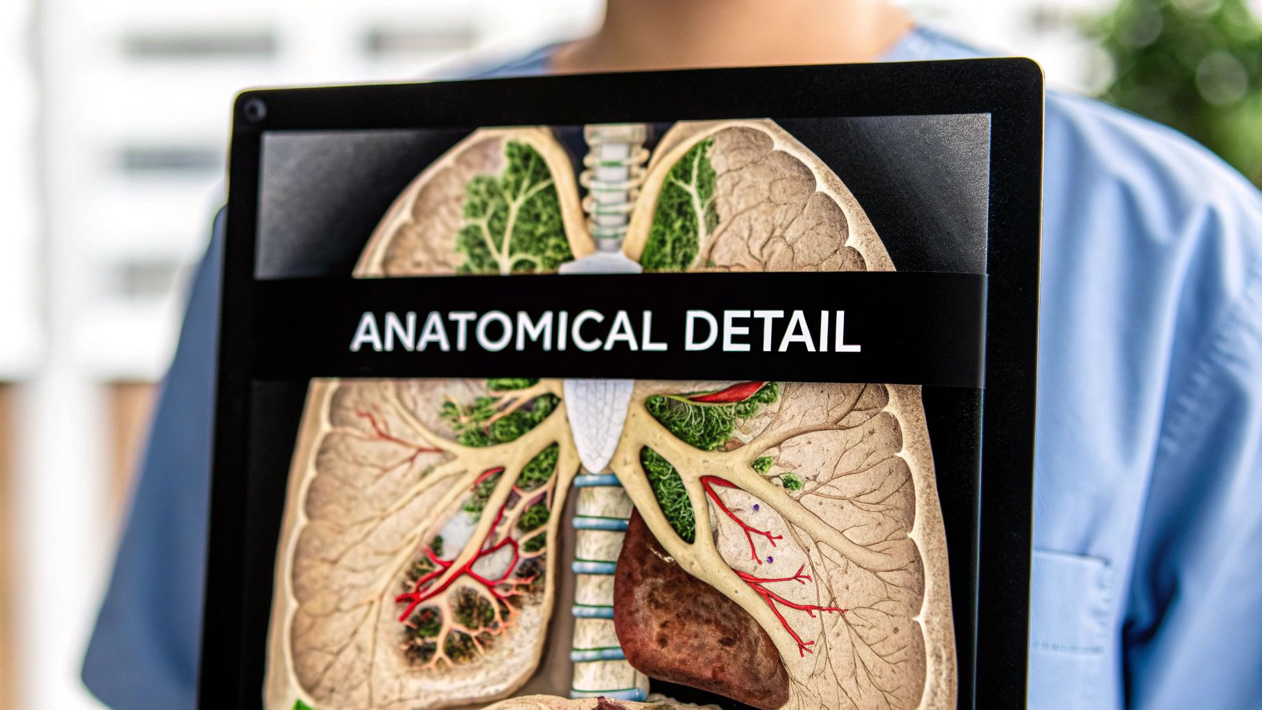
While textbook diagrams offer a simplified view of lung segment anatomy, the clinical reality is far more complex. Pinpointing individual lung segments presents substantial difficulties, even for experienced medical professionals. This complexity stems from the natural anatomical variations between individuals.
Variations in Vascular and Bronchial Patterns
One of the primary challenges lies in the unpredictable nature of vascular and bronchial structures. Textbooks often depict idealized versions of these structures. However, individual patients demonstrate a wide spectrum of variations, diverging from these standard anatomical maps.
For instance, the branching patterns of the pulmonary arteries and veins can differ significantly from one person to the next. This anatomical variability underscores the need for surgeons to meticulously evaluate each patient's unique structural makeup.
Additionally, the bronchial distribution, the foundation of segmental division, can also exhibit considerable variation. This anatomical variability can complicate procedures like segmentectomy, making it difficult to identify segmental boundaries. Surgeons must therefore adapt their surgical strategies based on intraoperative observations. These variations highlight the necessity of personalized surgical planning.
Addressing Challenges in Lingular Segment Identification
The lingular segments of the left lung present particularly complex challenges. Their proximity to vital structures and their variable anatomy make them a difficult area for surgical intervention. Surgeons rely on advanced techniques to accurately delineate the lingular segments, ensuring both safe and effective resection.
A significant challenge persists in identifying intersegmental planes during anatomic segmentectomy. A study involving 500 patients, which successfully reconstructed complete 3D segmental models, revealed a critical finding: the absence of distinct anatomical landmarks between segments. Read the full research here This lack of clear demarcation complicates precise identification of intersegmental planes, essential for successful segmental resections. Advanced imaging and surgical techniques, like ligation of segmental arteries followed by unilateral ventilation, can help visually distinguish the target segment by observing color changes caused by ischemia. However, further refinements are needed to improve the accuracy and efficiency of these procedures.
Accounting for Patient-Specific Differences
Advanced imaging technologies, such as 3D reconstruction from CT scans, are now vital for pre-operative planning. These techniques allow surgeons to visualize the patient's unique lung segment anatomy in detail, facilitating a more precise and personalized surgical approach.
Furthermore, intraoperative techniques, like indocyanine green (ICG) fluorescence imaging, offer real-time visualization of segmental perfusion. This real-time feedback helps surgeons confirm segmental boundaries during procedures. Combined with meticulous surgical technique, these tools enable a safer and more effective approach to segment lung surgery. Ultimately, this leads to improved patient outcomes and quality of life.
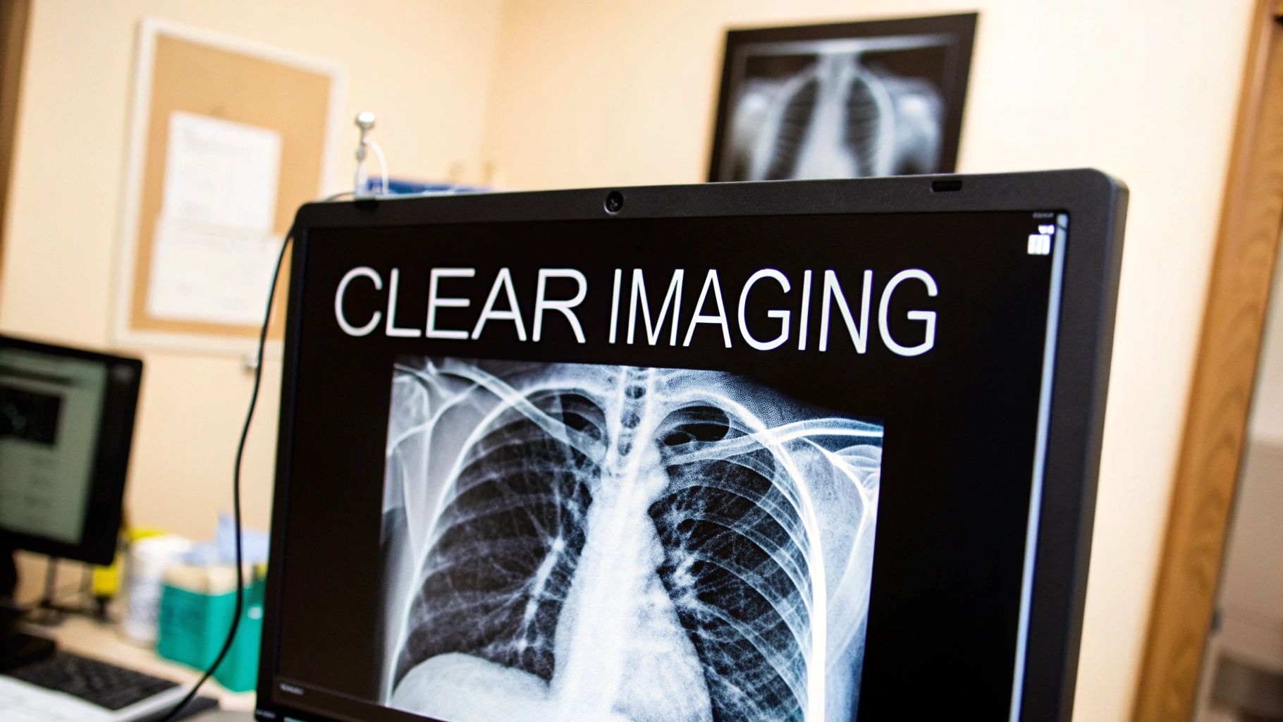
Advances in imaging technology have significantly improved our ability to visualize and analyze individual lung segments. This progress has made segment lung analysis a more precise medical science, enhancing diagnostic accuracy and treatment planning. Ultimately, these developments lead to better patient care by empowering clinicians to make more informed decisions.
Multi-Detector CT and Advanced 3D Reconstruction
Multi-detector computed tomography (MDCT) is crucial for modern segment lung analysis. These scanners produce high-resolution images, providing detailed views of lung structures. When combined with advanced 3D reconstruction techniques, MDCT offers a clear, three-dimensional lung representation, simplifying individual segment identification. This detailed visualization allows for accurate identification of segmental boundaries, a feat not possible with older imaging methods.
For instance, 3D reconstruction lets radiologists virtually navigate the bronchial tree, tracing each bronchus and clearly seeing the segment it supplies. This is particularly valuable in complex cases with anatomical variations.
Specialized Contrast Protocols and Respiratory Gating
Specialized contrast protocols further improve segmental anatomy visualization. These protocols use contrast agents to highlight vascular structures within each segment, differentiating their boundaries. This is similar to adding color to a grayscale image, revealing hidden details.
Respiratory gating techniques minimize motion artifacts, a common issue in lung imaging. By syncing image acquisition with the patient’s breathing, these techniques reduce blurring and deliver sharper images of the lung segments. This improved clarity is crucial for identifying subtle differences between segments.
Clinical Impact of Advanced Imaging
These advanced imaging techniques have a tangible impact on patient care, enabling segment-specific interventions previously impossible with conventional methods.
For example, in localized lung disease, these methods help surgeons precisely plan and perform segmentectomies, removing only the affected segment while sparing healthy tissue. This approach improves patient outcomes and minimizes complications.
These technologies also enhance diagnostic accuracy, leading to better treatment plans. This precision medicine approach is increasingly important for managing complex pulmonary conditions.
Imaging Modalities Comparison
To understand the advantages of modern imaging techniques, let's compare them to older methods. The following table illustrates the strengths and weaknesses of various imaging modalities for visualizing the segments of the lung.
Comparison of Imaging Modalities for Segment Lung Analysis
| Imaging Modality | Resolution Capability | Segmental Border Visibility | 3D Reconstruction Quality | Clinical Limitations |
|---|---|---|---|---|
| Conventional X-ray | Limited | Poor | Not applicable | Significant overlap of structures |
| CT Scan | High | Good | Good | Radiation exposure |
| MDCT with Contrast | Very High | Excellent | Excellent | Radiation exposure, contraindications to contrast |
| MRI | Moderate to High | Moderate | Good | Motion artifacts, less effective for visualizing air-filled spaces |
As the table shows, techniques like MDCT with contrast offer superior resolution and segmental border visibility, making them indispensable tools for modern pulmonologists and thoracic surgeons. The ability to create high-quality 3D reconstructions further enhances their clinical utility.
Mastering Modern Segment Lung Surgery Techniques

Modern segment lung surgery has evolved significantly beyond basic segmentectomy, leading to substantial improvements in patient outcomes. This article explores the key advancements that have shaped this field, from traditional open procedures to the latest minimally invasive options.
Minimally Invasive Approaches: VATS and Robotic-Assisted Surgery
Minimally invasive techniques have become the gold standard in segment lung surgery. Video-Assisted Thoracoscopic Surgery (VATS) and Robotic-Assisted Surgery (RATS) offer distinct advantages over traditional open thoracotomy.
These procedures typically involve smaller incisions, leading to reduced blood loss and faster recovery times. VATS utilizes a small camera (thoracoscope) and specialized instruments inserted through small incisions, providing surgeons with a clear view of the surgical field.
RATS enhances VATS by incorporating a robotic system controlled by the surgeon. This offers increased precision and dexterity, further minimizing trauma and improving patient comfort. However, both VATS and RATS require specialized training and expertise.
Identifying Intersegmental Planes: Key Techniques
Precise identification of the intersegmental planes, the boundaries between lung segments, is paramount for a successful segmentectomy. This highlights the critical role of surgical expertise.
Several techniques aid in this process:
- Inflation-Deflation Dynamics: This method involves inflating and deflating the lung to observe subtle differences in segment movement, aiding in boundary delineation.
- Indocyanine Green (ICG) Visualization: ICG, a fluorescent dye, is injected intravenously and visualized using a near-infrared camera. This technique highlights the vascular supply to each segment, enhancing intersegmental plane visibility.
- Ultrasound Guidance: Intraoperative ultrasound provides real-time visualization of the intersegmental planes, allowing for precise dissection.
These advanced techniques improve surgical precision, minimize the risk of resecting healthy tissue, and enable more complex procedures involving multiple segments.
Managing Complex Cases and Anatomical Variations
Thoracic surgeons often encounter complex cases with anatomical variations or multiple segment involvement. These scenarios require significant expertise and adaptability.
Variations in bronchial or vascular patterns can deviate from standard anatomy, making preoperative planning with 3D imaging crucial. This provides a personalized roadmap of the patient’s lung anatomy, allowing for more accurate intraoperative navigation. Surgeons must also be prepared to adapt their approach based on intraoperative findings.
Patient Positioning and Instrument Selection
Proper patient positioning, typically in a lateral decubitus position (lying on their side), is essential for optimal surgical access to the segment lung. This exposes the operative field and allows the surgeon to comfortably reach the targeted segment.
The selection of specialized instruments, such as minimally invasive staplers, is equally important. These instruments enable precise dissection and secure closure of bronchial stumps and blood vessels, minimizing complications like air leaks.
The combination of advanced surgical techniques, meticulous planning, and specialized instrumentation has dramatically improved outcomes in segment lung surgery. These advancements are crucial for preserving lung function while effectively treating localized diseases, underscoring their significance in modern thoracic surgery.
Real-World Applications of Segment Lung Expertise
The clinical impact of understanding segmental lung anatomy goes far beyond the operating room, influencing patient outcomes across a wide spectrum of respiratory conditions. This specialized knowledge is reshaping how medical professionals approach numerous pulmonary challenges.
Early Lung Cancer Management
Segment-specific approaches are significantly improving early lung cancer management. For patients diagnosed with small, localized tumors, segmentectomy offers a less invasive alternative to the traditional lobectomy. This targeted surgical approach preserves more healthy lung tissue, resulting in improved post-operative lung function and an enhanced quality of life for patients. This represents a major advancement compared to the more extensive lung removal required in a lobectomy.
Treating Challenging Conditions
Targeted segmental interventions are also demonstrating effectiveness in managing complex conditions such as localized bronchiectasis, fungal infections, and complications arising from tuberculosis. By precisely focusing treatment on the affected lung segment, physicians can minimize damage to the surrounding healthy tissue. This precision approach allows for the administration of higher medication doses or more targeted interventions, all while minimizing potential side effects. This targeted approach is akin to treating a single diseased plant in a garden rather than applying pesticides to the entire area.
Bronchoscopic Valve Placement and Targeted Therapeutic Delivery
Interventional pulmonologists leverage their knowledge of segmental anatomy for precise bronchoscopic valve placement. These valves, strategically positioned within the airways, redirect airflow away from diseased segments, enabling healthier areas of the lung to function more effectively. This intervention can significantly alleviate symptoms and improve overall lung function in patients with severe emphysema. This targeted strategy minimizes potential complications while maximizing treatment efficacy.
Furthermore, segmental anatomy plays a crucial role in guiding targeted therapeutic delivery, ensuring that medications reach the precise location where they are most needed. This localized approach enhances drug effectiveness and minimizes systemic side effects. This is particularly beneficial when delivering chemotherapy directly to cancerous segments, sparing healthy tissues from unnecessary exposure.
Radiation Oncology and Segment-Level Planning
Radiation oncologists utilize segment-level planning to optimize treatment efficacy and minimize damage to healthy lung tissue. By precisely targeting radiation to the diseased segment, they can administer higher doses while protecting the surrounding healthy areas. This approach results in improved tumor control while reducing the risk of radiation pneumonitis, a frequent side effect of lung radiation therapy.
Segment Lung Procedures by Clinical Indication
The following table illustrates how segmental expertise guides various clinical interventions. It provides examples of specific clinical conditions, the lung segments typically affected, the corresponding segment-specific interventions employed, and the anticipated outcomes.
| Clinical Condition | Affected Segments | Segment-Specific Intervention | Expected Outcomes |
|---|---|---|---|
| Early-stage lung cancer | Single segment | Segmentectomy | Preservation of lung function, improved quality of life |
| Localized bronchiectasis | Affected segment(s) | Targeted antibiotic delivery | Resolution of infection, minimized damage to healthy tissue |
| Severe emphysema | Hyperinflated segments | Bronchoscopic valve placement | Improved lung function, reduced shortness of breath |
| Lung abscess | Segment containing abscess | Percutaneous drainage | Drainage of infection, preservation of lung tissue |
As our understanding of segmental lung anatomy continues to advance, it promises to further enhance the precision of diagnosis and treatment, ultimately leading to improved outcomes for patients with a diverse range of respiratory conditions.
The Future of Segmental Lung Innovation
The field of segmental lung analysis and treatment is constantly advancing. New technologies are emerging with the potential to significantly improve respiratory care. These innovations offer exciting possibilities for enhanced diagnosis, treatment planning, and patient outcomes.
AI-Driven Segment Boundary Delineation
Artificial intelligence (AI) is playing a growing role in segmental lung analysis. Sophisticated algorithms are being developed to identify segment boundaries with remarkable precision, in some cases even exceeding human capabilities. This improved accuracy translates to more effective surgical planning and execution.
For instance, AI algorithms can analyze CT scans to generate detailed 3D lung models, automatically outlining individual segments. This provides surgeons with a clear visual guide for procedures like segmentectomy, potentially reducing surgical time and minimizing the risk of complications.
Advanced Intraoperative Navigation Systems
Intraoperative navigation is also seeing substantial progress. New systems are being developed that seamlessly integrate preoperative imaging data with real-time surgical visualization. This creates a dynamic surgical environment, giving surgeons unprecedented guidance.
Surgeons can now visualize the segmental lung in 3D, superimposed onto the actual surgical field. This real-time 3D mapping provides increased confidence and precision during complex procedures.
Molecular Imaging for Personalized Treatment
Molecular imaging techniques are transforming how we understand lung function at the segmental level. These methods enable visualization of functional differences between segments, providing insights beyond basic anatomical structure.
This personalized approach allows clinicians to tailor treatment strategies to each patient's individual lung characteristics. For example, in patients with COPD (Chronic Obstructive Pulmonary Disease), molecular imaging can pinpoint segments with the most significant airflow obstruction, guiding targeted interventions.
Segment-Specific Genetic and Microenvironmental Studies
Research is ongoing into the genetic and microenvironmental factors that influence disease within individual lung segments. This research holds great promise for the development of new targeted therapies for conditions like COPD and pulmonary fibrosis.
By understanding how these factors vary between segments, clinicians can develop more effective, targeted treatments. This granular approach addresses the underlying causes of disease at a much finer level.
This segment-specific approach represents a significant advancement toward truly personalized medicine in respiratory care. It facilitates the creation of therapies tailored to the unique genetic and microenvironmental profiles of diseased lung segments, maximizing treatment efficacy and minimizing side effects.
Ready to learn more about how AI can enhance your approach to medical imaging? Explore PYCAD's innovative solutions for improved diagnostic accuracy and operational efficiency.


