
The Evolution of Volumetric CT Scan Technology
Volumetric CT scanning has revolutionized medical imaging. It provides detailed, three-dimensional representations of the body's internal structures. This is a significant leap from traditional CT scans, which captured only individual slices. This evolution allows for diagnoses that were previously impossible with conventional imaging, giving healthcare professionals a more complete picture. Understanding this progress is key to appreciating the impact of volumetric CT scans on modern medicine.
The journey began in 1971 with Godfrey Hounsfield's introduction of the first practical CT scanner. This groundbreaking invention, the EMI Mark I, could scan the brain, but a single image took four and a half minutes. While limited compared to today's systems, it marked the beginning of volumetric imaging technology. Later advancements, such as multi-slice CT in the early 2000s, drastically reduced scan times. This allowed for faster coverage of larger areas and facilitated procedures like CT angiography, significantly broadening the application of CT scans. Learn more about the history of CT scans here.
Key Advancements in Volumetric CT
The development of spiral CT in the early 1990s further propelled this evolution. This technology allowed for continuous data acquisition. This eliminated the need for patients to hold their breath multiple times and reduced motion artifacts. The result? Clearer, more comprehensive images of the body, enabling precise three-dimensional reconstructions. This breakthrough paved the way for increasingly sophisticated applications of volumetric CT.
Multi-detector row CT dramatically increased the speed and detail of volumetric scans. These systems use multiple detectors to acquire more data simultaneously. This leads to higher resolution images, which has been essential for visualizing complex structures like coronary arteries. This advancement dramatically expanded the diagnostic capabilities of volumetric CT.
Impact on Diagnosis and Treatment
Volumetric CT scans have profoundly impacted patient care. These detailed 3D images provide essential information for diagnosing various conditions, from identifying small fractures to accurately staging tumors. Visualizing anatomical structures in three dimensions also allows for more precise surgical planning and minimally invasive procedures, often leading to improved patient outcomes. Looking forward, emerging technologies like AI and automation hold promise for areas like Conflict of Interest (COI) management. Learn more about The Future of COI Management: AI, Automation, and Compliance. Continued innovation in volumetric CT technology promises even more precise and personalized medical care.
Breakthrough Innovations in Volumetric CT Scan Development
Volumetric CT scans create detailed 3D images of the inside of the human body. This remarkable technology wasn't developed overnight. It's the result of continuous innovation in medical imaging. Each advancement, from single-slice images to today's sophisticated multi-detector systems, has built upon the previous ones, dramatically improving diagnostic capabilities. This evolution has been driven by the ongoing pursuit of faster scan times, clearer images, and increased patient comfort.
One of the most significant advancements was the development of spiral CT in the early 1990s. This innovation allowed the scanner's gantry to rotate continuously as the patient moved through the machine. This allowed for volumetric data acquisition in a single breath-hold.
This shift from traditional CT methods to volumetric imaging was significantly propelled by the introduction of spiral, or helical, CT. This single breath-hold capability significantly reduced motion artifacts. It laid the groundwork for the detailed 3D reconstructions now essential in modern medical diagnoses.
The transition from single-row detectors to systems with 16, 32, 64, or more rows by the early 2000s rapidly expanded the capabilities of CT scanners. This increase in detector rows directly improved both the speed and quality of volumetric scans. It enabled better visualization of organs and structures, such as coronary arteries, which were previously difficult to image clearly. Learn more about the evolution of CT scanning technology here.
The Rise of Multi-Detector CT
Building on spiral CT technology, the next significant advancement was multi-detector row CT (MDCT). These systems utilize multiple detector rows instead of a single detector. This allows them to acquire substantially more data simultaneously. The result is a significant increase in both the speed and resolution of volumetric scans, making it possible to visualize even the smallest details within the body.
Advanced Image Reconstruction Techniques
Improvements in image reconstruction techniques have also played a vital role in maximizing the value of the data captured by MDCT. For example, iterative reconstruction algorithms use complex mathematical models to enhance image quality. These algorithms reduce noise and artifacts while minimizing radiation dose. This offers clinicians the clearest possible images while prioritizing patient safety. These innovations have dramatically reshaped medical imaging, providing unparalleled insights into the human body.
The Technical Magic Behind Volumetric CT Scan Imaging
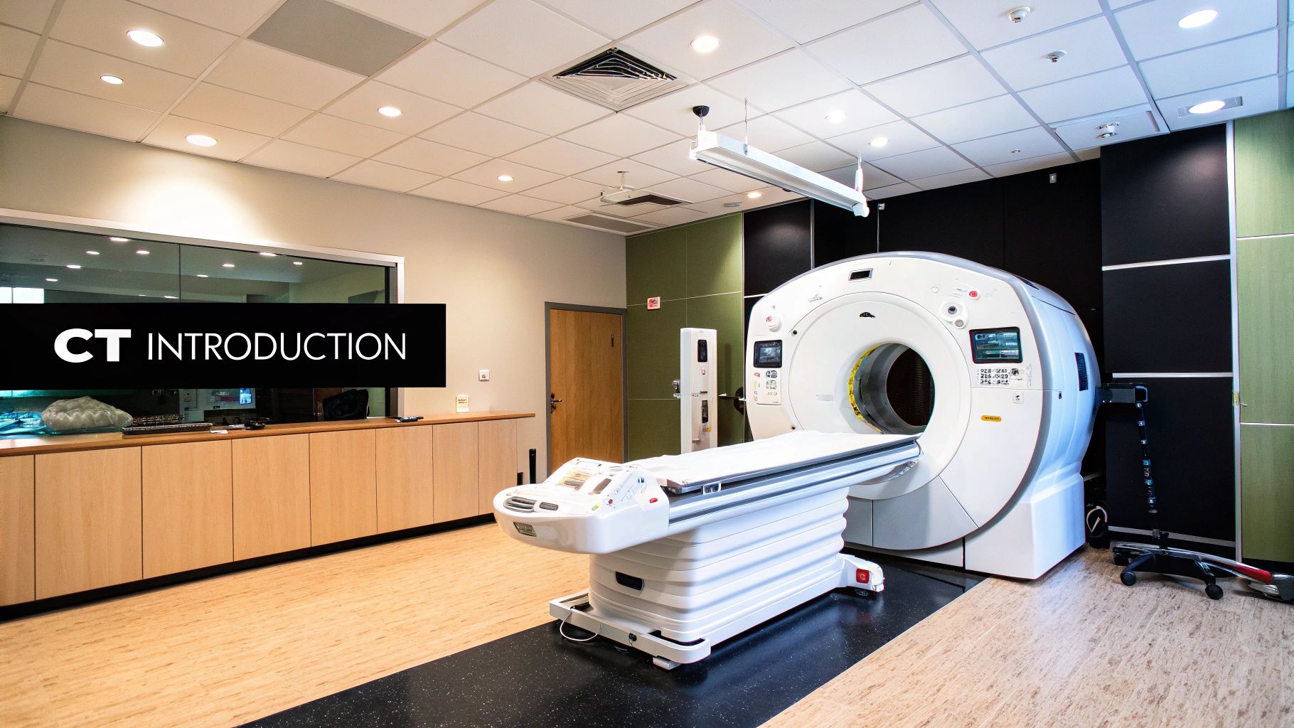
Volumetric CT scans create incredibly detailed 3D images of the inside of the human body. This is possible thanks to the complex interplay of several key components. These include advanced detector arrays, precisely calibrated X-ray tubes, and powerful processing systems. Understanding how these elements work together is crucial to appreciating the power of this imaging technique. It's about seeing how raw data is transformed into the visual representations doctors use for diagnoses.
The Role of Detectors and X-Rays
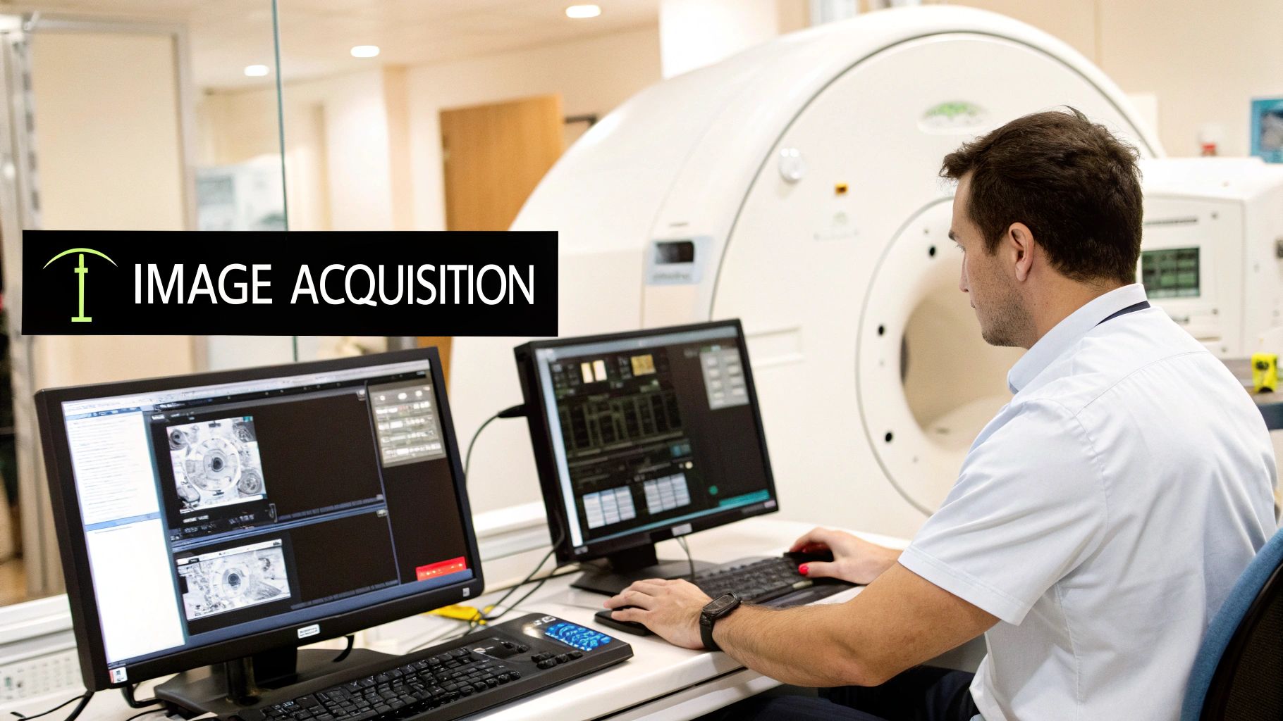
Think of the detector array as a digital camera. It captures multiple snapshots from different angles as the X-ray tube rotates around the patient. This coordinated movement allows the system to gather comprehensive data from all sides of the target area. Each detector in the array measures the amount of X-ray radiation passing through the body. This data is then used to reconstruct a detailed 3D image. The X-ray tube’s precision is vital for ensuring accurate measurements, as it emits the radiation that creates the images.
The speed of rotation also plays a critical role in image quality. Faster rotations minimize the effects of patient movement. This leads to sharper images. For example, a fast rotation is particularly beneficial in cardiac imaging. The beating heart could blur the image if the scan is too slow. This speed, combined with the number of detectors in the array, directly affects the resolution and clarity of the final 3D image.
Transforming Data into Images
The data captured by the detectors is just the starting point. Sophisticated algorithms reconstruct this raw data into clinically useful 3D images. It’s like assembling a complex jigsaw puzzle. Each piece of data represents a small part of the whole image. This reconstruction process is crucial. It transforms raw scanner data into the clear, high-quality images radiologists need for accurate diagnoses.
To illustrate the progress of CT technology, let's look at a table summarizing the evolution of volumetric CT scanners:
The following table shows the "Evolution of Volumetric CT Scanner Capabilities." It compares key technical specifications of volumetric CT scanners across different generations to show technological progression.
| Scanner Generation | Detector Rows | Rotation Time | Spatial Resolution | Coverage Per Rotation |
|---|---|---|---|---|
| 1st Generation | 1 | Several Minutes | Several Millimeters | Single Slice |
| 4th Generation | Hundreds | < 1 Second | Sub-millimeter | Multiple Slices |
| Modern MDCT | Thousands | Sub-second | < 125 Micrometers | Wide Volume Coverage |
This table highlights the dramatic improvements in speed, resolution, and coverage area that have occurred over time, leading to more detailed and informative scans.
One significant statistical improvement in volumetric CT scanning involves scan times and resolution. Modern CT scanners can complete a full rotation in less than a second. This is a drastic improvement from the minutes required by earlier machines. Recent advancements have led to a spatial resolution as low as 125 micrometers in some systems. Adjustable exposure parameters have also significantly lowered radiation doses. This makes volumetric scans safer and more efficient. For more detailed statistics, see this article. These advances are making volumetric CT scans increasingly important in modern medical practice.
Innovations in Image Reconstruction
Iterative reconstruction techniques further improve image quality and reduce radiation doses. These techniques use refined algorithms to enhance the image. They iteratively refine the image reconstruction process, which results in less noise and better image quality. Dual-energy CT scanning uses two different X-ray energy levels. This provides more information about tissue composition, allowing for more precise diagnoses and personalized treatment planning. These advancements highlight how ongoing technological refinements are constantly expanding the possibilities of volumetric CT imaging.
Clinical Applications of Volumetric CT Scans

Volumetric CT scans provide medical professionals with intricate 3D visualizations of the body. This advanced imaging technique has a significant impact on patient care, leading to earlier diagnoses and more precise treatment plans across various medical specialties. This detailed anatomical information enhances the way doctors approach complex medical cases.
The following table summarizes the key diagnostic advantages of volumetric CT scanning across different medical specialties.
| Medical Specialty | Key Applications | Diagnostic Benefits | Impact on Patient Care |
|---|---|---|---|
| Cardiology | Evaluation of coronary artery disease | Non-invasive visualization of coronary arteries, identification of blockages, assessment of disease extent | Informs treatment decisions (medication, angioplasty, bypass surgery), reduces need for invasive procedures |
| Oncology | Cancer staging | Precise determination of tumor size and location, assessment of metastasis to lymph nodes or other organs | Enables tailored treatment plans, maximizes treatment effectiveness, minimizes side effects |
| Emergency Medicine | Trauma assessment | Rapid identification of internal injuries, fractures, and bleeding | Enables immediate surgical intervention when necessary, crucial in life-threatening situations |
| Neurology | Diagnosis of neurological conditions | Visualization of brain abnormalities (strokes, tumors, aneurysms) | Aids in diagnosis and treatment planning |
| Surgery | Surgical planning | Detailed 3D anatomical information, visualization of complex anatomical relationships | Enables precise surgical approach, minimizes complications, improves surgical outcomes |
This overview demonstrates the diverse applications and benefits of volumetric CT across various specialties, highlighting its positive impact on diagnosis, treatment planning, and patient care.
Volumetric CT in Cardiology
In cardiology, volumetric CT scans are vital for evaluating coronary artery disease. These scans offer a non-invasive method to visualize the coronary arteries. This allows physicians to identify blockages and assess the severity of the disease. This detailed view informs treatment decisions, such as medication, angioplasty, or bypass surgery.
This information is crucial for determining the best course of action for each patient. The detailed imaging reduces the need for more invasive diagnostic procedures.
Volumetric CT in Oncology
Accurate cancer staging is critical for effective cancer treatment. Volumetric CT scans provide precise information about the size and location of tumors. They also show whether the cancer has spread to nearby lymph nodes or other organs. This helps oncologists personalize treatment plans for individual patient needs.
The 3D visualization is particularly useful for planning radiation therapy and complex surgical procedures. This precise approach maximizes treatment effectiveness while minimizing potential side effects.
Volumetric CT in Emergency Medicine
In emergency medicine, rapid diagnosis is essential. Volumetric CT scans offer fast, detailed assessments of trauma patients, helping physicians quickly identify internal injuries, fractures, and bleeding.
In cases of severe head trauma, for instance, a volumetric CT scan can immediately reveal intracranial bleeding. This rapid assessment allows for prompt surgical intervention, which can be life-saving in critical situations.
Volumetric CT in Neurology and Surgery
Neurologists use volumetric CT scans to visualize complex brain abnormalities, aiding in the diagnosis of conditions like strokes, tumors, and aneurysms. The 3D images offer a clearer understanding of these intricate brain structures, which is crucial for effective treatment planning.
Surgeons rely on volumetric CT scans, particularly for planning complex surgeries. The detailed 3D images offer comprehensive anatomical information, allowing surgeons to navigate intricate anatomical relationships and plan their surgical approach with greater precision. This pre-operative planning can minimize complications and improve patient outcomes.
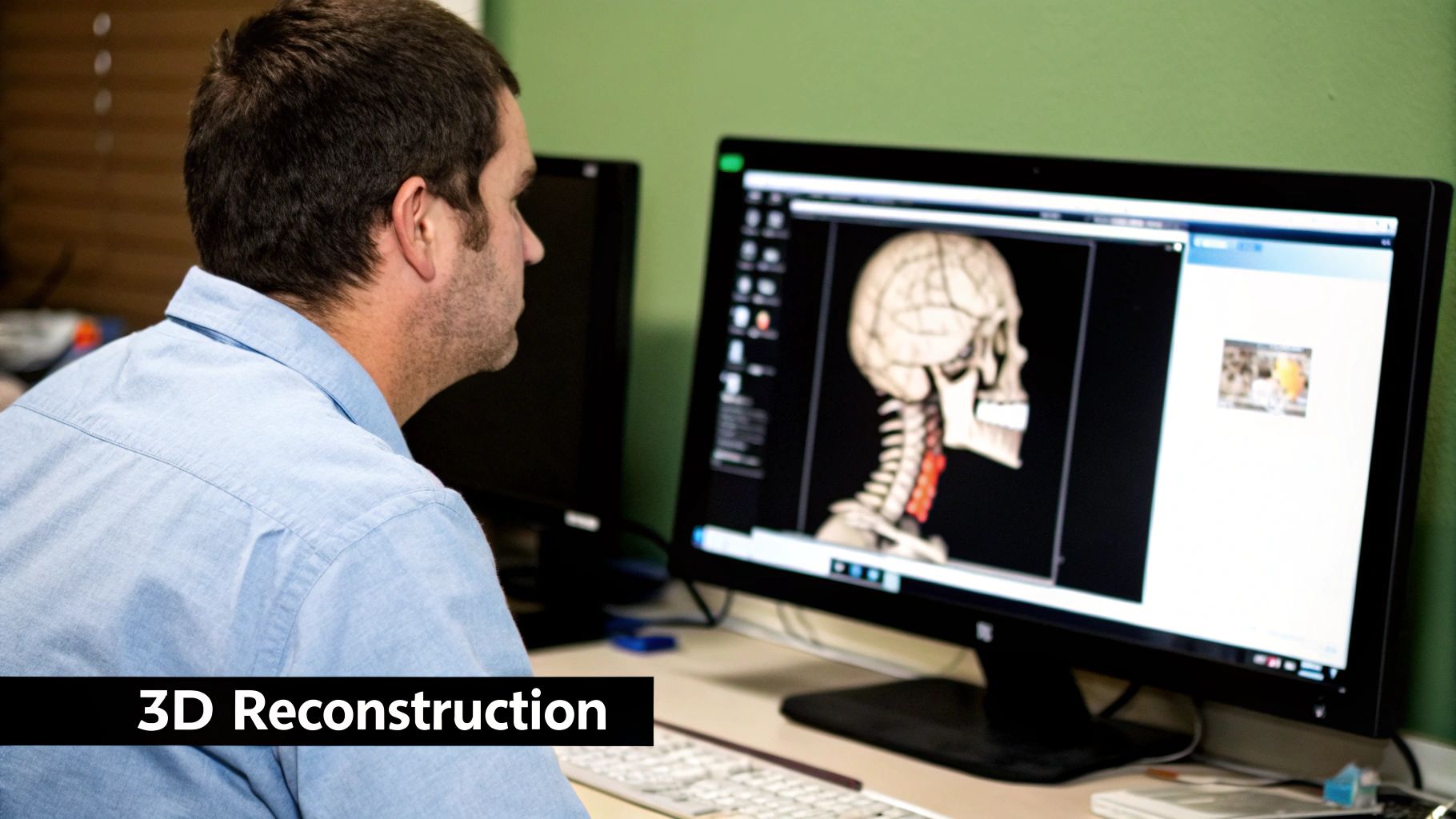
The Power of 3D
The ability to create 3D visualizations from volumetric data is invaluable. These visualizations enhance treatment planning and can be used to create 3D-printed models of organs or bones. These models allow surgeons to practice complex procedures, improving their precision and reducing the risk of complications. They also help doctors communicate complex medical information to patients and their families.
A key advantage of analytical models like SLIViT is their scalability and adaptability. They can analyze large datasets and be updated with new medical imaging data for future analysis. This can make expert-level image analysis more accessible, potentially leading to better patient outcomes. Research indicates that pre-training models on 2D scans and then fine-tuning them with 3D scan data can accurately identify disease biomarkers in 3D images. This method can even outperform models trained exclusively on 3D scans. Ongoing research, such as Google's CT Foundation tool, continues to explore data-efficient models for CT scan analysis.
Real-World Benefits and Challenges of Volumetric CT Scanning
Volumetric CT scanning provides medical professionals with detailed 3D images, offering several advantages over traditional imaging techniques. These detailed images facilitate more accurate diagnoses and enable the creation of personalized treatment plans. This leads to tangible benefits for patients, impacting everything from diagnosis speed to the duration of hospital stays.
Benefits of Volumetric CT Scanning
-
Reduced Diagnostic Uncertainty: Volumetric CT scans offer a comprehensive 3D view of the anatomy, reducing ambiguity and increasing diagnostic confidence. This clarity can eliminate the need for additional testing and procedures, saving time and resources.
-
Precise Interventions: The detailed visualizations provided by volumetric CT scans allow for more accurate surgical planning and guidance. This increased precision minimizes invasiveness and reduces the risk of complications, especially in complex procedures.
-
Faster Diagnoses and Reduced Hospital Stays: Volumetric imaging significantly accelerates the diagnostic process. Quicker interventions can lead to potentially shorter hospital stays, benefitting both patients and healthcare systems.
-
Improved Treatment Planning: The comprehensive 3D models generated by volumetric CT scans allow for a more personalized approach to treatment, optimizing outcomes for individual patients. This personalized approach is particularly important in treating conditions like cancer, where precision is critical.
Challenges of Volumetric CT Scanning
While volumetric CT scanning offers numerous advantages, it also presents certain challenges:
-
Radiation Exposure: CT scans utilize ionizing radiation. While individual scans are generally safe, repeated exposure can increase cumulative radiation levels. Therefore, careful consideration and strict adherence to safety protocols are necessary.
-
Accessibility: Volumetric CT scanners are expensive and require specialized staff and supporting infrastructure. This can limit their availability, particularly in resource-constrained environments. Addressing this disparity in access remains an important goal.
-
Data Management: Volumetric CT scans generate large datasets, necessitating substantial storage capacity and efficient management systems. Robust IT infrastructure and streamlined workflows are crucial for effectively handling this data.
Addressing the Challenges
The medical community is actively working to mitigate the risks and improve access to volumetric CT scanning:
-
Dose Reduction Techniques: Innovations in scanner technology and imaging protocols continuously strive to minimize radiation exposure. Techniques like iterative reconstruction can significantly reduce the radiation dose required to produce high-quality images.
-
Expanding Access: Initiatives are underway to increase access to volumetric CT technology, particularly in underserved communities. These initiatives include exploring more affordable scanner options and developing telehealth strategies.
-
AI-Driven Solutions: Companies like PYCAD, specializing in AI-driven medical imaging, are developing solutions to optimize CT analysis. AI can improve diagnostic accuracy, streamline workflows, and potentially expand access to expert image interpretation. For example, AI algorithms can help identify subtle abnormalities, accelerating the diagnostic process and assisting in treatment planning.
AI also plays a vital role in optimizing data management for volumetric CT. AI algorithms can compress large datasets without a significant loss of information. This improves storage efficiency and allows for faster data transfer and analysis.
The future of volumetric CT scanning is promising. Ongoing research into photon-counting detectors, spectral imaging, and AI integration holds the potential to further enhance diagnostic capabilities and enable even more personalized medical care. As technology continues to advance and become more accessible, volumetric CT scans are expected to play an increasingly important role in improving patient outcomes.
The Future of Volumetric CT Scan Technology
Volumetric CT scanning is constantly evolving. The driving forces behind this evolution are the desire for more detailed images, faster scan times, and lower radiation doses. These advancements hold the promise of significantly enhancing diagnostic capabilities and reshaping the future of medical imaging, impacting how doctors diagnose and treat a wide range of conditions.
Photon-Counting Detectors: A New Era of Image Quality
One of the most promising developments is the emergence of photon-counting detectors. Unlike conventional detectors that measure the total energy deposited by X-rays, these new detectors count individual X-ray photons and measure their energy. This allows for a more precise measurement of X-ray attenuation, leading to improved image quality, reduced noise, and better tissue characterization.
This finer level of detail is crucial for distinguishing subtle differences in tissue composition. It also allows for a significant reduction in radiation exposure, making volumetric CT scans safer for patients.
AI Integration: Enhancing Diagnostic Precision
Artificial intelligence (AI) is playing an increasingly important role in medical imaging. AI algorithms can analyze volumetric CT scans to identify subtle patterns and anomalies that might be missed by the human eye. This can lead to earlier and more accurate diagnoses, particularly for complex medical conditions.
For example, AI algorithms can be trained to detect small nodules in lung scans, which could indicate early-stage lung cancer. This early detection can be crucial for effective treatment and improved patient outcomes. AI is also being used to automate tasks like image segmentation and registration, freeing up radiologists to focus on more complex image analyses.
Spectral Imaging: Unlocking New Diagnostic Possibilities
Spectral CT, another area of active research, uses multiple X-ray energy levels to acquire information about the elemental composition of tissues. This detailed spectral information allows for more precise tissue characterization, going beyond traditional density-based imaging.
This advanced technique is particularly valuable in oncology, where it can help differentiate between cancerous and healthy tissue. It also shows promise in characterizing different types of kidney stones, leading to more targeted treatment strategies.
The Impact on Clinical Practice
These technological advancements have the potential to transform clinical practice in the coming years. The improved image quality, reduced radiation exposure, and enhanced diagnostic precision offered by photon-counting detectors, AI integration, and spectral imaging are all expected to significantly improve patient outcomes.
As these technologies mature and become more widely accessible, they will empower medical professionals to make more informed decisions, provide more personalized treatment plans, and ultimately enhance the quality of patient care.
Looking to optimize your CT analysis? PYCAD, specializing in AI-driven medical imaging solutions, offers comprehensive services from data handling and model training to deployment. Learn more about how PYCAD can enhance your medical imaging capabilities.