Demystifying CBCT: Understanding the Core Technology
Cone Beam Computed Tomography (CBCT) is a specialized X-ray technology producing detailed 3D images of teeth, jaws, and facial structures. Unlike traditional 2D X-rays, CBCT captures hundreds of images as a cone-shaped X-ray beam rotates around the head, creating a comprehensive 360-degree view.
Sophisticated software compiles these individual images into a precise 3D model. This allows dentists to examine anatomical structures from any angle, facilitating more accurate diagnoses.
This 3D imaging represents a significant advancement in dentistry. Imagine upgrading from a basic map to a detailed, interactive globe. Conventional X-rays offer a limited, flat perspective, whereas CBCT provides a complete three-dimensional representation, revealing details often obscured by traditional imaging. This is particularly valuable for accurate treatment planning for complex procedures such as dental implants or orthognathic surgery.
Core Components of a CBCT System
A CBCT system relies on three key components:
-
X-ray Source and Cone Beam: This component emits a cone-shaped X-ray beam, encompassing a wide area and gathering data from all angles as it rotates. This cone-shaped beam distinguishes CBCT from conventional CT scanners.
-
Detector: A flat panel detector, akin to a digital camera sensor, captures the X-rays passing through the patient. This detector registers the X-rays' varying intensities and converts them into digital data.
-
Computer and Software: Powerful computer software processes the digital data from the detector. The software reconstructs the individual 2D images into a comprehensive 3D model that can be viewed and manipulated from any perspective to assess the patient's anatomy.
History of CBCT Technology
The foundations of CBCT date back to the late 1990s. Dr. Yoshinori Arai in Japan and Dr. Piero Mozzo in Italy independently developed this technology for oral and maxillofacial radiology. The first commercial CBCT system became available in Europe in 1996 and in the US in 2001. Learn more about the history of CBCT.
CBCT offers extensive diagnostic information with significantly less radiation exposure compared to traditional CT scans. This balance of detail and safety has contributed to its growing popularity among dental professionals for various diagnostic and treatment planning applications.
The Evolution of CBCT Technology
From its initial development, Cone Beam Computed Tomography (CBCT) has become essential to modern dentistry. This journey is marked by constant improvement in image quality, reduced scan times, and minimized radiation exposure. Early CBCT machines were bulky, requiring scans that lasted minutes.
Today's systems capture detailed 3D images in a matter of seconds. This drastic improvement underscores the rapid progress of CBCT technology.
Advancements in Detector Technology
This progress wouldn't have been possible without key innovations. A crucial development lies in detector technology. Consider the evolution of cameras, from film to digital sensors. Early CBCT systems relied on less efficient detectors. This impacted both image quality and scan times.
The adoption of flat-panel detectors in the late 1990s was a major step forward. It allowed for faster scans and sharper images. This was significantly influenced by advancements in flat-panel detector technology itself.
In the 1980s, early CBCT implementations focused on high-contrast, high-resolution applications. However, these systems were limited by nonlinear imagers. The real breakthrough came with large-area digital x-ray detectors. These enabled compact flat panels suitable for high-resolution CBCT. Learn more about this technology. This evolution has improved visualization capabilities, with image quality now rivaling traditional CT scans in certain applications.
Enhanced Computing Power and Software Algorithms
Advancements in computing power and software algorithms have also played a crucial role. Increased computing power enables faster processing of the large datasets generated by CBCT scans. This contributes to shorter scan times and more detailed 3D reconstructions.
Sophisticated software algorithms also enhance image quality. They allow for clearer visualization of even the smallest anatomical details.
Improved Patient Outcomes
These technological advancements directly contribute to improved patient care. More precise images enable more accurate diagnoses and better treatment planning. For example, in implant dentistry, CBCT allows for accurate implant placement.
This precision minimizes complications and maximizes success rates. Procedures that were once lengthy and sometimes unpredictable are now more precise and efficient due to the detailed information CBCT provides. These continued improvements reinforce the value of CBCT scans, leading to better patient outcomes across various dental specialties.
What Is a CBCT Scan vs. Traditional Imaging: The Real Differences
Understanding the core differences between CBCT scans and traditional dental imaging is vital for both patients and dental professionals. This section breaks down those differences, highlighting key factors like radiation dose, image detail, and what each method can diagnose. Being informed helps everyone make the best decisions about using this advanced 3D imaging technology.
CBCT Scans vs. Panoramic X-Rays
Panoramic X-rays provide a broad 2D view of the entire mouth, useful for initial assessments. However, they lack the depth and detail of a CBCT scan. For instance, a panoramic X-ray might show an impacted tooth, but a CBCT scan pinpoints its 3D location relative to other teeth, nerves, and surrounding structures. This precise detail is crucial for planning procedures like extractions or other oral surgeries.
CBCT Scans vs. Bitewing X-Rays
Bitewing X-rays focus on the crowns of the teeth, primarily used for detecting cavities. They are highly effective for this purpose. But bitewings don't reveal information about the roots and supporting bone. CBCT scans offer a complete view of the tooth, from crown to root tip. This allows dentists to diagnose issues like root fractures or hidden canals often invisible on 2D images. Endodontists, specializing in root canals, commonly use CBCT scans for accurate diagnoses and treatment plans.
CBCT Scans vs. Conventional CT Scans
While both CBCT and conventional CT scans produce 3D images, there are important distinctions. Conventional CT scans offer greater resolution, making them suitable for diagnosing complex medical conditions. The downside is a significantly higher radiation dose. CBCT provides ample detail for most dental needs while using a much lower radiation dose, making it a safer choice for patients undergoing dental imaging.
Comparing Key Imaging Methods
To help understand these differences, let's look at a table summarizing the key features of each method:
CBCT vs. Other Dental Imaging Methods: A detailed comparison of CBCT against traditional dental imaging technologies based on key factors like radiation dose, image dimensionality, diagnostic applications, and cost considerations.
| Imaging Method | Radiation Dose | Dimensionality | Detail Level | Cost Range | Ideal Applications |
|---|---|---|---|---|---|
| Panoramic X-ray | Low | 2D | Low | Low | General overview, initial assessment |
| Bitewing X-ray | Very Low | 2D | Moderate | Low | Cavity detection, monitoring dental health |
| Conventional CT Scan | High | 3D | High | High | Complex medical diagnoses, trauma assessment |
| CBCT Scan | Low | 3D | Moderate to High | Moderate | Implant planning, endodontics, orthodontic analysis |
This table highlights the strengths of each imaging technique. While traditional methods are still valuable for specific purposes, CBCT offers enhanced diagnostics for many dental applications. It's a powerful tool for dentists who need precise information for accurate treatment planning. Conditions missed or misinterpreted on 2D images can be clearly seen with CBCT, ultimately leading to better outcomes for patients. This advantage is making CBCT increasingly vital in modern dental practices.
Clinical Applications of CBCT Imaging

Cone Beam Computed Tomography (CBCT) provides detailed 3D imaging that has significantly changed treatment planning in various dental specialties. This technology allows for more accurate diagnoses and ultimately, improved patient outcomes. Let's explore some key applications of CBCT in modern dentistry.
CBCT in Implantology
CBCT is an indispensable tool for implantologists. The precise 3D images offer accurate assessments of bone density and volume. This information is crucial for determining the ideal implant size and placement.
This level of detail ensures optimal implant stability and minimizes the risk of complications. CBCT scans precisely identify the location of vital structures such as nerves and blood vessels. This precision allows implantologists to plan surgeries with sub-millimeter accuracy.
CBCT in Endodontics
Endodontists, specializing in root canal treatments, also benefit significantly from CBCT technology. It reveals intricate details of root canal anatomy, including hidden canals or root fractures often missed by traditional X-rays.
Identifying these complex anatomical features is essential for successful root canal therapy and helps avoid complications. This increased accuracy reduces the need for retreatment and promotes long-term success.
CBCT in Orthodontics
Orthodontists use CBCT to gain a comprehensive view of tooth positioning, jaw relationships, and facial structures. This complete picture allows them to develop personalized treatment plans for each patient's unique needs.
For instance, CBCT can visualize the position of impacted teeth and their relationship to other dental structures. This detailed visualization facilitates more accurate planning for orthodontic interventions, leading to more effective and efficient treatments.
Emerging Applications of CBCT
The use of CBCT is expanding beyond these established applications into broader health concerns. One emerging application is airway analysis for sleep apnea patients. CBCT scans provide detailed images of the upper airway, helping identify obstructions that contribute to sleep apnea.
Additionally, CBCT is becoming increasingly valuable for TMJ evaluation. The detailed 3D images allow dentists to assess the temporomandibular joint and surrounding structures, aiding in diagnosis and treatment planning for TMJ disorders.
These emerging applications demonstrate the continued evolution of CBCT scanning and its potential to improve patient care across various fields. As technology advances, we can expect even more innovative uses of CBCT imaging in dentistry and beyond. From improved diagnostics to personalized treatment plans, CBCT is reshaping patient care, ultimately leading to better outcomes and improved patient experiences.
The Real Benefits and Limitations of CBCT Imaging
This section offers a balanced look at Cone Beam Computed Tomography (CBCT), exploring both its strengths and weaknesses. We'll examine what CBCT excels at and when other imaging methods might be a better fit. Let's explore the core advantages and disadvantages of this technology.
Advantages of CBCT
The primary advantage of CBCT lies in its ability to create true 3D representations of dental structures. This detailed view allows dentists to diagnose conditions that traditional 2D X-rays often miss. Think of the difference between a flat photograph and a detailed sculpture; CBCT reveals a far more complete picture. This is especially useful for treatment planning. For further insights into its application in implant dentistry, see Dr. Kaycee Walton’s course: CBCT and Implant Dentistry: Essential Tips and Tricks.
Accurate placement of dental implants relies heavily on understanding bone density and volume. CBCT provides the precision required for ideal implant positioning, maximizing stability and minimizing risks. This 3D imaging also improves endodontic treatment, helping specialists identify hidden canals and root fractures vital for successful root canal therapy, as discussed in: Four Ways CBCT Imaging Can Improve Endodontic Patient Outcomes.
CBCT also aids in the early detection of conditions like impacted teeth and jaw abnormalities that may be hidden in 2D images. Early detection allows for timely intervention and better long-term results.
Limitations of CBCT
While a powerful tool, CBCT has limitations. Radiation exposure, though lower than conventional CT scans, remains a consideration. This makes careful evaluation essential, especially for children, as highlighted in 7 Ways BeamReaders' ECAA Protocol Can Help Pediatric Patients Breathe Easier.
Another factor is cost. CBCT systems are a significant investment for dental practices, which can impact patient access. It’s important to determine when CBCT is truly necessary versus when traditional X-rays are sufficient.
CBCT may not be the best imaging choice in every situation. For basic cavity detection or routine checkups, bitewing and panoramic X-rays often provide enough information.
To help illustrate these points, let's look at a comparison table:
The following table provides a comprehensive overview of the advantages and disadvantages of CBCT technology in clinical practice, helping practitioners and patients understand when this imaging modality is most appropriate.
| Factor | Benefits | Limitations | Clinical Considerations |
|---|---|---|---|
| Image Detail | Provides 3D images for detailed anatomical information | Can generate artifacts that may complicate interpretation | Consider the diagnostic needs of the case. Is 3D information crucial? |
| Radiation Exposure | Lower radiation dose compared to medical CT scans | Still exposes patients to radiation | Carefully weigh the benefits against the risks, especially for children and pregnant women. |
| Cost | Facilitates accurate diagnosis and treatment planning, potentially reducing the need for revisions | Higher cost than traditional 2D radiography | Evaluate the cost-effectiveness of CBCT in relation to the specific clinical situation. |
| Diagnostic Capabilities | Enables visualization of complex anatomical structures, such as impacted teeth, jaw abnormalities, and nerve canals | Limited soft tissue contrast compared to MRI | Consider alternative imaging modalities if soft tissue evaluation is the primary objective. |
| Time | Quick scan acquisition time | Image processing and interpretation can require specialized software and training | Factor in the time required for image processing and interpretation. |
In summary, the table highlights that while CBCT offers superior image detail for complex cases, radiation exposure and cost must be carefully considered. Practitioners must weigh these factors against the diagnostic benefits when making clinical decisions.
Clinical Considerations
Dental professionals should carefully weigh the benefits and limitations of CBCT technology when determining its appropriateness for each patient. Factors include the complexity of the case, the potential diagnostic value of the CBCT scan, the patient's risk factors, and the availability of alternative imaging options. Selecting the right diagnostic tool is key to providing effective and safe patient care. This decision should be made collaboratively between the patient and the dental professional.
The Patient Experience: What To Expect During A CBCT Scan
Feeling apprehensive about your upcoming CBCT scan? It's perfectly understandable. Many patients experience uncertainty about the process. This guide will walk you through each step, from check-in to completion, clarifying what a CBCT scan entails and simplifying this straightforward procedure.
Before The Scan: Simple Preparation
The preparation process for a CBCT scan is remarkably simple. You'll be asked to remove any metal objects from your head and neck area, such as jewelry, eyeglasses, and dentures. These items can negatively impact the clarity of the resulting images. This minimal preparation ensures both efficiency and accuracy.
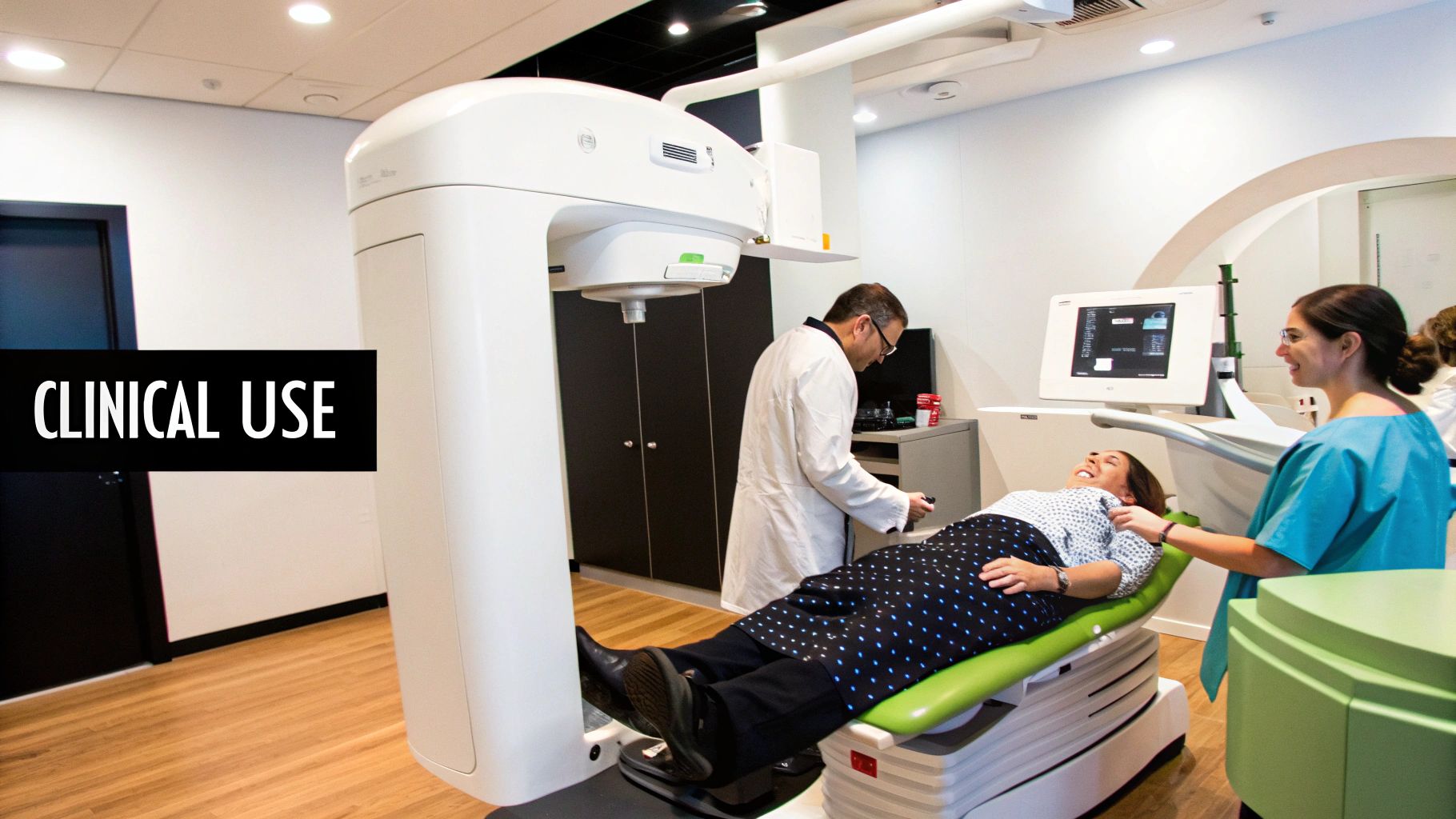
During The Scan: A Quick And Comfortable Procedure
The scan itself is brief, usually taking less than a minute. You'll be comfortably positioned in the CBCT machine, which typically features an open design. This helps avoid the enclosed feeling associated with some other medical scanners.
The technician will guide you into the correct position and ask you to remain still to capture the clearest possible images. The machine will rotate around your head, taking a series of X-ray images. You'll hear a gentle whirring sound during the rotation.
Patient Comfort Is Key
Patient comfort is a top priority throughout the entire scanning process. The open design of most CBCT machines helps reduce any potential claustrophobia. The scan itself is painless; you won't feel a thing. It's similar to a panoramic X-ray, but with 3D imaging. Feel free to address any questions or concerns to the technician. Their role is to ensure your comfort and ease during the procedure.
Addressing Common Questions
Many patients inquire about potential discomfort. Rest assured, the CBCT scan is entirely painless. The procedure is also very quick, typically lasting only 30 to 60 seconds.
While some individuals worry about claustrophobia, the open design of the CBCT scanner mitigates this concern. Most patients find the experience remarkably quick and easy.
After The Scan: Next Steps
After the scan, you can immediately resume your normal activities. No recovery time is necessary. Specialized software processes the images, which are then analyzed by a qualified radiologist or dental professional. The results are shared with your dentist, who will discuss them with you. Based on these results, your dentist will develop a personalized treatment plan if needed. This collaborative approach between dentist and patient promotes informed decisions and optimal treatment outcomes.
The Future of CBCT: Emerging Innovations
CBCT technology is constantly evolving, promising exciting advancements in dental diagnostics. What breakthroughs lie ahead? By examining current research and consulting with industry experts, we can gain insights into the future of CBCT and its potential to transform dental care.
Artificial Intelligence Integration
One of the most promising developments is the integration of artificial intelligence (AI). AI algorithms are being developed to automate the detection of pathologies, analyzing CBCT scans for potential problems like cavities, bone loss, and even tumors. This automation can significantly improve diagnostic accuracy by reducing the chance of human error.
Additionally, AI can streamline workflows, allowing dentists to dedicate more time to treatment planning and patient care. This shift in focus can enhance the overall patient experience.
Ultra-Low Radiation Protocols
Another key advancement focuses on ultra-low radiation protocols. While CBCT already uses considerably less radiation than traditional CT scans, researchers are continually working to minimize exposure even further.
New protocols and hardware are pushing the boundaries of low-dose imaging, improving the safety of CBCT for patients. This is particularly important for children and those requiring frequent scans. Initiatives like BeamReaders’ ECAA protocol exemplify this commitment to radiation reduction, especially for pediatric patients. Learn more about ECAA protocol.
Enhanced Software Algorithms
Beyond AI, improvements in software algorithms are allowing more diagnostic information to be gleaned from each scan. These advanced algorithms enhance image quality, highlighting subtle details that might be missed.
This increased clarity provides dentists with more comprehensive data, leading to more accurate diagnoses and more effective treatment plans. Ultimately, this enhanced precision benefits patients through better treatment outcomes.
Expanding Applications and Accessibility
These advancements are shaping the future of clinical practice. As CBCT becomes safer and more readily available, its applications could expand into routine dental care. This broader use could lead to earlier detection of dental issues, improving overall oral health outcomes.
Furthermore, innovations are enhancing the accessibility of CBCT technology in various practice settings. Smaller, more affordable units are becoming available, placing this powerful diagnostic tool within reach of a wider range of dental professionals.
This continuous evolution of CBCT technology demonstrates its significant impact on dental diagnostics and treatment planning. Both practitioners and patients benefit from the increasingly precise and comprehensive insights into oral health that CBCT provides.
Looking to integrate the power of AI into your medical imaging solutions? PYCAD offers comprehensive AI services for medical imaging, encompassing data handling, model training, and deployment. Learn how PYCAD can improve medical devices and enhance healthcare outcomes. Visit PYCAD for more information.


