Think of a DCM file not as just a picture, but as a complete digital folder for a medical scan. It’s the standard way hospitals around the world package a high-quality image, like an MRI or X-ray, along with all the critical patient information that goes with it. This single, secure file ensures a scan from one machine can be perfectly understood by any other, preventing the kind of data chaos that simply can't happen in healthcare.
The Two Halves of a DCM File
To really get what a DCM file is, you have to see it as two parts working together. First, it’s a container for the pixel data—the actual image itself. But this isn't your average JPEG or PNG. A DCM file often holds much more information, like multiple image "slices" from a CT scan that software can piece together into a 3D model.
The second part is just as important: the metadata. A DCM file is also a meticulous record-keeper, storing a huge amount of information right inside the file. This data, often called the "header," acts like the file's ID card, holding everything a doctor or radiologist needs to know.
More Than Just an Image
This marriage of image and data is what makes the DCM format so essential. Without that built-in metadata, a medical scan is just an anonymous picture—clinically useless and potentially dangerous. The DCM format solves this problem by permanently linking the what (the image) with the who, when, and how (the metadata).
To put it simply, every DCM file contains two fundamental parts. This structure is what makes it a comprehensive medical record, not just a picture.
Key Components of a DCM File at a Glance
| Component | Description | Example Information |
|---|---|---|
| Image Pixel Data | The high-fidelity visual information captured by the medical imaging device. | An X-ray image, a series of MRI slices, or ultrasound video frames. |
| Metadata Header | A rich set of text-based information that describes the patient, equipment, and study. | Patient name, study date, scanner model, and pixel dimensions. |
This two-part structure is the core of the entire system.
A DCM file is the reason a patient's CT scan from a machine in one hospital is perfectly readable and understandable by a radiologist's software in another, even if they use completely different equipment.
The format’s official name is DICOM, which stands for Digital Imaging and Communications in Medicine. It was born from a collaboration between the American College of Radiology (ACR) and the National Electrical Manufacturers Association (NEMA) back in 1983. Files using this standard have the .dcm extension and a very specific header structure that guarantees this universal compatibility. You can dive into the full history and technical details of the DICOM standard to see how it evolved.
Some of the key details stored in the header include:
- Patient Data: Name, ID, age, and sex.
- Study Information: Details about the procedure, like the date, time, and referring physician.
- Equipment Details: The manufacturer, model, and settings of the scanner.
- Image Parameters: Pixel dimensions, bit depth, and windowing information for ideal viewing.
Why the DICOM Standard Was Created
To really get why the DCM file is so important, you have to picture the chaotic world of medical imaging before it existed. In the early days, hospitals were wrestling with a huge, incredibly frustrating problem. Every imaging machine, whether it was from GE, Siemens, or Philips, spoke its own unique, proprietary digital language.
This created what we called "digital islands." An MRI scan from one machine was basically gibberish to a viewing station made by a competitor. Trying to share critical images between departments—or worse, between different hospitals—was a logistical nightmare. It meant dealing with custom software, messy data conversions, or even going back to printing physical films. This total lack of interoperability wasn't just inefficient and expensive; it put patient care at risk by causing delays.
The Push for a Universal Language
Seeing this crisis brewing, two major players decided to step in. The American College of Radiology (ACR) and the National Electrical Manufacturers Association (NEMA) formed a joint committee to tackle the problem head-on. Their mission was to build a universal standard—a common language that all medical imaging devices could finally speak.
The goal was ambitious, but the idea was simple: create one file format and one network protocol that worked for every machine, no matter who made it. This would finally allow information to flow seamlessly, from the scanner to the radiologist's desk and into a patient's permanent medical record.
The timeline below really captures the journey from a fractured, incompatible system to the connected one we have today.
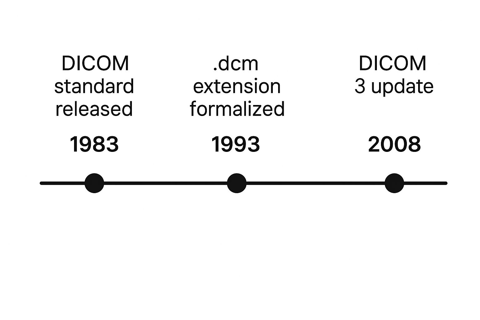
As you can see, the standard didn't just appear overnight. It evolved over a decade, adapting to new technology to keep up with the demands of modern medicine.
From Concept to Global Standard
The committee first got to work in 1983, laying the foundation for what would eventually become DICOM. Early versions proved the concept worked, and a big demonstration in 1990 showed it was truly possible, with six major companies successfully sharing images. But there was still a huge piece of the puzzle missing: a standard way to send these files over a network.
The real turning point came in 1993 with the release of DICOM 3.0. This version was a complete game-changer because it integrated network protocols like TCP/IP—the very same technology that powers the internet.
This was the crucial update that transformed DICOM from just a file format into a full-blown communication protocol. For the first time, you could not only store medical images in a universal format but also send them reliably across hospital networks and, eventually, the internet. You can dig into the complete history of DICOM's evolution to see all the technical steps.
This development finally tore down those digital islands, setting the stage for the connected healthcare systems we rely on today. The DCM file, backed by the powerful DICOM standard, became the key that unlocked a new era of collaboration in medical imaging.
A Look Inside a DCM File Structure
To really get a feel for what a DCM file is, we need to pop the hood and see what’s inside. It helps to think of a DCM file less like a simple picture file (like a JPEG) and more like a sophisticated digital folder—a self-contained package that holds everything related to a single medical scan.
This package is meticulously organized into individual components called Data Elements. Each one is a specific piece of information. Everything, from the patient’s birthdate to the X-ray tube current used during the scan, gets its own Data Element. This modular approach is what keeps the information clean, organized, and easy for computers to read.
So, how does a computer know what it's looking at? That’s where the real genius of the DICOM standard shines through.
Tags and Values: The DNA of a DCM File
Every single Data Element is built on a simple but powerful two-part structure: a Tag and a Value.
The Tag is essentially a unique label, a standardized code that tells you what kind of information you’re about to see. For instance, the tag (0010,0010) is the universal code for "Patient's Name." Because these tags are standardized, a medical imaging device in Germany can create a file that a specialist’s software in Canada can read perfectly, with no confusion.
The Value is the data itself—the content that corresponds to the tag. If the Tag is (0010,0010), the Value might be "Jane Smith." If the Tag is (0008,0020) (Study Date), the Value would be the specific date the scan took place.
This tag-value system is the bedrock of the entire DICOM format. It transforms a sea of disconnected information into a structured, searchable, and universally understood medical record.
This pairing of tags and values is what allows a radiologist's software to instantly pull up a patient's entire imaging history. The software can scan the tags to find all files for a specific person, sort them by date, and display them in chronological order for easy comparison.
This organized structure is absolutely critical in a clinical setting. It ensures the pixel data—the visual image itself—is never separated from the context that gives it meaning. To see this in action, let's look at a few of the most common tags you'll find in a DCM file.
Common DICOM Tags and Their Meanings
The table below breaks down a few essential DICOM tags. Think of it as a small sample of the hundreds of data points that can be stored, each giving crucial context to the medical image.
| DICOM Tag ID | Tag Name | Purpose in the File |
|---|---|---|
| (0010,0010) | Patient's Name | Stores the full name of the patient to ensure correct identification. |
| (0008,0020) | Study Date | Records the exact date the imaging procedure was performed. |
| (0008,0070) | Manufacturer | Identifies the company that built the imaging device (e.g., Siemens, GE). |
| (0028,0011) | Rows | Specifies the number of pixel rows, defining the image's vertical resolution. |
| (0028,0010) | Columns | Specifies the number of pixel columns, defining the image's horizontal resolution. |
By permanently bonding the image data with this rich, structured metadata, a DCM file guarantees that the right scan is always linked to the right patient, diagnosis, and medical history. This creates a complete and trustworthy record, which is exactly what modern healthcare demands.
How You Can Open and View DCM Files
So, you have a DCM file on your computer. If you've tried double-clicking it, you've probably already discovered that your standard photo viewer has no idea what to do with it. That's completely normal. A DCM file isn't just a picture; it's a rich container of data, and you need a specialized piece of software—a DICOM viewer—to unpack both the image and all the critical patient information tucked inside.
Fortunately, there’s a whole ecosystem of viewers out there, from simple tools for a quick peek to sophisticated platforms for medical research. For anyone thinking about a career in this field, getting hands-on with these tools is a fantastic start. Of course, professional work requires formal training, like an Access to Higher Education Diploma in Radiography, to truly master the science behind the images. Generally, DICOM viewers fit into three main camps, each designed for a different purpose.
Free and Lightweight Viewers
Let's say you're a patient who just got a CD with your latest MRI scan, a student learning about medical imaging, or anyone who just needs a quick, no-fuss way to look at a scan. A free, lightweight viewer is exactly what you need. These apps are built to do one thing well: open and display DCM files without any hassle. They’re usually a breeze to install and have straightforward, intuitive controls.
Great options to look into are RadiAnt DICOM Viewer and MicroDicom. They give you the essential tools—zooming, panning, and adjusting brightness and contrast—so you can inspect the image details without getting bogged down by the complex features of a full-blown clinical system.
The screenshot below of the RadiAnt DICOM Viewer shows just how user-friendly these tools can be.
You can see the interface is clean and uncluttered. The imaging tools are right there on the left, while the patient and study details are organized on the right, neatly presenting both the visual and the metadata.
Advanced Open-Source Platforms
When you move into the world of medical research, academics, or software development, you'll need something with a lot more horsepower. This is where advanced open-source platforms like 3D Slicer or the Mac-exclusive Horos come into play. These are far more than just viewers; they are powerful software suites packed with tools for deep image analysis, 3D modeling, and even scripting with languages like Python for custom research projects.
These platforms are the workhorses for tasks like:
- Image segmentation, where you might meticulously outline a tumor across hundreds of scan slices.
- Building stunning 3D models from a series of 2D images to visualize anatomy.
- Running computational analysis on huge datasets of images for a research study.
They definitely have a steeper learning curve, but for non-clinical and academic work, their power and flexibility are second to none.
Professional Clinical Systems (PACS)
Step inside any modern hospital or imaging clinic, and you won't find radiologists using standalone viewers. Here, DICOM files live within a Picture Archiving and Communication System (PACS). A PACS is the backbone of the entire medical imaging operation, handling everything from storage and retrieval to the management and distribution of scans.
Think of a PACS as the central command center for a radiology department. It's the library, the workbench, and the communication hub all in one. A radiologist uses it to pull up a patient's current scans alongside their entire imaging history, compare them instantly, and draft their diagnostic report.
These are enterprise-level systems built for security, reliability, and seamless integration with other hospital-wide information networks. They are the professional-grade solution for managing the immense volume of imaging data—often millions of DCM files—that a large healthcare facility generates.
The Journey of a DCM File in a Hospital
To really get a feel for why DCM files are so important, let's trace the path of one through a typical hospital workflow. Picture a patient, we’ll call her Sarah, coming in for a CT scan due to abdominal pain. The moment the technologist starts the scan, the DCM file's journey begins.
The massive CT scanner doesn't just capture hundreds of high-resolution image "slices" of Sarah's abdomen. It simultaneously packages those images with a treasure trove of critical metadata—her name, patient ID, the date and time, and specific scanner settings. All this information is bundled together into a single, brand-new DCM file, creating a complete and self-contained record of that one procedure.
From the Scanner to Central Storage
That freshly minted DCM file doesn't linger on the CT scanner's local drive. Instead, it’s immediately sent over the hospital’s secure internal network to a central hub. This digital fortress is known as a Picture Archiving and Communication System, or PACS.
You can think of the PACS as the digital heart of any modern radiology department. It’s a powerful server system built specifically to store, organize, and serve up millions of medical images from all kinds of machines—MRI, X-ray, ultrasound, you name it. It's essentially a high-security, specialized vault for a hospital's entire collection of patient imaging data.
The PACS is the single source of truth for a patient's imaging history. By keeping everything in one place, it prevents lost scans and ensures any authorized clinician can pull up the files they need, whether they're in the ER or a consultation room across the campus.
It’s this very system that lets a doctor compare a scan taken today with one from five years ago, side-by-side on the same screen, with just a few clicks.
Diagnosis and Continuity of Care
Now, somewhere else in the hospital, a radiologist gets an alert: Sarah's scan is ready for review. They log into their powerful PACS workstation and instantly retrieve her DCM file. The specialized viewing software doesn't just show the images; it reads the file's metadata, giving the radiologist crucial context. They can zoom in, adjust contrast, reconstruct the 2D slices into a 3D model, and pull up old scans for comparison—all possible because that rich data is baked right into the file.
This seamless access is the backbone of continuous patient care. After the radiologist makes their diagnosis, Sarah’s surgeon can view the very same DCM file to map out a potential surgery. Later, her oncologist might use it to track how well a treatment is working. The journey of this one file shows how the DICOM standard weaves an interconnected web of information, making patient care safer, faster, and more accurate across the entire hospital.
For anyone looking to dive deeper into these real-world applications, programs like the Access to Higher Education Diploma in Radiography offer the foundational knowledge behind these critical workflows.
How DCM Files Are Powering Medical AI
Artificial intelligence is making huge waves in medicine, and right at the center of it all is the unassuming DCM file. To build an AI that can make a real difference, you need more than just pixels—you need high-quality, structured data. A simple JPEG of a tumor is just a picture, but a DCM file is a complete patient story.
Let's use an analogy. An AI learning from JPEGs is like a medical student studying from flashcards. They might get good at recognizing a few conditions, but they're missing the critical context. An AI learning from DCM files, on the other hand, gets the whole textbook. It sees the image, but it also gets the patient's age, the specific scanner settings used, and even the radiologist's notes. It's this rich, contextual information that allows an AI to make genuinely smart connections.
Fueling a New Generation of Diagnostics
This powerful combination of image and metadata is exactly why DCM files are the perfect fuel for training advanced algorithms. Researchers can bring together massive, anonymized libraries of these files, sometimes numbering in the millions. By churning through this enormous dataset, an AI model can start to pick up on incredibly subtle patterns—the kind that are often invisible to the human eye.
This is already leading to some incredible real-world results in diagnostics. For instance:
- Early Disease Detection: AI models trained on DCM files can spot the earliest signs of diseases like cancer or Alzheimer's, often long before a human radiologist might notice them.
- Workload Reduction: Think of AI as a first-pass filter. Algorithms can automatically scan and flag exams with potential abnormalities, allowing busy radiologists to focus their attention where it's needed most.
- Increased Accuracy: By offering a consistent, data-backed second opinion, AI helps reduce the chances of diagnostic errors and improves overall precision.
The Importance of Standardized Data
The success of AI in medicine completely depends on the quality and consistency of the training data. Because the DICOM standard is a global benchmark, researchers can pool anonymized data from different hospitals, regions, and even countries. This creates far more robust and diverse datasets.
This consistency is key. It ensures an AI model trained on data from a hospital in one part of the world can perform just as reliably when analyzing data from somewhere completely different.
The standardized structure of a DCM file is what makes large-scale medical AI possible. It provides a universal language for both the image and its context, allowing algorithms to learn from a global well of medical knowledge.
The seamless integration of DCM files with artificial intelligence supports sophisticated AI-powered decision-making, ultimately leading to better diagnoses and personalized treatment plans. This synergy is redefining what's possible in patient care, making diagnostics faster, smarter, and more reliable than ever before.
A Few Common Questions About DCM Files
Diving into medical imaging, especially for the first time, naturally brings up a few questions. Let's clear up some of the most common points of confusion about DCM files and how they work.
Is DICOM the Same as a DCM File?
In practice, yes, they're two sides of the same coin. Think of DICOM (Digital Imaging and Communications in Medicine) as the "language" or the standard itself—it's the universal rulebook for how medical images and their related data should be structured, stored, and shared.
A DCM file (the one with the .dcm file extension) is the actual file you see on your computer. It's the physical container built following all the rules laid out by the DICOM standard. So, DICOM is the blueprint, and the DCM file is the house built from that blueprint.
Can I Convert a DCM File to a JPEG?
Technically, yes, you can. But it's almost always a bad idea, and here's why: you'll lose a massive amount of critical information.
When you convert a DCM file to a simpler format like JPEG or PNG, you're essentially just taking a screenshot. You keep the picture, but you throw away all the rich metadata baked into the original file—patient ID, the date of the scan, the exact machine settings, the radiologist's notes, and more.
For a quick presentation slide or a non-clinical illustration, a JPEG might do the job. But for any kind of diagnostic or analytical work, that conversion makes the image medically useless. Always, always keep the original DCM file as the source of truth.
Why Are DCM Files So Big?
If you've ever been surprised by the size of a DCM file, you're not alone. They are often much larger than a typical photo, and for good reason.
- Raw Image Quality: To ensure a radiologist can make an accurate diagnosis, the image data is either completely uncompressed or uses a lossless compression method. Every single pixel detail is preserved.
- Greater Bit Depth: Your phone takes 8-bit photos. Medical images often use 12-bit or 16-bit grayscale, which captures a vastly wider and more subtle range of tones. This is essential for spotting faint anomalies.
- Image Stacks: A single DCM file doesn't always contain just one picture. It can hold an entire series of images, like the hundreds of individual "slices" that make up a full CT or MRI scan.
At PYCAD, we don't just see large files; we see rich data waiting to be unlocked. We specialize in building powerful AI solutions by working directly with the complex information inside DCM files. From managing massive datasets to deploying accurate diagnostic models, we help innovators turn medical imaging into meaningful results. See how we can help your project at https://pycad.co.


