
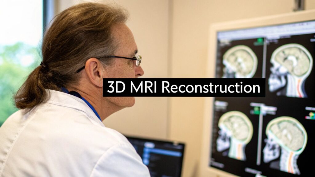
An MRI does not take a picture. It records radio signals from protons in a magnetic field and stores them in k-space. Reconstruction is the step that turns that frequency data into slices you can read, and then into a volume you can rotate.
This page is about that MRI-specific path: how k-space is filled, why 2D multi-slice and true 3D acquisition are not the same job, what Fourier / compressed sensing / AI actually do, and where the volume is useful (brain, joints, heart). For CT projection math and rendering, see 3D reconstruction from CT.
K-space is not an image
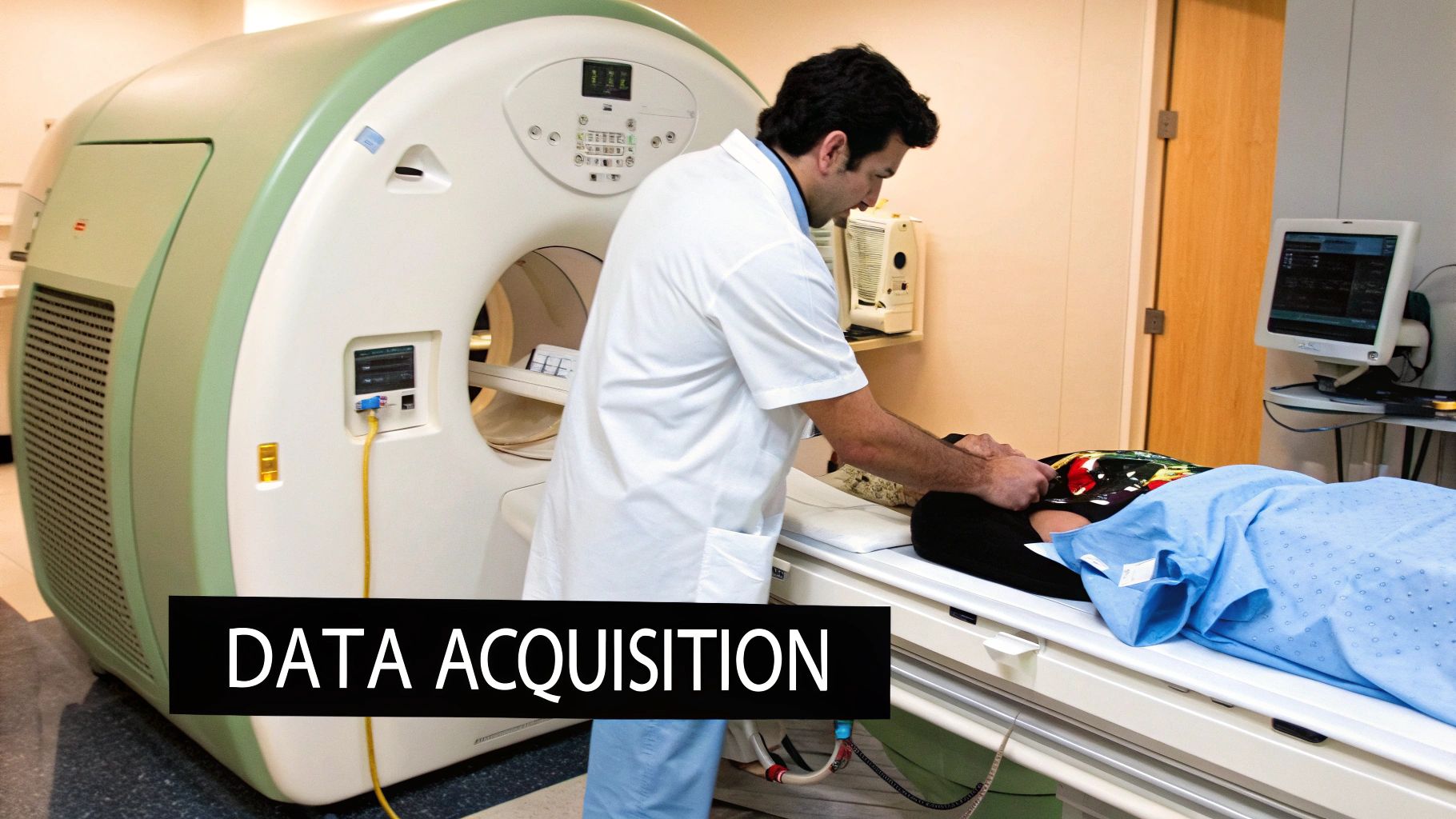
The scanner aligns hydrogen protons with a strong static field, knocks them with radiofrequency pulses, and listens as they relax. Coils record those faint signals. The raw matrix is k-space: frequency and phase, not pixels.
- The center of k-space holds low spatial frequencies (contrast, overall brightness).
- The edges hold high frequencies (edges, fine detail).
- How you walk through that grid (the trajectory) is the pulse sequence. It sets scan time, contrast, and what artifacts you will fight later.
A 3D model cannot be sharper than the k-space you collected. Undersample the edges and you lose detail. Undersample the center and contrast collapses. Reconstruction algorithms can recover a lot, but they cannot invent a signal that was never measured.
2D multi-slice vs 3D volumetric acquisition
Two different ways to fill a volume. Stacking 2D slices is not the same as exciting a slab.
| Characteristic | 2D multi-slice | 3D volumetric |
|---|---|---|
| Data collection | One slice at a time, then stack | Excites a whole slab; encodes the third axis in k-space |
| Voxels | Usually anisotropic (slice thicker than in-plane) | Can be isotropic cubes, which reformats cleanly |
| Gaps | Often small gaps between slices | No inter-slice gap |
| SNR | Lower for a given time | Higher: the whole volume is excited repeatedly |
| Time | Faster for large FOV surveys | Often better when you need high-res of a smaller region |
| Best for | Routine work, motion-prone patients, fast looks | Neuro, MSK, MRA, anything you will reformat or render in 3D |
Pulse sequence still matters. Spin echo is slower and more forgiving. Gradient echo is faster and the usual choice for true 3D, but more sensitive to susceptibility and metal. If the goal is a usable 3D model, start with a volumetric GRE (or a 3D spin-echo variant) and isotropic voxels. Stacking old 2D slices and interpolating the gaps gives stair-steps that fail for measurements and print.
Fourier, iterative reconstruction, compressed sensing
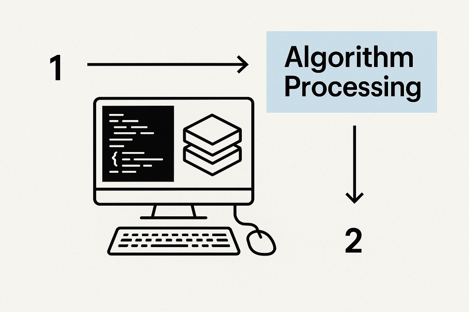
The default decoder is the Fourier transform (in practice an FFT). It maps k-space into the spatial domain. That is still the core of clinical MRI reconstruction.
Two extras show up when you cannot, or will not, collect a full k-space:
- Iterative reconstruction. Start with a guess, compare simulated k-space to what was measured, update, repeat. Each pass knocks down noise and some artifacts. Slower than a single FFT.
- Compressed sensing. Medical images are sparse in some transform (wavelets, total variation). If you undersample k-space in a way that does not alias into that sparse domain, you can recover a full image from far fewer lines. That is how many 3D exams drop from “too long to tolerate” to a few minutes.
Two failure modes the algorithms exist to fight:
- Motion. Breathing, a swallow, a twitch corrupt k-space while it is being filled. Correction either tracks motion during the scan or fixes inconsistencies after.
- Noise. Fast scans have less signal. Iterative and learning-based denoisers try to remove grain without smearing anatomy.
What AI actually changes
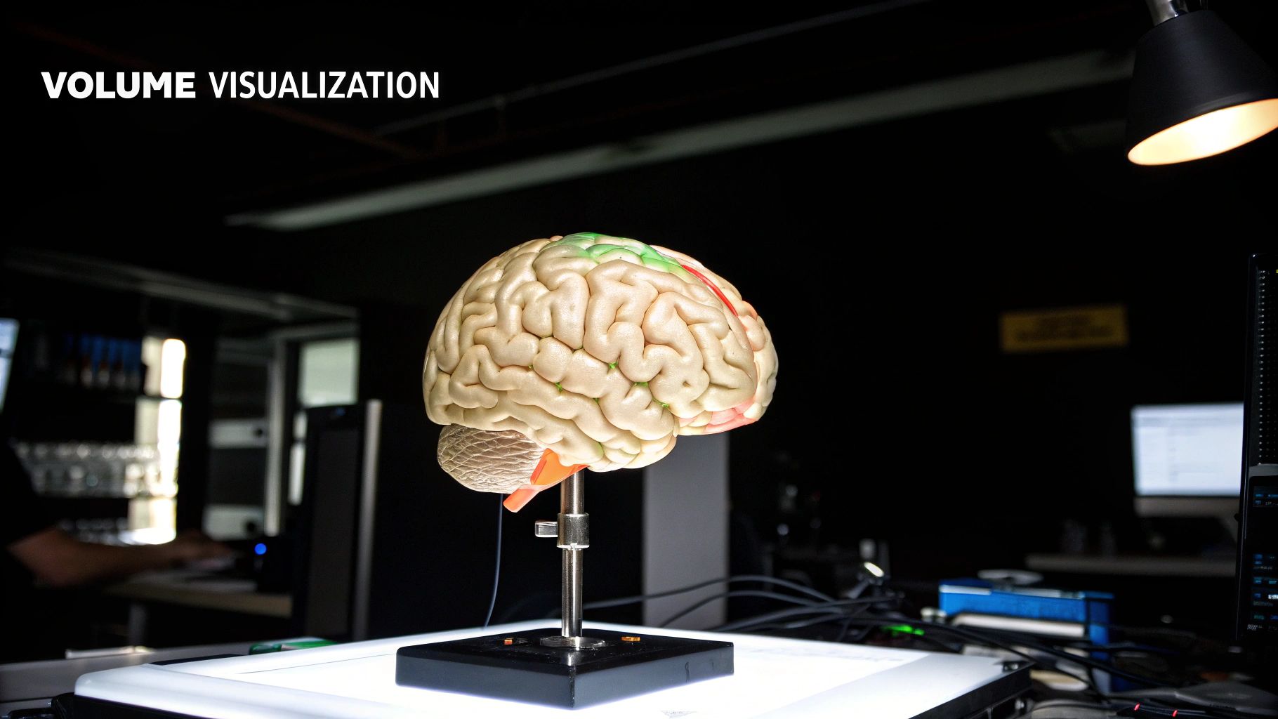
Deep networks (usually CNNs, now also transformers) are trained on paired full and undersampled k-space, or on noisy/clean images. Three jobs they take in a 3D MRI pipeline:
- Faster scans. Reconstruct a diagnostic volume from sparse k-space. Same idea as compressed sensing, learned from data instead of a hand-picked sparsity prior.
- Denoise / de-artifact. Separate anatomy from grain and some motion ghosts. Over-aggressive models invent texture; that is the clinical risk.
- Segmentation. Once you have a volume, someone still has to mark the tumor, ventricle, or cartilage. A trained model does that in minutes instead of hours, with less inter-observer drift. A person still has to check the contours before anyone cuts or prints.
Reconstruction itself does not add radiation. MRI uses magnets and RF, not X-rays. The reconstruction step is software, after the patient has left the magnet.
Where the 3D volume is used
- Neurosurgery. Tumor size, shape, and distance to vessels or eloquent cortex. The same volume can load into a neuronavigation system.
- Orthopedics. Ligaments, cartilage, and bone in a knee or shoulder are hard to hold in your head from a stack of 2D slices. A volume shows tear location and graft path before incision.
- Cardiac / congenital. Chambers, valves, and great vessels in kids with structural defects. Follow-up volumes are how you watch the repair, not only the anatomy on day one.
Cine / 4D MRI adds time (a beating heart, a breathing lung). Quantitative maps (T1, T2, fat fraction, perfusion) can be overlaid on the same anatomy. Those are extra channels, not a replacement for a clean isotropic acquisition.
If you need a mesh you can print or drop into CAD, that is a later conversion step (threshold or AI segment → surface → STL). The CT version of that workflow is CT scan to 3D model. Photo-based 2D-to-3D is a different problem: how to turn a 2D image into a 3D model.
PYCAD builds custom web DICOM viewers with MPR and volume rendering for teams that already have the scan and need it inside a real workflow. Case studies.