At its heart, a Picture Archiving and Communication System (PACS) is the engine that drives modern radiology. It’s a sophisticated medical imaging technology that securely stores and provides immediate access to images from all kinds of sources, like X-rays, CT scans, and MRIs.
Think of it as the ultimate digital library for a patient's entire imaging history, completely replacing the old, cumbersome world of physical film jackets and light boxes.
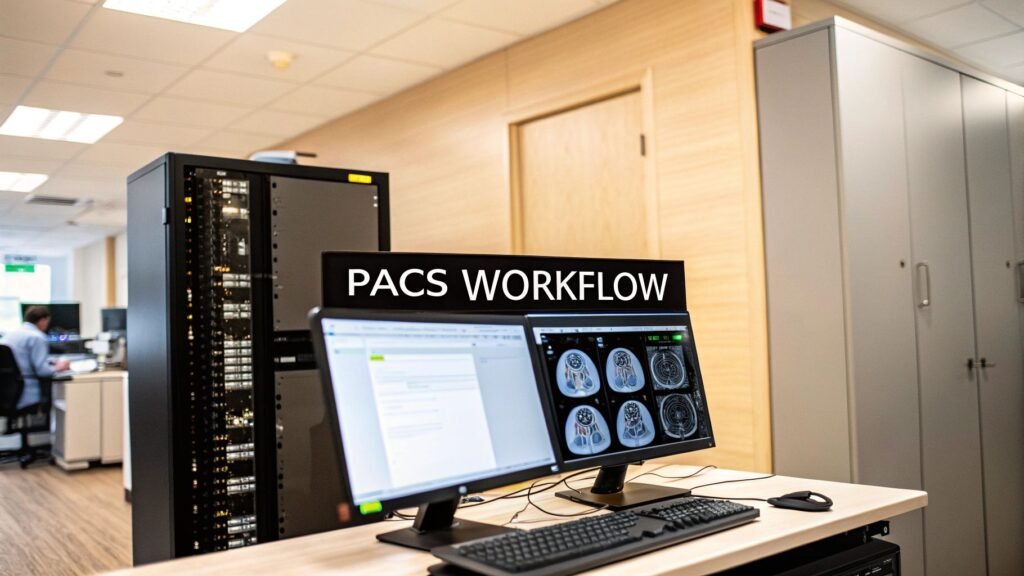
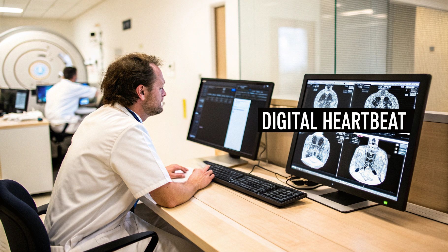
The Digital Heartbeat of Modern Radiology
Before PACS became the standard, radiology departments were literally drowning in film. Radiologists and physicians had to navigate a slow, frustratingly manual process of hunting down, transporting, and reviewing bulky X-ray films. It was a system riddled with delays, lost records, and staggering storage costs—all of which stood in the way of timely and effective patient care.
The introduction of PACS wasn't just an upgrade; it was a complete reinvention. It moved medical imaging from a physical, analog process into an entirely digital world. This system acts as a central hub, the very heartbeat that pumps critical imaging data throughout a healthcare organization.
From Film Jackets to Digital Files
Let’s put this into perspective. Imagine a patient comes into the ER needing an urgent CT scan. In the pre-PACS era, this involved developing the film, physically filing it, and then hand-carrying it to a specialist for review.
With PACS, those images are captured and sent directly to a secure digital archive in seconds. A radiologist—who might be in another building or even another city—can instantly pull up the images on a high-resolution monitor and make a diagnosis.
This isn't just about convenience. It's about better medicine. It leads to faster diagnoses, seamless collaboration between specialists, and a much quicker path to treatment. The change is as profound as swapping a library's dusty card catalog for a powerful search engine.
This leap forward from film to digital truly reshaped the entire field. The differences are night and day.
How PACS Transformed Radiology From Film to Digital
This table highlights the fundamental shift from traditional film-based radiology to a modern, efficient PACS-driven workflow, showcasing key differences and benefits.
| Aspect | Traditional Film-Based System | Modern PACS Environment |
|---|---|---|
| Storage | Bulky physical rooms filled with film jackets | Secure digital servers (on-premise or cloud) |
| Image Access | Manual retrieval, one person at a time | Instant, simultaneous access from anywhere |
| Distribution | Physical transport (hand-delivery or mail) | Electronic transfer over secure networks |
| Collaboration | Specialists must be in the same location | Remote collaboration between doctors is easy |
| Data Integrity | Risk of lost, damaged, or misfiled films | Centralized, backed-up, and secure data |
| Workflow Speed | Slow and labor-intensive | Fast, automated, and highly efficient |
The move to a PACS environment didn't just improve old processes; it created entirely new possibilities for patient care.
The Power of Integration and Customization
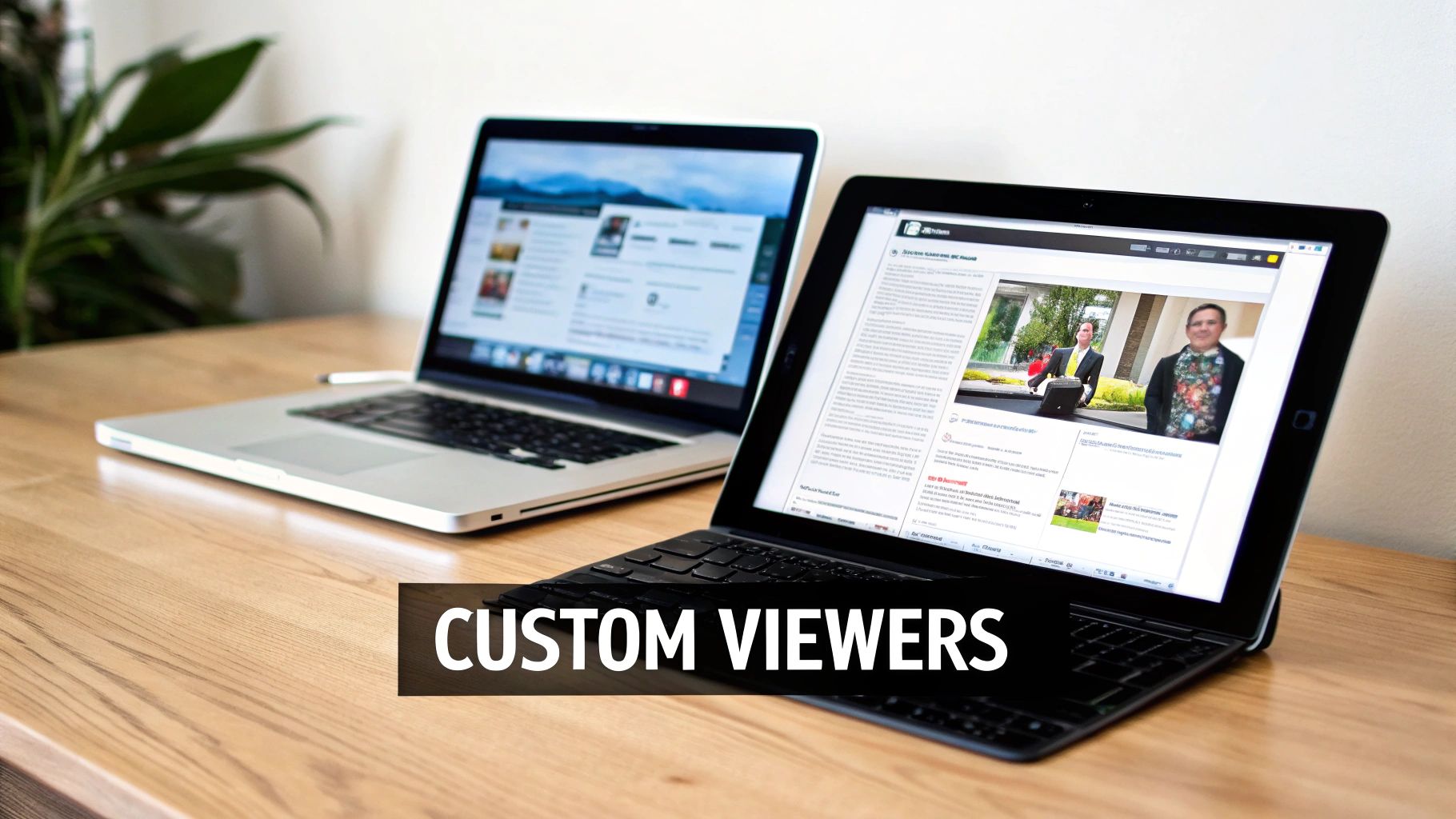
While standard PACS setups offer incredible value, the real magic happens when you create integrated and customized viewing experiences. We at PYCAD, know that a one-size-fits-all solution rarely works in the complex world of healthcare. That’s why we build custom web DICOM viewers and integrate them into medical imaging web platforms.
This approach gives healthcare providers viewing tools designed for their specific workflows, whether they’re conducting research, managing clinical trials, or providing direct patient care. By building tailored solutions, we put the exact tools medical professionals need right at their fingertips. You can see how these custom integrations are making a difference by exploring our work in our portfolio.
This shift to digital isn't just about making things more efficient today. It’s about building the foundation for the future, opening the door for innovations like AI and remote diagnostics to elevate patient outcomes even further.
Cracking Open the PACS Hood: What’s Inside?
To really get what a PACS is, you have to look under the hood. It’s not just one piece of software; it’s a living, breathing ecosystem built on four core pillars. When these parts work in harmony, they create a seamless flow of medical information that’s truly powerful.
Think of it like a high-performance pit crew at a racetrack. You have the crew chief, the tire changers, the fueler, and the driver. Each has a specific job, but it's their flawless coordination that wins the race. In the same way, the components of a PACS come together to deliver a clear, accurate, and lightning-fast diagnostic picture.
Image Acquisition: Where It All Begins
The journey starts with image acquisition modalities. These are the "eyes" of the medical world—the incredible machines that capture the images in the first place.
This is where you find the heavy-duty equipment, each built for a specific diagnostic mission. The most common ones you'll see are:
- Computed Tomography (CT) Scanners: These machines give us those amazing cross-sectional "slices" of the body.
- Magnetic Resonance Imaging (MRI) Machines: Using powerful magnets and radio waves, MRIs paint incredibly detailed pictures of our organs and soft tissues.
- X-ray Machines: The classic workhorse of radiology, still essential for looking at bones and certain tissues.
- Ultrasound Systems: These use sound waves to generate real-time moving images, like seeing a baby on a monitor.
The moment an image is captured, it’s instantly converted into a standardized digital file, ready to start its journey through the system.
The DICOM Network: The Medical Data Superhighway
Okay, so we have a digital image. Now what? We need to move it, and fast. That’s where the DICOM network steps in. If the scanners are the eyes, the network is the secure, high-speed highway connecting every part of the hospital.
This network runs on the Digital Imaging and Communications in Medicine (DICOM) protocol. You can think of DICOM as the universal language for medical imaging. It’s the reason a GE scanner can talk to a Siemens workstation without any hiccups. This standardization is everything; without it, we’d be back in the digital dark ages, trying to make incompatible systems talk to each other.
It wasn't always this simple. The idea for PACS emerged in the 1970s, but it took until 1993 for DICOM to be established. It was a massive collaboration between the American College of Radiology and the National Electrical Manufacturers Association, and it completely changed the game.
At its heart, a PACS needs a powerful and resilient network to handle the sheer volume of data. Having a robust network infrastructure isn't just a nice-to-have; it's absolutely critical for the system to work without a hitch.
The PACS Server: The Digital Fort Knox
Once an image zips across the network, it arrives at its home: the PACS server and archive. This is the system’s Fort Knox—a secure, centralized vault where every single medical image and all its associated data are stored.
This server manages both short-term storage for immediate access and long-term archiving for a patient's entire medical history. It has to be incredibly reliable, with layers of backups to prevent data loss. A lost scan isn't just a lost file; it's a gap in a patient's story that can have serious consequences. If you want to dive deeper into the protocols that make this possible, check out our guide on https://pycad.co/dicom-standards/.
Viewing Workstations: The Radiologist's Cockpit
The final piece of the puzzle, and the one most people see, is the viewing workstation. This is the radiologist's command center—a high-performance computer armed with specialized software and crystal-clear, medical-grade monitors. This is where the magic of diagnosis happens.
From this cockpit, a radiologist can do things that were once pure science fiction with old-school film. They can zoom in on the tiniest details, tweak the brightness and contrast to reveal subtle issues, build interactive 3D models from 2D scans, and pull up a patient’s prior images for instant comparison.
But radiologists aren't the only ones who need to see these images. Surgeons, oncologists, and other specialists need access too, often with very specific tools. That’s why at PYCAD, we don't just stop at the standard setup. We build custom web DICOM viewers and integrate them into medical imaging web platforms, giving every specialist the exact view and tools they need to do their best work. You can explore our work on our portfolio page.
How PACS Transforms the Radiology Workflow
To really get a feel for the magic of a PACS, let's walk through the life of a medical image. We'll see how it completely flips the old-school radiology workflow on its head. This isn't just a minor tech upgrade; it’s a radical rethinking of how we deliver patient care, trading slow, manual steps for a fluid, digital dance of information.
Think back to how things used to be. A patient gets a CT scan. What happens next? A technician hustles off to a darkroom to develop the film, slides it into a massive file jacket, and then someone physically carries it over to the radiology department. The radiologist is stuck waiting, then has to find an open lightbox to hang the films. The entire process was defined by one thing: waiting.
An Instantaneous Flow of Information
Now, let's step into a modern, PACS-driven clinic. The moment that CT scan is finished, the digital images are captured and beamed across a secure network to the central PACS archive. No film. No chemicals. No couriers. The journey from the scanner to the radiologist's screen takes mere seconds.
This immediate access is where everything changes. A radiologist—who could be just down the hall or working from home hundreds of miles away—gets a notification that a new study is ready to be read.
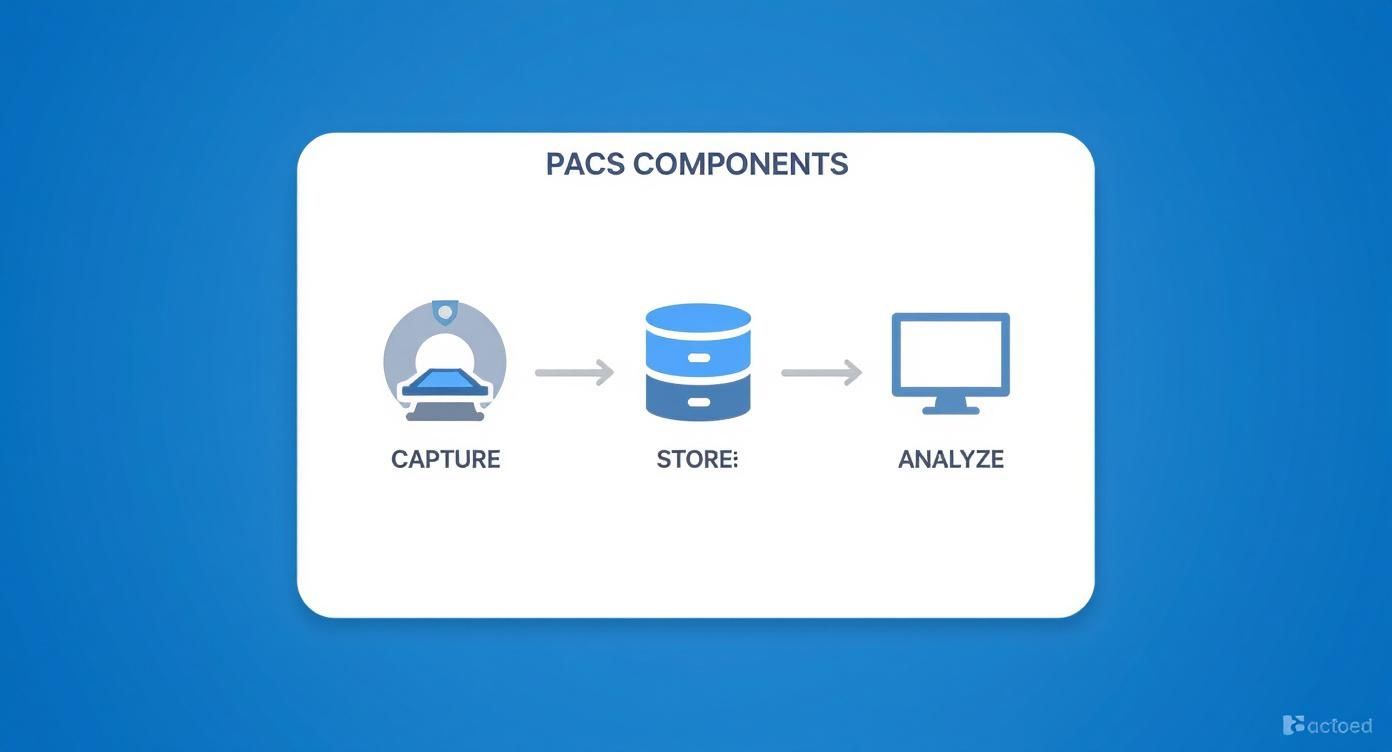
This simple but powerful three-step process is the heartbeat of every PACS workflow: capture, store, and analyze. It’s what breaks down the physical barriers and creates a seamless highway for critical patient data.
Unlocking Deeper Diagnostic Insight
With a single click, the radiologist pulls up the new CT scan. But they get so much more than just today’s images. The PACS automatically fetches the patient's entire imaging history and displays it right alongside the new study. This is where the real diagnostic artistry begins.
Being able to make side-by-side comparisons is a game-changer. A radiologist can instantly pull up an MRI from last year or an X-ray from five years ago to compare against the current scan. This historical view is absolutely essential for tracking how a disease is progressing, seeing if a treatment is working, or catching subtle new developments.
A landmark study in the American Journal of Roentgenology revealed that radiologists using PACS consulted far more historical images than their counterparts working with physical film. And it didn't slow them down. In fact, it armed them with the context needed to write richer, more accurate reports. You can dive into the full research on diagnostic accuracy to see the data for yourself.
This ability to see the whole story, not just a single snapshot, leads to stronger, more confident diagnoses.
Fostering Collaboration and Tailored Solutions
The workflow doesn't stop with one person. What if the scan shows something complex that needs a surgeon’s eye? The radiologist can instantly share the images with the entire surgical team. Multiple specialists can hop on, view the same study from different locations, and map out a treatment plan together in real-time. This level of collaboration was simply a dream in the age of film.
Of course, different doctors need different tools. The standard viewer a radiologist uses might not have the precise measurement or 3D modeling functions an oncologist or orthopedic surgeon relies on.
That’s where customization becomes so incredibly important. We at PYCAD, specialize in this. We build custom web DICOM viewers and integrate them into medical imaging web platforms, making sure every clinician has the exact toolkit they need to do their best work. By crafting these bespoke solutions, we give healthcare providers the power to interact with medical images in a way that makes sense for their role, which ultimately drives better patient outcomes.
To get a sense of what this looks like in practice, take a look through our portfolio.
The Strategic Gains of a Modern PACS
Bringing a PACS into your healthcare facility isn't just about going digital; it's a fundamental shift in how you operate. Think of it less as an IT upgrade and more as a complete operational overhaul that breathes new life into diagnostics and patient care. It’s the move that unlocks a new tier of efficiency, diagnostic precision, and collaboration.
By leaving the cumbersome world of physical film behind, a PACS injects some much-needed speed into the entire diagnostic workflow. This isn't a minor tweak—it's a game-changer that frees up your clinical and support staff to concentrate on what they do best: caring for patients.
Putting Efficiency and Productivity into Overdrive
Remember the days of hunting down a patient's old X-ray folder or waiting for films to be physically shuttled from one department to another? A PACS makes that world obsolete. It automates the entire process of finding and sharing images, which dramatically slashes report turnaround times.
Radiologists can suddenly interpret more studies in a day, and referring physicians get the insights they need in a fraction of the time. This acceleration isn't just a number on a spreadsheet; it means critical treatment decisions can be made faster, which can have a profound impact on a patient's journey.
Boosting Diagnostic Accuracy and Confidence
One of the most powerful, yet sometimes overlooked, advantages of a PACS is the instant access to a patient’s full imaging history. With just a few clicks, a radiologist can pull up a CT scan from today and compare it directly against an MRI from two years ago. This provides crucial context that was often a nightmare to assemble with physical files.
This historical view is absolutely essential for tracking how a disease is progressing, seeing if a treatment is working, or catching those subtle changes that might otherwise be missed. Plus, the digital tools on a modern viewing workstation—zoom, pan, contrast adjustments—allow for an incredibly detailed analysis that a simple lightbox could never offer. The result is a diagnosis delivered with far greater confidence and precision.
"The ability for a system to provide economical storage, rapid retrieval, and simultaneous access to medical images has driven its global adoption. This transition has led to significant cost savings, with some estimates showing that digital storage can cut film and related expenses by up to 70%."
The market reflects this undeniable value. The global PACS market was already valued at over $4 billion in 2020 and has only continued its upward climb. You can get a better sense of this trend from the full research on PACS adoption.
Making Seamless Collaboration a Reality
PACS simply demolishes the physical barriers that used to keep medical specialists apart. A radiologist in one city can now consult in real-time with a surgeon across town or an oncologist on another continent. They can all look at the exact same images, at the same time, discussing findings and mapping out a patient’s care plan together.
This ability to collaborate is indispensable for complex cases and fosters a truly unified approach to medicine. Of course, protecting patient data is non-negotiable in this environment. For a great primer on understanding HIPAA compliance in digital platforms, this resource is a must-read.
Unlocking Real, Long-Term Cost Savings
While there’s an upfront investment, a thoughtfully implemented PACS pays for itself many times over. The most immediate win is waving goodbye to film and all the associated costs—chemicals, processing equipment, and massive storage rooms.
But the savings run much deeper than that:
- Reduced Administrative Burden: Your team spends far less time managing, pulling, and filing physical records.
- Fewer Redundant Scans: Lost or damaged films often meant re-imaging a patient, a costly and unnecessary step that PACS helps eliminate.
- Better Staff Productivity: When clinicians can get more done in less time, you’re making the most of your most valuable resource—your people.
All these factors compound to lower your cost per study and strengthen the financial health of your entire imaging department. At PYCAD, we believe the best system is one that’s built for your specific goals. That’s why we build custom web DICOM viewers and integrate them into medical imaging web platforms. Our goal is to ensure the technology doesn't just work, but works for you. Take a look at our portfolio to see what we mean, or dive into our guide on a successful PACS integration.
7. Beyond Off-the-Shelf: Building Custom Solutions for Modern Imaging
While standard PACS workstations are undeniably powerful, the reality of modern healthcare is that a one-size-fits-all solution just doesn't cut it anymore. We've moved past simple image viewing. Today’s clinical world is a dynamic web of systems—radiology departments, electronic medical records (EMRs), telehealth platforms, and groundbreaking AI tools all need to speak the same language.
This is where the real innovation begins. Off-the-shelf software often hits a wall when faced with these complex, interconnected demands. That’s why specialized development is so crucial. It’s about pushing past the traditional boundaries of PACS to build something that fits a real-world workflow, not the other way around. The future isn't just about looking at scans; it's about weaving that visual data into the very fabric of every clinical decision.
More Than Just a Viewer
Think about a cardiologist reviewing an angiogram. Instead of a generic viewer, what if their tool was built directly into the patient's EMR, automatically flagging potential blockages and giving them exact measurements on the spot? Or picture a surgeon in the operating room, manipulating a patient's 3D CT scan on a tablet to plan their very next move.
These aren't daydreams. This is what's possible when we build solutions with a purpose. This approach opens up a world of possibilities:
- Workflows That Just Work: Creating viewers and tools that mirror the specific processes of a cardiology unit or an orthopedic surgery team.
- Intuitive by Design: Crafting interfaces that feel natural and efficient, cutting down on clicks and giving clinicians back their most valuable asset—time.
- Specialized Toolkits: Adding unique features that standard viewers simply don't have, like advanced 3D modeling, AI-powered analysis, or specific measurement tools for niche procedures.
We at PYCAD, live and breathe this. We build custom web DICOM viewers and integrate them seamlessly into medical imaging web platforms. By creating these tailored solutions, we give healthcare professionals the power to interact with medical images in a way that’s genuinely meaningful for their specific role. A great system doesn't just present data; it anticipates what the user needs next. You can always check our portfolio page to see our work in action.
The Power of a Connected Platform
Real breakthroughs happen when systems communicate effortlessly. A custom DICOM viewer that pulls patient history from an EMR and sends diagnostic insights to a telehealth portal creates a single, unified story for that patient. This demolishes the data silos that have held healthcare back for decades, giving every provider a complete, holistic view.
This deep integration is also the backbone of flexible and scalable medical data storage solutions that can adapt as an organization grows.
By building viewers that work on any device—from the radiologist's high-resolution monitor to a surgeon's tablet in the OR—we make critical imaging data accessible wherever and whenever it's needed most. This is the key to delivering care that is timely, collaborative, and truly centered on the patient.
The ultimate goal is to make the technology fade into the background, so clinicians can focus on what truly matters: their patients. To see how these custom-built solutions are already shaping the future of diagnostics, we invite you to explore our portfolio and discover what’s possible.
The Future of PACS with AI and Cloud Technology
The world of medical imaging is on the cusp of a major shift, one driven by two powerful forces: artificial intelligence and the cloud. We're not just talking about incremental upgrades here. This is about completely rethinking what a PACS can be—transforming it from a digital filing cabinet into an active, intelligent partner in patient care.
We're moving toward a smarter, more connected future for radiology. The goal is a system that can anticipate a clinician's needs, erase geographical boundaries, and deliver powerful insights right to their fingertips, wherever they happen to be.
AI as a Diagnostic Co-Pilot
For years, PACS was all about storage and retrieval. Now, Artificial Intelligence (AI) is giving it a brain. Think of AI as a co-pilot for the radiologist, one that helps manage the ever-growing workload and sharpens diagnostic focus.
Imagine smart algorithms working quietly in the background, analyzing every single image that flows into the system. Here’s what that looks like in practice:
- Flagging Potential Issues: AI can spot subtle anomalies that are easy to miss on a first look, guiding the radiologist’s eyes to areas that need a closer inspection.
- Automating Routine Tasks: Mundane but critical jobs, like measuring organs or calculating tumor volume, can be handed off to AI. This frees up the radiologist's valuable time for what truly matters—complex interpretation and clinical judgment.
- Prioritizing Urgent Cases: By pre-screening studies for critical findings, AI can automatically bump the most urgent cases to the top of the queue, ensuring life-threatening conditions get the immediate attention they demand.
The Cloud Revolution in Medical Imaging
At the same time, the migration to cloud-based PACS is completely changing the game for image management and access. Instead of being locked away on servers in a hospital basement, medical images are moving to the cloud, unlocking a new level of flexibility and security.
This move frees healthcare organizations from the endless, costly cycle of buying and maintaining on-site hardware. It enables secure, on-demand image access from literally anywhere with an internet connection, making seamless teleradiology and collaboration a reality for everyone.
A cloud-native system also means better disaster recovery and the ability to scale up or down as needed, without massive upfront investments.
We at PYCAD, see this as an incredible opportunity. We build custom web DICOM viewers designed for the cloud, and we integrate them into medical imaging web platforms that are both powerful and intuitive. Our goal is to make sure those advanced, AI-driven insights are available to clinicians on any device, anywhere.
The future of what is PACS in radiology is predictive, universally accessible, and deeply intelligent. To see how these modern, integrated solutions are already making a difference, we invite you to explore our portfolio.
Your Top Questions About PACS, Answered
Diving into medical imaging technology can feel like learning a new language, filled with acronyms and complex systems. As PACS becomes more and more vital to modern healthcare, it's natural to have questions. Whether you're a radiologist, an IT pro, or a hospital administrator, getting clear answers is key.
Let's break down some of the most common questions about PACS in radiology and clear up the confusion.
What’s the Real Difference Between PACS and DICOM?
This is easily the most common point of confusion, but the distinction is actually quite simple. The best way to think about it is with an analogy: DICOM is the language, while PACS is the library and the librarian combined.
DICOM (Digital Imaging and Communications in Medicine) is the universal standard—the common tongue that all imaging equipment speaks. It’s what allows a CT scan from a Siemens machine to be read perfectly on a GE workstation. Without DICOM, we'd have a digital Tower of Babel.
A PACS (Picture Archiving and Communication System), however, is the whole ecosystem. It’s the sophisticated system that receives, stores, organizes, and displays those DICOM files. It’s the library where all these medical images are meticulously archived and made available for review.
Is PACS Actually Secure Enough for Patient Data?
Absolutely. Security isn't just a feature of PACS; it’s the foundation it's built on. These systems are designed from the ground up to meet and exceed stringent privacy regulations like HIPAA.
A PACS doesn't just have one lock on the door. It has multiple layers of defense: strong user authentication, granular access controls (so people only see what they’re supposed to see), powerful data encryption, and detailed audit trails that record every single interaction with patient data. This ensures that sensitive information stays private and protected.
Can Doctors Really Access PACS from Outside the Hospital?
Yes, and this is one of the most powerful capabilities of any modern PACS. Radiologists and referring physicians can securely access images and reports from anywhere with an internet connection—whether it's their home office, another clinic, or even on the go.
This remote access is what makes teleradiology possible and dramatically speeds up diagnoses, especially in emergency situations. This is a core focus for us at PYCAD. We build custom web DICOM viewers and integrate them into medical imaging web platforms, giving clinicians the freedom to work securely from any device.
What Exactly Is a Vendor Neutral Archive (VNA)?
Think of a Vendor Neutral Archive, or VNA, as a universal storage vault for medical images. It's a smart evolution in how we handle long-term archiving. A VNA stores images in a standardized format, completely separate from the PACS viewing software.
Why does this matter? It breaks the chains of "vendor lock-in." It means a hospital can switch or upgrade its PACS viewer in the future without the nightmare of a massive, complicated data migration. The images remain in the VNA, accessible to whatever new system comes along, giving healthcare organizations true freedom and flexibility.
We at PYCAD, see a future where medical imaging is more connected, intelligent, and accessible than ever before. We're dedicated to building the platforms that make this vision a reality.
Want to see how our custom solutions are helping healthcare providers innovate? We invite you to explore our portfolio.