
DICOM, which stands for Digital Imaging and Communications in Medicine, is the global standard for medical imaging. It's the framework that governs how medical images are stored, shared, and transmitted, creating a common language for everything from MRI machines to CT scanners.
The Universal Language of Medical Images
Picture a time before a shared standard existed. Every medical scanner spoke its own unique, proprietary language. An MRI from one company couldn't be viewed on software from another, creating silos of vital patient information. This digital Tower of Babel was the chaotic reality of early medical imaging, making collaboration a nightmare and hindering patient care.
DICOM was born to fix this. Think of it as the universal translator for the world of medical imaging. Just like JPEG became the go-to format for digital photos, DICOM became the essential foundation for every device that produces an image in healthcare, from X-rays and ultrasounds to complex nuclear medicine scans.
More Than Just a Picture
A DICOM file isn't just a picture; it's a comprehensive, self-contained data package. It intelligently bundles the visual information with a rich header full of metadata, ensuring that every scan is forever tied to its critical context.
This makes every DICOM file an incredibly detailed record containing:
- Pixel Data: The raw, high-resolution image itself, preserving every subtle detail needed for an accurate diagnosis.
- Patient Information: Securely embedded details like the patient's name, ID number, and date of birth.
- Study Details: Essential context about the exam, including the date, time, and the specific equipment used.
- Equipment Data: Technical specifications of the scanner that generated the image.
This powerful combination of image and data makes DICOM indispensable. By standardizing the entire package, it guarantees that a CT scan taken in one country can be flawlessly opened, read, and interpreted by a specialist on the other side of the world. You can explore the nitty-gritty of these rules by diving into the official DICOM standards.
To make this even clearer, here’s a quick breakdown of what makes DICOM tick.
DICOM at a Glance
| Component | Function | Simple Analogy |
|---|---|---|
| File Format (.dcm) | Stores the image and its metadata in a single file. | A digital manila folder holding a patient's photo and their complete medical chart. |
| Network Protocol | Defines how to send and receive files over a network (like the internet). | The postal service for medical images, ensuring secure and reliable delivery. |
| Data Structure | Organizes the metadata using standardized "tags" (e.g., Patient Name, Study Date). | The organized fields on a form, where every piece of information has a specific place. |
| Conformance | Ensures different devices and software can communicate without errors. | A shared grammar book, making sure everyone is speaking the same language correctly. |
This elegant structure is what allows for the seamless flow of information that modern healthcare relies on.
How Collaboration Forged a Healthcare Revolution
Imagine a time, not so long ago, when medical imaging was the digital Wild West. Every manufacturer had its own rules. An MRI from one company couldn’t talk to a CT scanner from another, and specialized viewing software was shackled to specific hardware. This created digital islands, making it impossible to share vital patient information efficiently and safely.
This chaos was a roadblock to progress. Visionaries across the medical field knew that if digital imaging was ever going to reach its full potential, everyone needed to start speaking the same language. The challenge was massive: get fierce competitors and leading medical organizations to put aside their differences and work toward a common good.
The Alliance That Changed Everything
The big breakthrough happened in the 1980s. Two powerful groups decided it was time to build a bridge across the digital divide: the American College of Radiology (ACR) and the National Electrical Manufacturers Association (NEMA). They formed a joint committee with a goal that was as simple as it was ambitious: create one universal standard for medical imaging.
This wasn't just about writing code or defining file types. It was a mission to build a future where a patient's medical history could travel with them, securely and instantly, no matter what machine created the images. Here at PYCAD, we carry that spirit of innovation forward. We build custom web DICOM viewers and integrate them into medical imaging web platforms to create powerful, connected solutions.
Before DICOM, interoperability was a dream. The standard’s creation was a monumental step, uniting a fragmented industry under a single, powerful vision for connected healthcare.
A New Standard Is Born
The path wasn't a straight line. An early version, the ACR-NEMA standard, came out in 1985, but it had its limits. It was designed for simple, point-to-point connections, which wasn't enough for the increasingly connected world of healthcare.
The real game-changer arrived in 1993 with DICOM 3.0—the version that became the global standard we rely on today. This release was built for a networked world, using the same TCP/IP protocols that power the internet to let medical devices communicate seamlessly. For a deeper dive, you can explore the rich history of this incredible evolution.
This milestone laid the foundation for the Picture Archiving and Communication Systems (PACS) that are the heart of every modern hospital. It proved that when people are driven by a shared purpose, they can overcome any technical hurdle and reshape an entire industry for the better. This spirit of building connected solutions is what drives our work, and you can see the results in our client successes on the PYCAD portfolio page.
Cracking Open a DICOM File: What’s Inside?
To really get what makes DICOM special, you have to look under the hood. A DICOM file isn't just another image format like a JPEG or PNG. It’s a sophisticated, self-contained package of information, meticulously designed for the high-stakes world of medicine.
Imagine it's less like a simple photograph and more like a detailed patient file and an X-ray fused into one. This single file holds both the picture and the story behind it.
This "smart file" is built from two main parts: a comprehensive metadata header and the raw pixel data that forms the image itself. The header is the magic—it contains hundreds of standardized data fields, called tags, that describe everything imaginable. We're talking patient ID, the date of the scan, the type of machine used, and even the precise settings on that machine. This structure ensures that a medical image never becomes an anonymous picture; its vital context is locked in, traveling with it wherever it goes.
The Building Blocks of DICOM Communication
So, how does DICOM manage all this complexity and ensure a CT scanner in Germany can talk to a viewing station in Brazil? It all comes down to a few core concepts that create a universal language for medical imaging.
Here are the key components that make it all work:
- Information Object Definitions (IODs): Think of these as blueprints. An IOD spells out exactly what information must be included for a specific type of image. For example, the IOD for an MR scan dictates a different set of required tags than one for a dental X-ray, guaranteeing that no critical data is ever missing.
- SOP Classes (Service-Object Pair): If IODs are the blueprints (the "what"), SOP Classes are the action plans (the "how"). A SOP Class links an IOD (like a "CT Image") with a specific task (like "Store" or "Query"). The "CT Storage SOP Class," for instance, defines the exact rules for successfully sending a CT image from a scanner to a PACS server.
The genius of a DICOM file is in its all-in-one structure. By embedding a rich, detailed story directly within the image file, it creates a single, portable source of truth that is unbelievably reliable for patient care.
These concepts form the logical foundation that has made DICOM the powerhouse it is today. While it might sound technical, learning how to read DICOM files pulls back the curtain and shows you just how elegant and organized the system truly is.
This standardized approach is precisely what allows different devices, from different manufacturers, to communicate without a hitch. It's also what empowers us at PYCAD to develop custom web-based DICOM viewers. We specialize in building solutions that can dive into this intricate data, parse it in the blink of an eye, and display it on an intuitive web platform. Check out our portfolio page to see some of these powerful viewers in action.
DICOM in Action: From Scanner to Specialist
This is where the standardized world of DICOM truly comes alive, transforming a simple patient scan into a journey of critical data. Picture this: a patient is undergoing a CT scan. The moment the scanner captures an image, the real magic begins. That picture is instantly converted into a powerful piece of medical intelligence.
From that first second, the image is wrapped in a rich layer of DICOM metadata. The scanner doesn’t just spit out pixels; it creates a complete digital file. It tags the image with the patient’s ID, the study date, and the specific technical parameters of the scan itself. This complete package is then sent to the hospital’s central digital library—a Picture Archiving and Communication System, or PACS.
This process highlights how a DICOM file is built by merging raw image data with its essential metadata.
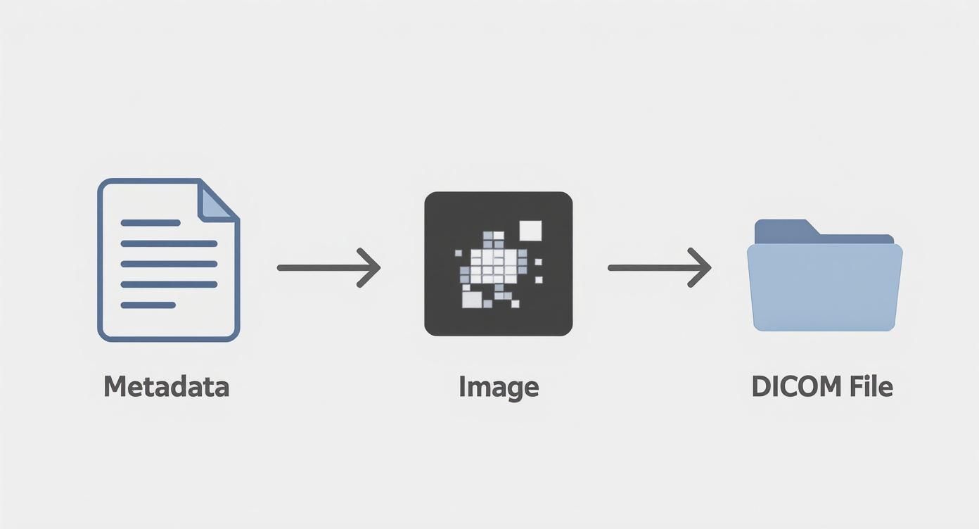
The takeaway here is simple but profound: an image alone is just a picture. But an image paired with its metadata becomes a reliable, trustworthy medical record.
Powering a Smarter Workflow
One of the most powerful DICOM services in this entire process is the Modality Worklist. Think about the old way: a technician would have to manually type a patient's name and other details into the CT scanner. It was a system just begging for human error.
The worklist service changes all that. It automatically pulls the scheduled patient information directly from the hospital's information system. This simple yet brilliant feature dramatically reduces the risk of misidentification—a critical leap forward for patient safety in any clinic.
This isn’t just a recent development; it’s part of a long journey of innovation. DICOM gained network support way back in 1993, and by 1995, it was already being used for ultrasound and nuclear medicine. The Modality Worklist arrived in 1996, and by 2003, the standard was handling complex multi-frame formats for 3D MRI and CT.
Today, DICOM is a giant, supporting over 100 imaging modalities and more than 1,500 information objects, covering everything from radiology to digital pathology. It’s an incredible story of growth. If you're curious about how this all came to be, you can dive deeper into the DICOM communication protocol.
Enabling Care Without Boundaries
Once that CT scan is securely stored on the PACS, DICOM’s true collaborative power is unleashed. A radiologist, potentially hundreds of miles away, can instantly query the PACS, retrieve the scan, and view it on their specialized workstation.
This is the heart of teleradiology. It means a patient in a small, rural clinic can get a life-saving diagnosis from a world-class specialist in a major city, all without leaving their community.
While DICOM excels at managing these complex images, the broader challenge of organizing comprehensive medical records for all aspects of patient care remains a crucial piece of the puzzle. DICOM provides the solid foundation, ensuring every person involved—from the technologist running the scanner to the remote specialist—has access to the exact same high-fidelity information.
Making Medical Imaging Accessible on the Web

For decades, viewing a DICOM file meant being chained to a specific, high-powered workstation inside a hospital. This created a frustrating bottleneck. Specialists couldn't easily consult from afar, collaboration was clunky, and getting imaging data into modern, cloud-based health records was a constant struggle.
So, how do we break these powerful images out of their digital silos and bring them into the flexible, interconnected world of the web?
The answer is the web DICOM viewer. Think of it not just as a tool, but as a translator—a gateway that converts the complex language of DICOM into something any standard web browser can display. It liberates medical imaging from the confines of the radiology department, making it securely accessible from anywhere with an internet connection.
This single shift is igniting a new era in healthcare, one where critical data flows freely and securely to the people who need it most, right when they need it.
The Power of Web-Based Viewers
A web DICOM viewer does so much more than just put a picture on a screen; it completely reimagines the diagnostic workflow. By moving these crucial images into a browser, we unlock incredible possibilities for collaboration, research, and patient care.
These platforms are becoming absolutely essential for:
- Remote Diagnostics: A specialist can now review an urgent scan from their home office, delivering a life-saving diagnosis without having to be physically on-site.
- Collaborative Annotations: Imagine multiple doctors viewing the same image at the same time, adding notes and measurements that everyone on the care team can see instantly.
- Seamless EHR Integration: Viewers can be embedded directly into Electronic Health Record (EHR) systems, putting a patient’s full imaging history right where it belongs—alongside their other medical data.
The real magic of a web DICOM viewer is its ability to democratize access. It transforms a static, isolated file into a dynamic, collaborative asset for modern medicine.
Of course, none of this works without trust. To make this level of accessibility a reality, it's absolutely crucial to operate within secure cloud environments for storing and sharing DICOM files. This security is the bedrock that reliable remote access is built upon, protecting sensitive patient data at every turn.
Building the Future of Medical Imaging
At PYCAD, we are right in the thick of this movement. We don't just use these tools; we build them. Our team creates custom web DICOM viewers from the ground up and integrates them into powerful medical imaging web platforms.
Our passion is creating solutions that don’t just solve today’s problems but also anticipate tomorrow’s challenges. We're driven by the goal of empowering healthcare providers, researchers, and AI innovators with tools built for a modern, interconnected world.
We believe in building platforms that push the boundaries of what’s possible in digital health. Our work is all about creating fluid, intuitive, and powerful experiences that elevate patient care and accelerate medical discovery. To see our vision in action, we invite you to explore our portfolio page.
Answering Your Lingering DICOM Questions
As we’ve journeyed through the world of DICOM, from its early days to its role in modern medicine, a few common questions tend to pop up. Let's tackle them head-on to clear up any lingering confusion and really cement your understanding.
Is DICOM a File Format or a Protocol?
This is a classic question, and the answer is one of the most elegant things about the standard: it's both.
Think of it this way: DICOM defines a specific file format (you'll often see them with a .dcm extension) that brilliantly packages a medical image with all its vital metadata. But it doesn't stop there.
It also lays out the rules of the road—a network communication protocol—that dictates how these files move between different systems. It’s how an MRI machine talks to a central server, ensuring the data is understood at both ends. This two-in-one design is what makes DICOM so robust; the data is standardized whether it's sitting still or on the move.
What’s the Difference Between DICOM and PACS?
This one is best explained with an analogy. Imagine DICOM is the universal language spoken by all books in a library, while PACS is the library itself.
- DICOM is the standard that governs how each "book" (the medical image and its data) is written, structured, and understood.
- PACS (Picture Archiving and Communication System) is the entire infrastructure—the building, the shelves, the librarians, and the delivery system—that stores, organizes, and shares those books.
So, PACS is the system that manages all the imaging files, and DICOM is the specific type of file it was built to manage. One can't really function without the other in a modern hospital.
Why Can’t I Just Open a DICOM File Like a JPEG?
You can open a JPEG in almost any program because it’s just a picture. A DICOM file, on the other hand, is a sophisticated, self-contained medical record packed with Protected Health Information (PHI).
Its structure is far more complex. The file includes a huge header brimming with patient details, study parameters, and even the settings of the machine that captured the image. This data is woven directly into the pixel data itself. Your standard photo viewer has no idea how to read all of that critical information, which is why you need a specialized DICOM viewer.
This is exactly the problem we solve here at PYCAD. We build custom web DICOM viewers and integrate them into medical imaging web platforms, transforming this complex data into something intuitive and accessible for clinicians. You can see examples of our work on our portfolio page.
DICOM's structure is its greatest strength, ensuring that a medical image is never just a picture but always a complete, actionable piece of patient data. This design choice is fundamental to patient safety and diagnostic accuracy.
Is DICOM Only for Radiology Images?
It certainly started there, but not anymore! DICOM's roots are deeply planted in radiology—think CT scans, MRIs, and X-rays. Over the years, however, it has blossomed.
Today, the standard has been adapted to handle images from nearly every corner of medicine. We're talking about cardiology, digital pathology, ophthalmology, dentistry, and even dermatology. This incredible flexibility has cemented DICOM's status as the true backbone of our interconnected healthcare world.
At PYCAD, our passion is building the next generation of medical imaging tools. We specialize in creating secure, web-based platforms with advanced DICOM viewers and integrated solutions that empower healthcare professionals across the globe. To see how we're making medical data more accessible and actionable, take a look at our portfolio of work.


