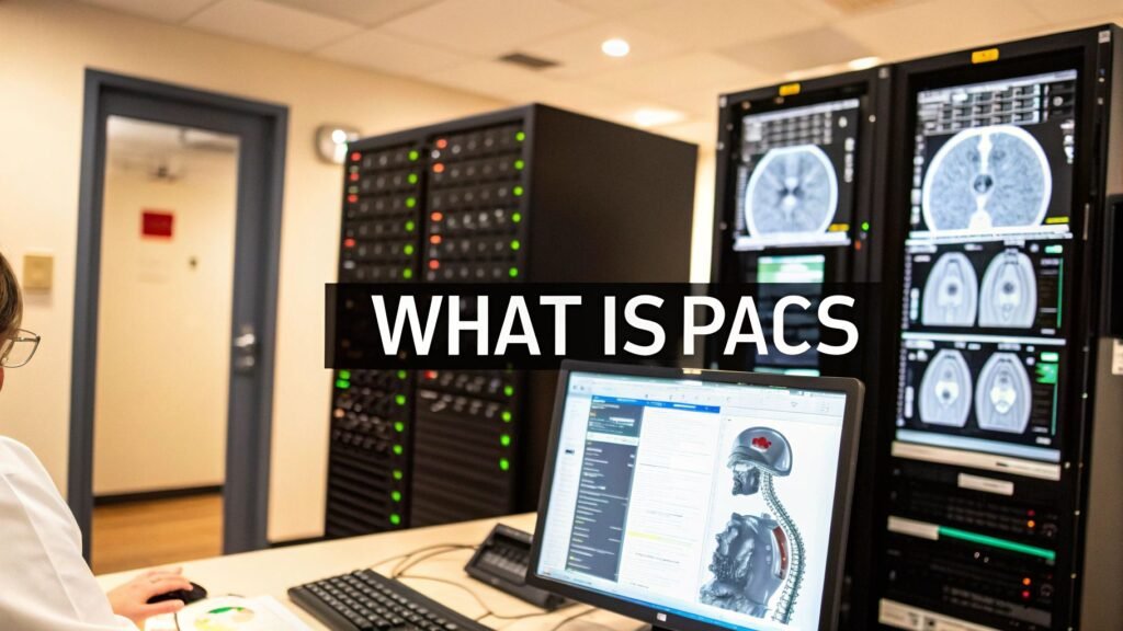
In a world of instant information, healthcare once lagged far behind, shackled by bulky film jackets and endless rows of physical archives. Put simply, a Picture Archiving and Communication System (PACS) is the secure digital library for all medical images. It’s the central nervous system for modern diagnostics, making patient scans instantly available to any authorized doctor, anywhere in the world.
The Digital Heartbeat of Modern Medicine
Let's rewind to a hospital’s radiology department before PACS. It was a world of darkrooms, smelly chemical processors, and vast libraries filled with heavy, cumbersome film jackets. Finding a patient's previous X-ray could take hours of searching. Sharing that scan with a specialist across town? That meant physically shipping the films.
This old way of doing things was painfully slow, expensive, and riddled with potential for human error. Films could be lost, damaged, or misfiled—small mistakes that could have a huge impact on patient care.
PACS didn't just improve this system; it completely dismantled it. Think of it as upgrading from an old-school library with a card catalog to a powerful, cloud-based streaming service. Instead of hunting through shelves, clinicians can now search a digital database and pull up a patient's entire imaging history in seconds. This move from physical to digital laid the foundation for the connected, efficient hospitals we see today.
Before we dive deeper, let's look at a quick comparison to truly grasp the shift.
Imaging Workflows Before and After PACS
| Feature | Traditional Film-Based System | Modern PACS System |
|---|---|---|
| Storage | Bulky physical film archives, prone to loss and damage. | Secure digital servers or cloud storage. |
| Image Access | Manual retrieval from archives; one person at a time. | Instant access on any authorized computer. |
| Sharing | Films physically copied and mailed to other specialists. | Images shared electronically in seconds. |
| Workflow | Slow, linear process: acquire, process, view, report, file. | Parallel, collaborative workflow. |
| Cost | High costs for film, chemicals, storage space, and couriers. | Reduced costs for supplies and physical storage. |
This table really just scratches the surface of the change. It wasn't just a minor upgrade; it was a fundamental reinvention of the diagnostic workflow.
A Revolution in How Doctors Work
The impact was immediate and profound. Suddenly, caregivers had tools they could only have dreamed of before.
- Instant Access: Clinicians can view crystal-clear, high-resolution images on secure workstations just moments after they are captured, completely wiping out the delays of film processing.
- Collaborative Care: Specialists from different departments—or even different continents—can review the exact same images at the same time, leading to better-informed and much faster diagnoses.
- Reduced Errors: Digital archiving all but eliminates the risk of losing patient films, ensuring a complete and accurate medical record is always just a click away.
The rapid adoption of this technology marks a huge step forward in healthcare's digital journey. The global PACS market was valued at $4.5 billion in 2024 and is projected to skyrocket to $8.5 billion by 2025, a crystal-clear sign of its essential role. This growth isn't just about new tech; it reflects the entire industry's shift toward solutions that offer real flexibility and save money. You can find more insights on this market expansion on Archive Market Research.
At PYCAD, we live and breathe this stuff. We're passionate about building on this digital foundation. We at PYCAD, build custom web DICOM viewers and integrate them into medical imaging web platforms. Our goal is to make this critical data even more accessible and genuinely useful for the clinicians on the front lines. By designing these seamless viewing experiences, we help hospitals unlock the full potential of their digital imaging archives. To see how we put these ideas into action, take a look at our portfolio.
How a PACS System Actually Works
To really get a feel for what PACS brings to the table in healthcare, it helps to peek under the hood. Imagine a high-tech, incredibly efficient digital mailroom designed just for medical images. In this system, every piece of the puzzle has a specific job, making sure critical information zips from the scanner to the specialist in an instant, all while keeping it secure.
This whole operation is a beautifully orchestrated dance, creating a smooth and seamless experience for everyone involved in making a diagnosis.
This infographic breaks down the incredible evolution of medical imaging—from the days of clunky, physical film to today's globally accessible PACS data.
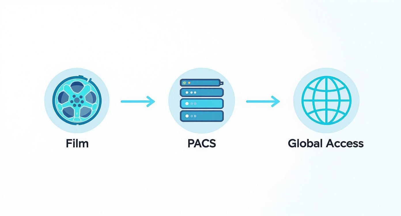
You can see the journey laid out visually, moving from cumbersome physical media to a single, central hub. It's a massive leap forward.
The Image Acquisition Process
It all starts at the source: the imaging modalities. These are the powerful machines we're all familiar with—CT scanners, MRI machines, and X-ray equipment. In our mailroom analogy, these are the folks creating and sending the digital packages.
To make sure every other part of the system can read these "packages," each image is created using a universal format. This common language is absolutely essential, allowing different types of machines and software to communicate without a hitch. If you want to dive deeper into the nuts and bolts, we have a complete guide that explains DICOM standards and why they are so vital.
The Central Archive and Server
As soon as an image is captured, it’s whisked away over a secure network to the PACS server. This is the heart of the entire system, acting as the central sorting facility and warehouse.
The server grabs the incoming image, reads the patient information attached to it, and files it away in a highly organized and secure digital archive. This archive is built for both immediate access and long-term storage, keeping a patient's complete imaging history safe and sound, ready to be called up at a moment's notice.
A PACS archive is more than just digital storage; it’s an intelligent library. It uses a powerful database to manage millions of images, letting a doctor pull up a specific patient’s scan from years ago in seconds.
This ability to retrieve images instantly is what makes modern diagnostics so powerful. No more digging through dusty film rooms.
The Display and Interpretation Workstations
The final stop on this journey is the display workstation. These aren't your average desktop computers. They are high-powered machines with crystal-clear, high-resolution monitors where radiologists and other doctors view and interpret the images.
These workstations come loaded with specialized software, giving clinicians the tools they need to zoom in, measure anomalies, and add notes directly onto the image to build an accurate diagnosis.
Here at PYCAD, this is where we shine. We at PYCAD, build custom web DICOM viewers and integrate them into medical imaging web platforms, creating intuitive and powerful interfaces for clinicians. Our goal is to make sure specialists can interact with medical images from anywhere, securely and efficiently. These workstations are the windows into the PACS, turning raw data into life-saving insights. Check out our portfolio to see our work in action.
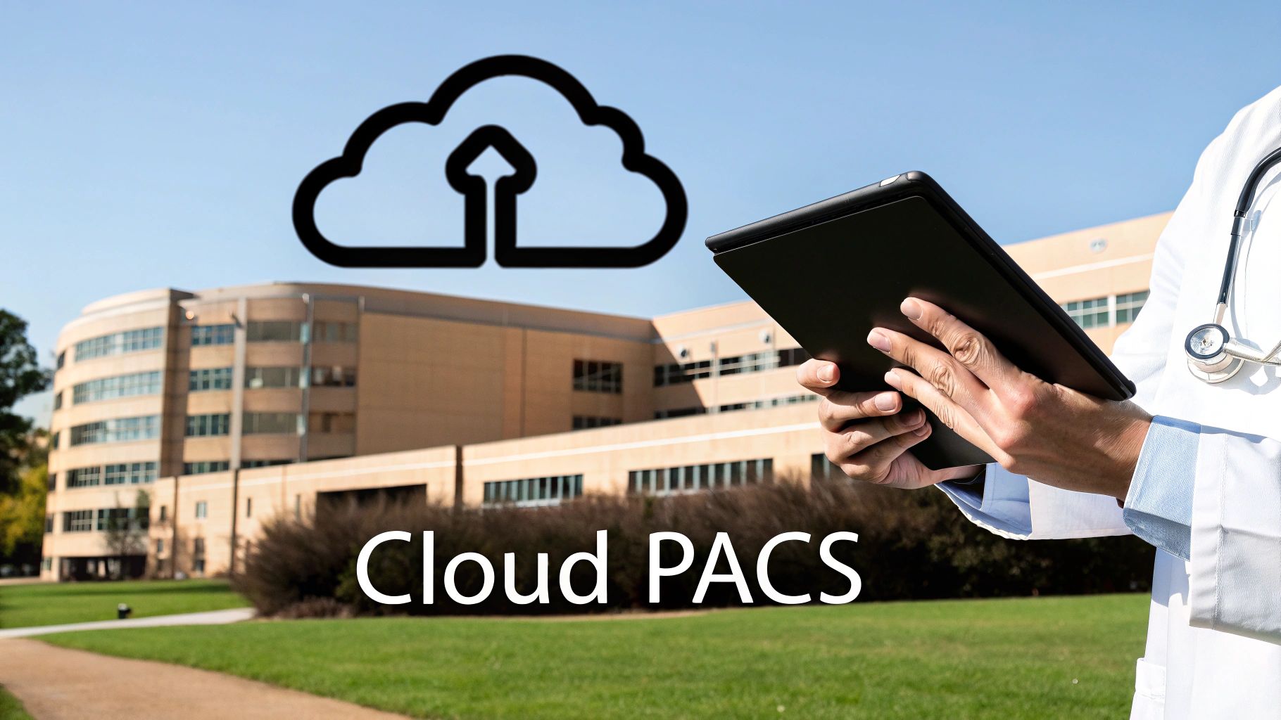
The Shift to Cloud-Based PACS Technology
Medical imaging is no longer chained to the hospital server room—it's moving to the cloud. This isn't just a simple storage upgrade; it's a fundamental shift in how we think about accessing, scaling, and collaborating with critical patient data. It’s a leap forward in healthcare's digital journey.
Think about the old way: on-premise systems demanded huge upfront costs for hardware, a dedicated IT team for round-the-clock maintenance, and a lot of physical space. Need more capacity? That meant buying more servers—a slow and expensive cycle. Cloud-based PACS completely turns that model on its head, offering a far more flexible and financially sane alternative.
This isn't just a niche trend; it's rapidly becoming the new standard. The Imaging in the Cloud 2024 Report from KLAS revealed that nearly two-thirds of healthcare organizations are already using the cloud for image viewing and storage. That kind of momentum sends a clear signal: the industry is moving toward smarter, more efficient systems. You can dive deeper into this trend in the full 2024 outlook report.
Unlocking New Possibilities in Patient Care
Moving to the cloud gives healthcare facilities capabilities that were once exclusive to massive hospital systems. It’s a great equalizer, putting enterprise-level imaging tools within reach of smaller clinics and practices, which in turn sparks innovation everywhere.
Here’s what that looks like in the real world:
- Lighter IT Load: Hospitals can hand off the heavy lifting of server maintenance, security patches, and data backups to cloud experts. This frees up their internal IT teams to focus on what really matters—initiatives that directly improve patient care.
- Bulletproof Disaster Recovery: Cloud platforms are built with redundancy in mind. Patient data isn't just stored in one place; it's securely copied across multiple locations. This means that even if a local disaster strikes, that critical information is safe and accessible.
- Borderless Collaboration: Imagine a specialist halfway around the world securely viewing a high-resolution scan in real-time to offer a life-saving second opinion. That's the power of the cloud—creating global teleradiology networks that directly improve patient outcomes.
The Technical Journey to the Cloud
Of course, migrating a system as vital as PACS isn't a weekend project. It requires meticulous planning and a steady hand. You're moving enormous volumes of sensitive patient data from local servers into a secure, compliant cloud environment. For anyone interested in the broader principles of this kind of move, this technical guide on how to migrate to the cloud offers a great overview.
Here at PYCAD, we live and breathe the cloud. We specialize in building tools that unlock its full potential for medical imaging. We at PYCAD, build custom web DICOM viewers and integrate them into medical imaging web platforms, giving clinicians the fast, secure access they need, wherever they are. We’re experts in using powerful platforms like the Google Cloud Healthcare API to create these sophisticated, scalable solutions.
Want to see how our custom viewers are already changing the game for medical imaging workflows? Take a look at our portfolio.
When One Size Doesn't Fit All: Tailoring PACS for Medical Specialties
Medicine isn't a single entity; it's a rich tapestry of specialized fields, each with its own language, tools, and diagnostic puzzles. So why would we expect a single, generic piece of technology to serve them all equally? While a general-purpose PACS is a fantastic starting point, its real power is unleashed when it’s meticulously crafted for the unique demands of a specific medical discipline.
This is the world of specialty PACS. These aren't just slightly modified systems; they are ground-up redesigns for fields like cardiology, mammography, pathology, and orthopedics. They move beyond simple image storage to offer workflows and visualization tools that feel like they were built by specialists, for specialists.
Different Jobs Demand Different Tools
Think about the day-to-day work of a cardiologist versus a radiologist. A radiologist might spend their day examining static X-rays or CT scans, hunting for structural clues. A cardiologist, on the other hand, needs to see the heart in motion. They work with dynamic echocardiograms—videos of a living, beating heart—and need tools that can loop playback, measure blood flow, and precisely track valve function as it happens.
A generic system would simply buckle under that pressure. A cardiology-specific PACS, however, is built for exactly that.
The philosophy behind specialty PACS is beautifully simple: technology should bend to the clinician’s will, not the other way around. When you give an expert a tool designed for their specific craft, you empower them to work faster, diagnose with greater confidence, and ultimately, improve patient lives.
This purpose-built approach makes a world of difference across the board:
- Mammography: These systems are designed for comparison, letting specialists view prior scans side-by-side with ease. They handle advanced tomosynthesis (3D mammography) and come with reporting templates that meet strict regulatory standards like BI-RADS.
- Pathology: Digital pathology PACS is a game-changer, managing enormous whole-slide images. Pathologists can pan and zoom across a digital tissue sample just like with a physical microscope, but with the added power of digital annotation and AI-driven analysis.
- Orthopedics: For a surgeon planning a joint replacement, precision is everything. Orthopedic PACS provides meticulous measurement tools for calculating angles and overlaying implant templates onto images, which is absolutely critical for pre-operative success.
A Growing Demand for Precision
This shift toward specialized systems isn't just a small trend—it's a major market movement. The global specialty PACS market was valued at an impressive $3.74 billion in 2024 and is on track to hit $6.57 billion in the near future. This explosive growth is a clear signal that the future of healthcare lies in more precise, specialized diagnostic tools. Discover more insights about these market trends.
At PYCAD, we live and breathe this need for specialization. We at PYCAD, build custom web DICOM viewers and integrate them into medical imaging web platforms, creating interfaces that are tailor-made for the specific challenges of different medical fields. By building these focused solutions, we help clinicians unlock the true diagnostic potential hidden within their medical images. You can see examples of our specialized work in our portfolio.
Integrating PACS with Web DICOM Viewers and AI
A PACS is powerful on its own, but its true genius is unlocked when you start connecting it to other technologies. When you link a PACS with other vital healthcare tools, it transforms from a digital filing cabinet into an intelligent, dynamic ecosystem that actively supports clinicians and pushes the boundaries of medicine.
This all starts with DICOM, the universal language that lets all medical images talk to each other. But the real magic happens when you bring modern web interfaces and artificial intelligence into the mix, completely changing how doctors and specialists interact with this vast ocean of data.

A Modern Window Into the Archive
Web DICOM viewers are that modern window. These aren't just simple picture viewers; they're sophisticated applications that let clinicians pull up high-fidelity, diagnostic-quality images on any device. It could be a high-resolution workstation in the reading room or a tablet during rounds. That kind of accessibility is a complete game-changer for collaboration and teleradiology.
Think of a web DICOM viewer as a secure, high-performance streaming service for medical images. It gives authorized users instant access to the entire PACS library without needing to install heavy software. This empowers faster, more flexible diagnostic workflows.
This is exactly where we live and breathe. Here at PYCAD, our specialty is that we at PYCAD, build custom web DICOM viewers and integrate them into medical imaging web platforms. Our mission is to create intuitive, powerful tools that make imaging data not just accessible, but genuinely actionable. See what's possible by visiting our portfolio.
The Dawn of AI-Powered Diagnostics
The most exciting frontier in PACS integration is definitely the fusion with artificial intelligence. AI algorithms are now being trained on the massive datasets stored in a PACS, serving as a powerful assistant to augment the incredible skills of human radiologists.
These AI tools are already performing some remarkable tasks:
- Early Disease Detection: AI can be trained to spot incredibly subtle patterns in images that might be missed by the human eye. This flags potential abnormalities for review and can lead to much earlier detection of conditions like cancer or neurological disorders.
- Workflow Automation: Imagine AI handling the routine, time-consuming tasks like measuring anatomical structures or triaging urgent cases. This frees up radiologists to focus their deep expertise on the most complex interpretations where they're needed most.
- Predictive Analytics: By analyzing a patient's entire imaging history alongside other clinical data, AI models can begin to predict disease progression or how a patient might respond to a particular treatment.
This synergy between human expertise and machine intelligence is truly shaping the future of medicine. As AI becomes more deeply embedded in medical imaging, it's even changing how we interact with patients, which is clear when you look at the growing role of healthcare chatbots in the field.
Bringing these technologies together isn't just a technical upgrade; it's a fundamental leap toward a more proactive, precise, and personalized standard of care. If you want to dive deeper into the nuts and bolts of making these systems work together, you'll find our guide on PACS integration to be a helpful resource.
The Future of Connected Medical Imaging
Think about how far we've come. We've journeyed from clunky, cumbersome film jackets to an intelligent, deeply interconnected imaging ecosystem. PACS has been at the very heart of that incredible transformation.
We've seen what PACS is in healthcare—so much more than just digital storage. It’s the central nervous system for medical images, the hub that connects everything from the moment an image is captured to the instant a specialist views it on a screen halfway across the world.
Now, with the move to the cloud and game-changing integrations with AI and advanced DICOM viewers, we're watching its next chapter unfold in real time. The potential for smarter diagnostics and truly collaborative care is exploding.
PACS isn't just a digital filing cabinet; it’s the launchpad for medical innovation. It provides the structured data that AI needs to learn and the universal access required for global collaboration, completely reshaping how we diagnose and treat patients.
At PYCAD, we live and breathe this stuff. We're on the front lines, building the tools that turn this incredible data into decisive action. We at PYCAD, build custom web DICOM viewers and integrate them into medical imaging web platforms, bringing this future to life for our partners.
Curious to see what this looks like in practice? Take a look at our portfolio to see some real-world examples of our work.
Your PACS Questions, Answered
Jumping into the world of medical imaging technology can feel like learning a new language, filled with acronyms and complex systems. As PACS becomes the backbone of modern healthcare, it’s natural to have questions. Whether you're an IT pro or a clinician on the front lines, getting a handle on the details is crucial.
Let's clear up some of the most common points of confusion. We'll unpack the differences between related systems, explain the core standards that make it all work, and touch on the all-important topic of security.
What’s the Difference Between PACS and RIS?
It helps to think of them as two specialists working together on the same case. The PACS is the master archivist—its entire job is to manage the images themselves. The Radiology Information System (RIS), on the other hand, is the workflow coordinator, managing all the patient data and processes around those images.
Think of the RIS as the operational brain of the radiology department. It handles things like:
- Scheduling a patient's CT scan
- Managing billing and insurance details
- Tracking report dictation and sign-off
The PACS is the vast, secure library where the actual CT images are stored, retrieved, and viewed. The two systems are in constant communication. An appointment scheduled in the RIS triggers an order; once the scan is complete, the images land in the PACS. The radiologist’s final report, created in the RIS, is then linked back to those images in the PACS, creating a complete, seamless patient story.
How Does DICOM Fit into PACS?
If PACS is the library, then DICOM (Digital Imaging and Communications in Medicine) is the universal language all the books are written in. It’s the global standard that ensures a CT scanner from one company can send an image that a viewing station from a completely different company can perfectly understand and display.
A PACS is designed from the ground up to be fluent in DICOM. This allows it to effortlessly receive, store, and share medical images from any source. DICOM files are more than just pictures; they contain a treasure trove of critical patient information embedded right within the file itself. Without this shared standard, the interconnected world of medical imaging we rely on simply wouldn't exist.
Is a Cloud-Based PACS Secure Enough for Patient Data?
Yes, and in many ways, it's even more secure. Top-tier cloud PACS providers are bound by stringent regulations like HIPAA and build their entire business on a foundation of trust and security. They deploy sophisticated security protocols that often go far beyond what a single hospital's IT department can realistically manage on-site.
It's a common misconception that "on-premise" means safer. In reality, the security architecture of a major cloud provider is often far more robust and resilient than a local server room, offering superior protection against data breaches, system failures, and even physical disasters.
These platforms use powerful tools like end-to-end data encryption, granular access controls to lock down who can see what, and comprehensive audit trails that log every single action taken within the system.
At PYCAD, our expertise lies in creating the secure, high-performance tools that bring this data to life. We at PYCAD, build custom web DICOM viewers and integrate them into medical imaging web platforms, giving clinicians the intuitive interfaces they need to provide the best care. To see how we put these principles into practice, take a look at our portfolio.


