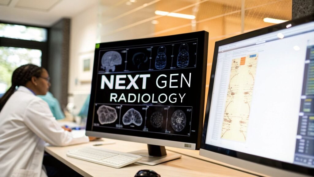
We're standing on the brink of a profound change in medical imaging. The old way—isolated radiologists, manual processes, and cumbersome data sharing—is giving way to something far more dynamic and intelligent. This is the world of next-generation radiology, an interconnected ecosystem where AI-powered analytics, secure cloud platforms, and advanced tools work together to make diagnostics faster, smarter, and more collaborative.
The Dawn of a New Era in Medical Imaging

Imagine stepping into a diagnostic environment where the limitations of physical location simply disappear. This isn't science fiction; it’s the reality of next-generation radiology today. We're moving beyond just looking at images on a screen and into a space where patient data flows freely and securely, generating critical insights almost instantly.
This isn't just a minor upgrade. It’s a fundamental rethinking of the entire diagnostic workflow, built on a few powerful pillars that completely change the game. We're breaking free from the constraints of localized hardware and embracing the boundless potential of the cloud, making vital information accessible whenever and wherever it's needed most.
The Core Pillars of Modern Radiology
This new era in medical imaging is supported by several key technologies working in harmony:
- AI-Powered Diagnostics: Think of AI as an expert partner, tirelessly scanning images to flag subtle patterns or anomalies the human eye might miss. It doesn't replace the radiologist's skill but amplifies it, leading to quicker, more confident diagnoses.
- Secure Cloud Platforms: These are the digital highways that connect specialists, no matter where they are. They tear down the walls of traditional radiology departments, fostering real-time teamwork on complex cases from opposite sides of the globe.
- Advanced Web DICOM Viewers: The web DICOM viewer is the radiologist's primary lens into a patient's story. A truly high-performance viewer is more than just an image display; it’s a sophisticated toolkit for deep analysis, untethered from any single workstation.
As imaging technologies become more complex, a solid grasp of different modalities is key. For example, understanding the nuances of procedures through resources like a friendly guide to heart echocardiograms helps paint a fuller picture of this evolving field. This isn't just academic—it's driving serious momentum in the market.
The global radiology market was valued at about US$ 33.52 billion in 2024 and is expected to climb to an incredible US$ 83.42 billion by 2033. This growth is a direct result of these technological leaps and the growing need for advanced diagnostics for chronic diseases.
Here at PYCAD, we live and breathe this innovation. We specialize in crafting custom web DICOM viewers and integrate them into medical imaging web platforms. Our goal is to bring these powerful next-generation elements together seamlessly. Take a look at our portfolio of successfully executed projects to see what this looks like in action.
Think of AI not as a replacement for a radiologist, but as an incredibly sharp, tireless co-pilot. This isn't science fiction anymore. It's the reality of next generation radiology—a powerful partner that helps navigate massive datasets, spot subtle patterns, and bring a new level of precision to every single scan.
AI algorithms are trained on vast libraries of anonymized medical images, learning to detect anomalies with a speed and consistency that beautifully complements human expertise. This powerful collaboration frees radiologists from the more repetitive aspects of their work, allowing them to pour their focus into complex cases where their critical thinking is needed most.
At its heart, AI serves as a powerful analytical engine. In a high-pressure clinical setting, an AI tool can act as an intelligent gatekeeper, automatically triaging incoming studies. It might flag a potential brain bleed or a collapsed lung for immediate review, ensuring the most critical patients get attention first. This simple shift can have a profound impact on patient outcomes.
Before we dive deeper, let's look at the bigger picture of how radiology is evolving. The shift is about more than just one new piece of technology; it's a fundamental change in workflow, data management, and the very tools of the trade.
Evolution from Traditional to Next-Generation Radiology
This table breaks down the key shifts that are defining the future of medical imaging:
| Aspect | Traditional Radiology | Next Generation Radiology |
|---|---|---|
| Image Analysis | Manual, human-driven interpretation | AI-assisted detection, quantification, and triage |
| Data Access | Localized servers, PACS-dependent | Cloud-native, secure web-based access from anywhere |
| Workflow | Linear and manual report generation | Automated report structuring, task automation |
| Collaboration | Primarily within the radiology department | Seamless, platform-based multidisciplinary collaboration |
| Patient Data | Siloed imaging data | Integrated view of imaging, EMR, and pathology data |
| Predictive Power | Retrospective analysis | Predictive analytics for disease progression |
This evolution from a static, localized process to a dynamic, intelligent, and interconnected ecosystem is the core of next-generation radiology. It’s about creating systems that empower clinicians instead of hindering them.
From Detection to Prediction
AI's role is quickly moving beyond just finding what’s already there. Advanced models are starting to predict disease progression, handing clinicians a crystal ball for proactive patient care.
These predictive tools can analyze subtle shifts in tissue density or lesion size across a series of scans—changes that are often imperceptible to the human eye. By identifying these early trends, AI helps forecast how a condition might progress, opening the door for earlier, more effective interventions.
This is a game-changer in oncology. We know that clinicians often spend 1.5 to 2.5 hours per patient just preparing for multidisciplinary tumor boards. AI agents can slash this prep time by synthesizing imaging data, pathology reports, and clinical notes into a single, coherent summary.
Automating the Mundane to Elevate Expertise
One of the most immediate wins with AI is its ability to automate the monotonous tasks that bog radiologists down. This includes everything from meticulously measuring nodules to structuring and pre-filling radiology reports.
“Your mind is split in two. You're reading images, yet simultaneously wrestling with unnatural dictation tools. It's mentally exhausting and prone to error.”
This quote from a radiologist hits the nail on the head. It captures the daily friction that AI is designed to eliminate. Imagine a radiologist simply speaking their findings naturally while a generative AI system instantly organizes that dictation into a flawless, structured report.
This shift allows radiologists to get back to what they do best: interpreting images and piecing together the patient’s story. Instead of battling clunky software, their cognitive energy is dedicated to diagnosis. The road ahead is even more exciting, with innovations like autonomous systems emerging among the agentic AI use cases in healthcare that promise an even deeper partnership.
At PYCAD, we live and breathe this dynamic. We don't just build software; we architect intelligent ecosystems. Our specialty is to build custom web DICOM viewers and integrate them into medical imaging web platforms.
Ultimately, AI isn't here to replace the radiologist. It's here to augment their vision, sharpen their insights, and lift the administrative weight that contributes to burnout. By doing so, it elevates the entire practice, making diagnostics faster, more precise, and fundamentally more human. For a closer look, our guide on artificial intelligence for radiology offers a deeper exploration of these powerful applications.
The Power of Integrated Cloud Platforms and DICOM Viewers
If AI is the brilliant mind behind next-gen radiology, the integrated cloud platform is its central nervous system. This is what connects every tool, every scan, and every specialist into one cohesive whole. It’s a monumental leap from the old world of siloed departments and dusty server rooms.
Think about how banking has changed. We used to be tied to a physical branch with set hours. Now, we manage everything from a secure app, collaborating and accessing services from anywhere. Cloud platforms do the same for medical imaging, completely freeing it from physical walls and creating a truly borderless environment for diagnostics.
This shift to the cloud is a game-changer. It’s about more than just storage; it’s about creating a unified, collaborative workspace for the modern care team.
Why Cloud-Native Platforms Are Non-Negotiable
The sheer amount of medical imaging data being generated is mind-boggling, and it’s only growing. Traditional on-premise Picture Archiving and Communication Systems (PACS) just weren't built for this kind of scale. They often become bottlenecks, demanding costly hardware upgrades and creating frustrating access issues.
A cloud-native platform is designed from the ground up to solve these problems. It offers practically infinite scalability, growing right alongside a healthcare organization’s data needs. This elasticity also flips the financial model, turning massive capital expenses into predictable operational costs. You only pay for what you use.
Even better, these platforms democratize expertise. A specialist in a major city can instantly pull up the same high-resolution scan as a rural clinician, offering a consult in real-time. This is how we break down geographic barriers and get expert eyes on a case, no matter where the patient is.
The infographic below shows how AI tools, which live on these platforms, are supercharging radiology in detection, prediction, and workflow automation.
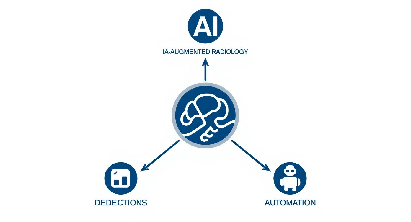
This really drives home how an integrated platform isn’t just a backdrop—it's the foundation that makes these incredible AI capabilities accessible and truly effective.
The DICOM Viewer: The Critical Window to Diagnosis
If the cloud platform is the engine, the web DICOM viewer is the cockpit. It’s the single most important interface where a clinician interacts with imaging data. A clunky, slow, or limited viewer adds friction, slows down diagnoses, and burns out your users. But a powerful, intuitive viewer? It can make the entire workflow feel effortless.
A state-of-the-art platform is worthless if its main user interface is a roadblock. The DICOM viewer isn't just a feature; it's the absolute cornerstone of the user experience in modern radiology.
Today’s diagnostic challenges demand a viewer with a few key traits:
- Zero-Footprint Access: Clinicians need to securely open and work with full-fidelity images from any device with a web browser. No installs, no plugins, no fuss.
- Advanced Visualization Tools: We’re talking about tools like 3D rendering, multi-planar reconstruction (MPR), and specialized measurement instruments that allow for a deep, nuanced analysis of the anatomy.
- Seamless Integration: The viewer has to play nicely with everything else. It must connect smoothly with AI algorithms, EMRs, and reporting software to create one uninterrupted workflow.
This is exactly where we live and breathe at PYCAD. We build custom web DICOM viewers and integrate them into medical imaging web platforms for medtech companies. Our entire focus is on creating that fluid, powerful, and intuitive experience that lets clinicians do their best work.
Ultimately, the goal is to build a digital environment where the technology just melts into the background. It should allow a clinician to focus solely on the patient's story hidden within the images. The right combination of a robust cloud platform and a high-performance DICOM viewer makes that a reality. For a closer look at what goes into a great viewer, check out our guide on the essential features of a modern DICOM viewer.
Pioneering the Future with Interventional and Digital Radiology
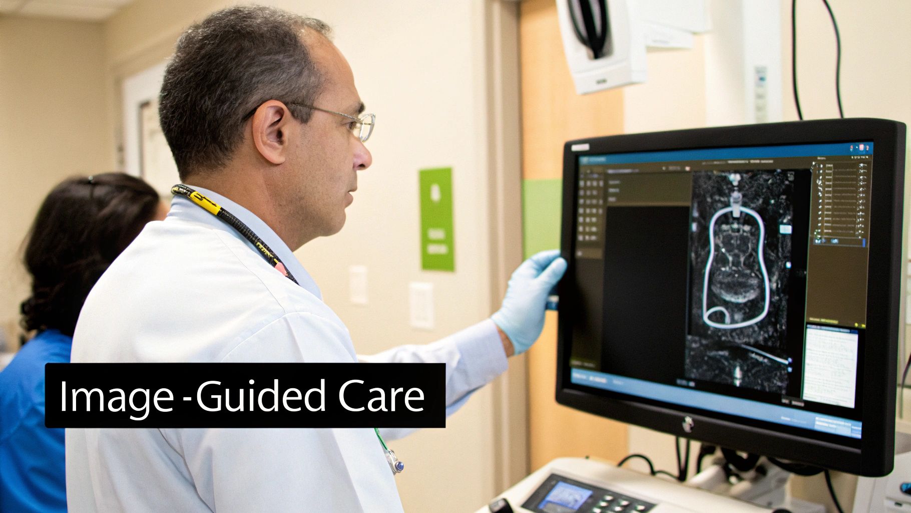
The world of next generation radiology is stretching far beyond the traditional diagnostic suite and stepping into the operating room. This is where radiology gets hands-on, becoming a powerful tool for active treatment. The most exciting frontier in this journey is interventional radiology—a field where radiologists become surgeons, leveraging their mastery of imaging to perform procedures with incredible precision.
Often called "image-guided surgery," this discipline uses real-time imaging from CT scans, fluoroscopy, and ultrasound as a live map to navigate inside the human body. Instead of making large incisions, specialists guide tiny instruments through blood vessels or small entry points to fix problems right at the source. It’s the very definition of minimally invasive, meaning patients experience faster recoveries, less pain, and lower risk than with conventional open surgery.
And this isn't just a niche specialty. It's a rapidly growing force in medicine. The market for interventional radiology hit an estimated USD 28.8 billion in 2024 and is on track to reach nearly USD 49.4 billion by 2035. That steady climb is a direct result of the growing demand for effective, less invasive treatments that get people back to their lives faster. You can discover more insights on the interventional radiology market growth and what's driving it.
The Digital Leap from Film to Pixels
Hand-in-hand with these interventional breakthroughs is a fundamental shift in how we capture and manage images: the move from analog to digital. This transition is as profound as the jump from old-school film photography to today’s digital cameras, and its impact on speed, collaboration, and diagnostic quality is simply massive.
Remember what it was like to use a film camera? You had to take the picture, carefully handle the roll, process it in a darkroom with chemicals, and only then could you finally see the result. It was slow, clunky, and left no room for error. Early radiology worked much the same way, with physical films that had to be developed, archived in enormous file rooms, and physically carried from one doctor to another.
Digital radiography completely replaces that cumbersome chain with instant electronic capture. A detector plate converts X-ray energy directly into a high-resolution digital image that appears on a monitor in seconds.
The shift to digital isn't just about convenience—it's a complete reimagining of the diagnostic workflow. It collapses time, eliminates physical barriers, and unlocks a new level of image clarity and analytical power.
This immediate feedback lets technicians verify image quality right away, cutting down on the need for patient recalls. Even more importantly, these digital files are dynamic. Radiologists can zoom in, adjust the contrast, and apply filters to highlight subtle details that would be completely invisible on a physical film.
An Interconnected Diagnostic Ecosystem
These two monumental advances—interventional and digital radiology—aren't happening in isolation. They are deeply connected parts of a bigger picture, working together with AI and cloud platforms to forge a new standard of patient care.
A digital scan today isn't just saved on a local computer; it’s uploaded to a secure platform, making it instantly and widely accessible.
- For Interventional Radiologists: They can review a patient’s high-resolution scans from anywhere, meticulously plan a procedure, and then use live digital imaging to guide their instruments with pinpoint accuracy during the treatment itself.
- For AI Algorithms: Clean, high-quality digital images are the perfect fuel for AI. These models can analyze scans, flag areas of concern, and provide hard data that helps inform both the diagnosis and the treatment plan.
This seamless flow of information is what truly defines modern radiology. Here at PYCAD, we specialize in building the digital backbone that makes this all possible. We build custom web DICOM viewers and integrate them into medical imaging web platforms, creating the infrastructure for this new era of diagnostics and treatment. Explore our portfolio to see how we bring these powerful systems to life.
Building Your Next Generation Radiology Solution
So, you're ready to create a radiology platform that stands apart. This is where the vision for a smarter, more connected diagnostic experience starts to take real shape. For any medtech innovator or forward-thinking healthcare provider, building a custom solution is the ultimate step—it’s about architecting an ecosystem that truly empowers clinicians and transforms patient outcomes.
The journey from an idea to a fully functional platform is a deliberate one. It’s not about just piecing together different technologies; it’s about crafting a digital environment with purpose. This requires a crystal-clear roadmap, a deep dive into the real-world needs of your users, and an absolute commitment to security and a seamless experience. Get these foundational choices right, and you're on your way.
Start by Defining Your Clinical and Operational Needs
Before you even think about code, you need to listen. What’s holding your current workflow back? Are radiologists wasting precious minutes waiting for images to load? Do referring physicians struggle with clunky reporting systems? Are collaboration tools non-existent? Pinpointing these very real frustrations is your starting line.
Take the time to map out the entire diagnostic journey, from the moment an image is captured to the second a report is delivered. You need to understand the critical requirements for everyone involved—the radiologist, the referring doctor, and even the administrative team. The goal isn't to build a platform with a long list of features; it's to build one that solves actual problems.
Choosing the Right Technology Stack
Once you have a clear picture of your clinical needs, it's time to select the tools for the job. This is where you make crucial decisions about the platform’s core foundation, like its cloud infrastructure, how it will manage vast amounts of data, and what the user-facing applications will look like. The tech stack you choose has to be scalable, secure, and ready to evolve with what’s next.
You'll inevitably face the classic "build versus buy" debate, especially for key components. While off-the-shelf software might seem like a quick fix, it rarely delivers the truly integrated and unique experience you’re aiming for. This is especially true for the single most important part of the entire system: the web DICOM viewer.
The DICOM viewer is the heart and soul of the user experience. Dropping in a generic, third-party viewer can create a clunky, inefficient workflow that completely undermines the rest of your custom-built platform. A bespoke viewer, on the other hand, feels like a natural extension of your entire solution.
This is exactly where having an expert partner makes all the difference. At PYCAD, we build custom web DICOM viewers and integrate them seamlessly into medical imaging web platforms. We believe this critical tool shouldn't be an afterthought—it should be a core element, designed to work perfectly with your system from day one. You can explore our successfully executed projects at our portfolio page: https://pycad.co/portfolio
Fusing Regulatory Compliance with a World-Class User Experience
In medicine, security and compliance aren't just features—they're the bedrock of trust. Any next generation radiology platform has to be built on an ironclad framework that meets regulations like HIPAA head-on. That means robust data encryption, strict access controls, and detailed audit trails to protect sensitive patient information without compromise.
But compliance alone isn't enough. The user experience (UX) is what will make or break your platform. A powerful system is worthless if clinicians find it clunky or confusing. The interface must be intuitive, minimizing clicks and making common tasks feel effortless. A truly great design lets a radiologist focus their brainpower on the diagnosis, not on wrestling with the software. For a deeper dive, check out our guide on medical imaging software development, where we cover these very considerations.
The market is clearly moving in this direction. The digital radiography sector was valued at around USD 1.6 billion in 2024 and is on a steady growth path. This isn't just a trend; it's a fundamental shift as healthcare providers adopt digital systems to slash costs, boost image quality, and keep up with ever-increasing diagnostic loads. To get a better sense of this momentum, you can learn more about these digital radiography market trends.
Building your own platform is a bold move, but it's one that pays off. It gives you the freedom to create a solution that perfectly matches your vision—one that’s secure, compliant, and genuinely enjoyable to use. With a clear strategy and the right partner at your side, you can build a system that not only solves today's challenges but is ready for the future of imaging.
Common Questions About Next-Generation Radiology
Whenever we talk about a major shift in how medicine is practiced, questions are bound to bubble up. And that's a good thing. Stepping into the world of next-generation radiology is a big move, and getting clear on the fundamentals—the technology, the benefits, and the real-world impact—is the first step toward making a truly smart decision for your future.
Let's dive into some of the questions we hear most often from clinics and medtech leaders who are ready to make the leap.
What Is the Most Crucial Part of a Next-Gen System?
It's easy to get focused on the dazzling AI algorithms or the massive cloud storage, but the real heart of the entire system is the integrated web platform. Think of it as the digital command center for your entire diagnostic world. It’s the single place where every scan, every report, and every specialist can connect and collaborate seamlessly.
And if the platform is the command center, the web DICOM viewer is the cockpit. It’s where your clinicians do their most important work, day in and day out. A fast, intuitive, and powerful viewer isn't just a nice-to-have feature; it’s the very foundation of the user experience and, ultimately, the success of the whole platform.
How Does AI Actually Help a Radiologist's Workflow?
The goal of AI in radiology isn't to replace the radiologist—it's to give them a superpower. It acts as an incredibly diligent and precise assistant, taking over the repetitive, time-consuming tasks that lead to fatigue and freeing up specialists to focus on what humans do best: critical thinking and complex problem-solving.
Here’s what that looks like in practice:
- Automating Measurements: An AI can measure a nodule or track a lesion's growth in a fraction of a second, with perfect consistency every time. This alone saves countless hours of manual clicking and measuring.
- Pre-populating Reports: Imagine an AI that analyzes an image and drafts a structured report with preliminary findings. The radiologist then steps in to review, refine, and add their expert interpretation. It’s about collaboration, not replacement.
- Flagging Urgent Cases: In a busy queue, an AI can act as a vigilant gatekeeper, instantly identifying studies with signs of a stroke or pulmonary embolism and pushing them to the top of the list for immediate review.
This human-AI partnership allows radiologists to work at the absolute peak of their abilities, bringing more focus and energy to every single case.
Are Cloud-Based Radiology Platforms Secure?
This is one of the most important questions, and the answer is a resounding yes—as long as they are engineered correctly. Modern healthcare cloud platforms are built from the ground up with security as the number one priority, not a feature tacked on at the end. They use a layered, defense-in-depth strategy to guard patient data at every possible point.
Security in a modern cloud environment isn't just about a single firewall. It's a comprehensive architecture of encryption, strict access controls, and continuous monitoring designed to meet and exceed the toughest regulatory standards like HIPAA.
These systems rely on heavy-duty, end-to-end encryption, which means data is essentially scrambled and unreadable whether it's being sent over the internet or sitting in storage. On top of that, strict, role-based access rules ensure that only the right people can see specific patient information. With constant monitoring and regular security audits, these platforms are often far more secure than an old server sitting in a closet down the hall.
Can Smaller Clinics Afford These Advanced Systems?
Absolutely. In fact, the shift to cloud-based systems is what makes this technology more accessible than ever before. The old days of needing a massive, six-figure capital investment for on-site servers and software are quickly becoming a thing of the past.
Today's best platforms run on subscription-based models (often called Software-as-a-Service, or SaaS). This changes the game completely, turning what was once a huge upfront purchase into a predictable, manageable operating expense. A smaller clinic can start with a plan that fits its current patient load and budget, and then easily scale up as it grows.
This approach delivers an incredible long-term return on investment (ROI). When you factor in the massive gains in efficiency, diagnostic speed, and collaborative power, the subscription cost becomes a smart, strategic investment for practices of any size.
At PYCAD, we don’t just talk about these systems; we build them from the ground up. We specialize in creating the very heart of modern radiology: custom web DICOM viewers integrated into secure, high-performance medical imaging platforms. We’re passionate about crafting the tools that empower clinicians to do their best work.
To see how we turn these ideas into reality, we'd love for you to take a look at our work.


