So, you have a .dcm file and need to open it. Forget trying to use your standard photo app—it just won't work. You'll need a dedicated DICOM viewer, like RadiAnt or Horos for your desktop, or a web-based tool if you want to avoid installation altogether.
These viewers are built specifically to decode the rich, complex structure of a DICOM file, which is far more than just a picture.
Beyond Pixels: Why DCM Files Are a Different Breed
When you open a .dcm file, you're not just looking at a static image; you're accessing a complete patient narrative. A JPEG or PNG is simply a grid of pixels—it shows you what something looks like. A DICOM file, however, tells you the who, what, when, where, and how behind that image. This is the crucial difference that makes DICOM the foundation of modern medical imaging.
Think of it this way: a JPEG is a postcard. A DICOM file is that same postcard, but with the entire patient's medical chart securely attached to the back. This rich, structured data is exactly why you can't just double-click and open it in Paint.
More Than Just an Image
The true power of the DICOM standard is how it bundles everything into one self-contained, portable record. Every scan is a complete story.
- Patient Information: Name, ID, age, and other essential demographics are embedded right into the file.
- Acquisition Data: You get the full context—details about the scanner (MRI, CT, etc.), the exact settings used, and even the operator's notes.
- Spatial Context: The file knows the image's orientation and position relative to the patient’s body, which is critical for accurate interpretation.
- Pixel Data: This is the actual image, but it's often captured with a much higher bit depth than a consumer photo, revealing subtle details vital for diagnosis.
This all-in-one approach is what moved medical imaging from a tangled mess of proprietary formats into the connected, universal ecosystem we have today. Here at PYCAD, we live and breathe this stuff; we build custom web DICOM viewers and integrate them into medical imaging web platforms. Seeing this data come to life is what fuels everything we do. Check out our portfolio to see some of these solutions in action.
The need to open and interpret DCM files is a non-negotiable in modern healthcare. By 2023, over 90% of hospitals in the United States and Western Europe were running on PACS that depend entirely on the DICOM standard. You can dive deeper into this widespread adoption on the Medicai blog.
This guide is for everyone. Whether you're a clinician needing to make a quick diagnostic call or a researcher sifting through thousands of scans for a study, you'll find the right tool for the job here. And if you're just starting out, our guide on what a DCM file is is a great place to get your bearings.
Choosing Your Desktop DICOM Viewer
Finding the right tool to open a .dcm file can feel like unlocking a new level of understanding, transforming a complex dataset into a clear, actionable story. When you move beyond just viewing an image and step into the world of diagnostic-grade desktop software, you're not just looking at pixels—you're mastering them.
The first step is picking a viewer that truly fits what you do every day. Are you a busy clinician who needs lightning-fast performance? A researcher digging deep with specialized analytical tools? Or maybe a student just getting your feet wet in medical imaging? Your answer will point you to the perfect software.
Understanding Your Workflow Needs
Before you download the first viewer that pops up, pause and think about your primary goal. This simple decision tree is a great way to frame your choice based on your role, whether you're on the front lines of clinical care, pushing the boundaries of research, or building the next big thing in med-tech.
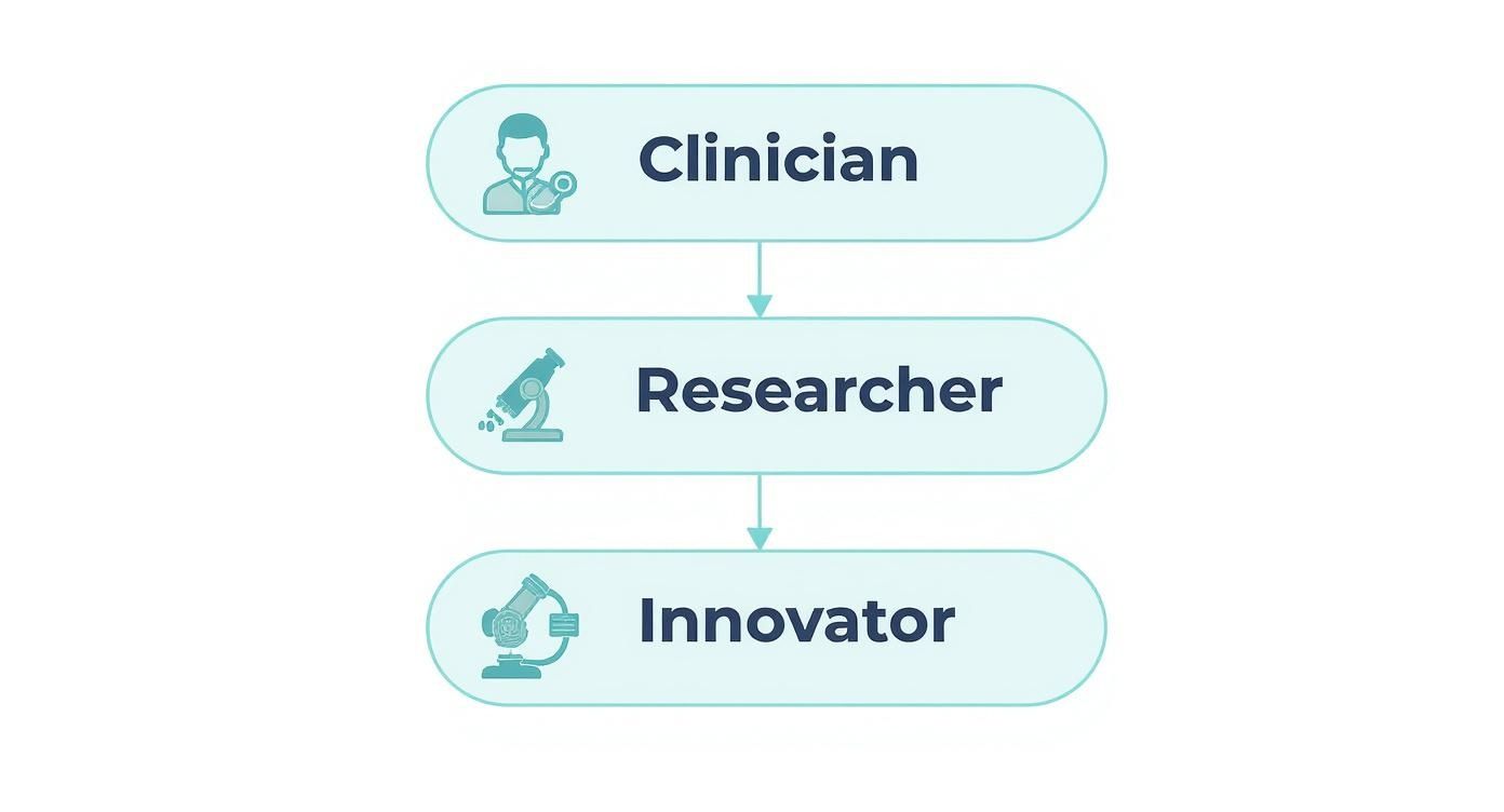
It’s clear that the "best" tool isn't a one-size-fits-all solution. It's about matching powerful features to your specific tasks to get the job done right.
Desktop viewers are your workhorses. They offer a robust, offline solution that puts immense power right at your fingertips. Unlike their web-based cousins, they use your computer's full processing capability, which is a lifesaver when you're dealing with large, multi-frame series without any lag. This is where the real work of diagnosis, analysis, and discovery happens.
The challenge with opening a .dcm file often comes down to its rich metadata and the sheer size of the image data. A single CT scan file might be a manageable 2 MB, but a full MRI series can easily blow past 100 MB. In fact, a 2020 study found that 78% of institutions ran into trouble opening DICOM files because of metadata inconsistencies or missing tags, which really drives home the need for a capable viewer.
Top Contenders for Your Desktop
Let's dive into some of the most respected free desktop viewers out there, each with its own unique strengths.
-
RadiAnt DICOM Viewer (Windows): Known for its breathtaking speed, RadiAnt is a favorite in high-volume clinics. Picture a radiologist needing to fly through dozens of CT series a day; RadiAnt's ability to load huge datasets almost instantly isn't just a nice-to-have—it's a critical workflow accelerator. Its clean interface makes tools like windowing, measurements, and 3D rendering easy to find and use.
-
Horos (macOS): For Mac users, especially those in research, Horos is a game-changer. As an open-source platform, it's built on a vibrant community that creates powerful plugins for specialized tasks, from advanced segmentation to quantitative analysis. Imagine a researcher using a custom plugin to analyze tumor volumes across hundreds of MRI scans for a study. Horos provides that flexible, extensible environment needed for innovative work.
-
MicroDicom (Windows): Simplicity is MicroDicom's greatest strength. It’s a fantastic starting point for students, patients, or anyone who needs a straightforward, no-frills tool to open a .dcm file and take basic measurements. It’s lightweight and easy to install, making it incredibly approachable.
A Quick Comparison of Free Desktop DICOM Viewers
To make the choice even clearer, here’s a side-by-side look at these popular options. This table breaks down their key features to help you find the best fit for your operating system and daily tasks.
| Viewer | Operating System | Key Features | Best For |
|---|---|---|---|
| RadiAnt DICOM Viewer | Windows | Blazing-fast performance, 3D rendering, intuitive multi-panel view | Clinicians & high-volume environments |
| Horos | macOS | Open-source, extensive plugin library, advanced research tools | Researchers & Mac users needing customization |
| MicroDicom | Windows | Lightweight, simple interface, basic measurement tools | Students, patients, & beginners |
Ultimately, the best tool is the one that feels like an extension of your own expertise, helping you work faster and more effectively.
Choosing a desktop viewer is the first step toward true data fluency. Mastering its tools—learning the shortcuts for window level adjustments, perfecting your measurement techniques, and knowing how to access hidden metadata—is what empowers you to extract every bit of value from a scan.
While these tools are fantastic for individual use, integrating these kinds of capabilities into a larger healthcare platform is a whole different ballgame. At PYCAD, that’s exactly what we specialize in. We build custom web DICOM viewers and integrate them into medical imaging web platforms, creating seamless, secure, and powerful solutions for our clients. For a deeper dive, check out our detailed guide to the best free DICOM viewers for even more options.
Accessing Medical Images From Any Browser
Picture grabbing a laptop, opening a browser, and diving straight into a .dcm file without fussing over installs. That’s the beauty of web-based DICOM viewers—they bring imaging studies to your fingertips, wherever you are. Collaboration feels seamless, and patients can engage with their own scans in a way that was once impossible.

I remember a time when a patient called me late at night, eager to review her MRI images before a second opinion appointment the next day. With a web viewer, she logged in from her home PC and I could guide her through windowing adjustments in real time. That moment highlighted just how empowering zero-footprint tools can be.
When Web Viewers Shine
Browser-based viewers excel when speed and simplicity matter most. No installation headaches, no OS hurdles—just instant access from any device with internet.
Here are a couple of favorites for quick, non-diagnostic checks:
- FViewer: Drag, drop, and you’re in. Perfect for a single .dcm file when you need to confirm image integrity.
- DICOM Web Viewer (DWV): Open-source and surprisingly robust. Windowing tweaks, linear measurements, even basic overlays—all in-browser.
These lightweight tools open doors for students, researchers, and curious patients. Yet, they also come with trade-offs in security and integration depth. For clinical-grade needs—think encryption, audit logs, and seamless EHR connectivity—you’ll want something more solid.
Free viewers crack open the door, but healthcare platforms require strict security, reliable performance, and advanced features. That’s where we step in at PYCAD.
The Professional-Grade Difference
When imaging is at the heart of a clinical workflow, viewers must do more than display pixels. They need to integrate, secure, and empower.
Imagine a telehealth session where a specialist annotates a CT scan live, shares insights instantly with a primary care doctor, and updates the patient’s record without skipping a beat. That’s the level of interaction we build into our custom solutions.
Key differentiators:
- Deep Integration: Syncs flawlessly with your EHR or PACS, auto-fetching patient context and pushing back structured reports.
- Advanced Toolsets: Multi-planar reconstruction (MPR), 3D rendering, volumetric tools, and precision measurements.
- Unyielding Security: End-to-end encryption, role-based access controls, and full HIPAA compliance.
Turning a basic viewer into a comprehensive diagnostic hub transforms workflows and propels patient care forward. At PYCAD, we thrive on crafting these sophisticated experiences. We build custom web DICOM viewers and integrate them into medical imaging web platforms. Curious to see them in action? Check out our portfolio for real-world case studies and demos.
Opening DCM Files Programmatically with Python
Let's be honest—manually clicking through medical scans is a dead end for any serious project. When you need to process thousands of images, you have to bring them into your code. This is where Python, and specifically the pydicom library, becomes your best friend. It’s how you turn a mountain of DICOM files into a structured, analyzable dataset.
Imagine you're tasked with prepping 10,000 scans for a new deep learning model. Doing that by hand would take weeks, if not months. With a simple Python script, you can extract all the pixel data and metadata you need in a matter of minutes. That’s not just efficient; it’s repeatable and far less prone to human error.
Reading the DCM File
First things first, you need to get the right tools in your environment. A quick pip install pydicom numpy will get you set up. From there, loading a DICOM file is astonishingly simple.
Just a couple of lines of code, and you're in.
import pydicom
ds = pydicom.dcmread('patient_scan.dcm')
That one line pulls the entire DICOM file—header, metadata, pixel data, everything—into a dataset object. A standard 5 MB CT slice, for instance, typically loads in about 0.1 seconds on a modern machine. That speed is absolutely critical when you're chewing through terabytes of imaging data.
Extracting Metadata Tags
Every DICOM file is a treasure trove of information beyond just the image. The metadata tags tell the story: who the patient is, when the scan was taken, and what kind of machine was used. Accessing this data feels just like working with a Python dictionary.
patient_id = ds.PatientID
modality = ds.Modality
study_date = ds.StudyDate
You can pull out all sorts of crucial information this way. Some of the most common tags you'll work with are:
- PatientID: Essential for anonymizing data and tracking subjects.
- StudyDate: Lets you build timelines or analyze changes over time.
- Modality: Helps you instantly separate CT scans from MRIs or X-rays.
- PixelSpacing: Absolutely vital for taking accurate physical measurements from the image.
Metadata provides the context that turns a simple picture into a piece of clinical evidence. It’s what ensures your analysis or AI model is grounded in reality.
For a deeper dive, our guide on programmatic DICOM access at https://pycad.co/how-to-read-dicom-files/ has more examples and tips to get you started.
Converting Pixel Data for Visualization
Of course, you need to see what you're working with. Before you feed thousands of images into a model, you should always run a few sanity checks. The pydicom library plays nicely with matplotlib to make this easy.
You can grab the raw pixel data as a NumPy array and display it in just a few lines.
import matplotlib.pyplot as plt
image = ds.pixel_array
plt.imshow(image, cmap='gray')
plt.show()
This little snippet is my go-to for quickly verifying that a file isn't corrupted or that the orientation is correct. It’s an invaluable step for spotting artifacts or other issues before they contaminate your entire dataset.
Automating Batch Processing
No one works with a single DICOM file. The real power comes from processing an entire directory—or an entire study—at once. A simple loop is all it takes to get started.
Here’s a function I often use to load all the DICOM slices in a folder into a single 3D volume.
import os
import numpy as np

def load_all(path):
    arrays = []
    for fname in os.listdir(path):
        if fname.endswith('.dcm'):
            ds = pydicom.dcmread(os.path.join(path, fname))
            arrays.append(ds.pixel_array)
    return np.stack(arrays)
Just be mindful of your system's memory here. A series of 100 high-resolution scans can easily take up 20 GB of RAM, so plan accordingly.
Performance Comparison Table
To give you a feel for what to expect, here’s a quick benchmark of read times on a typical developer workstation. This helps in estimating how long a large batch job might take.
| File Size | Load Time | Pixel Conversion |
|---|---|---|
| 1 MB | 0.02 s | 0.03 s |
| 5 MB | 0.10 s | 0.12 s |
| 10 MB | 0.25 s | 0.30 s |
Tips for Robust Pipelines
Building a script is one thing; building a reliable data pipeline is another. From experience, here are a few practices that save a lot of headaches down the road:
- Always validate metadata. Make sure required tags like
PatientIDorModalityactually exist before you try to use them. - Wrap your code in try-except blocks. Some DICOM files can be malformed. Don't let one bad file crash your entire batch job.
- Log everything. Keep track of which files were processed, any errors encountered, and how long it took. It’s a lifesaver for debugging.
Automating your DICOM workflows is a game-changer. It frees you from tedious manual work and dramatically reduces the potential for errors in large-scale medical imaging projects.
And if you’re looking to take your automation skills to the next level, understanding how to apply these concepts to more complex tasks is key. This article on building AI agents with Python offers great insights that can complement your work with medical data.
At PYCAD, this is exactly what we do. We specialize in building custom web DICOM viewers and integrating these kinds of programmatic workflows directly into medical platforms. Whether it's for research, clinical trials, or AI development, we help teams turn raw DICOM data into actionable insights. You can see examples of our work in our portfolio.
Handling DICOM Data Responsibly And Securely
Every DICOM file can carry sensitive Protected Health Information (PHI). Treating these images with care isn’t just best practice—it’s a legal and ethical necessity. When you secure .dcm files properly, you safeguard both patient privacy and your institution’s reputation.
We at PYCAD build custom web DICOM viewers and integrate them into medical imaging web platforms. Explore our work at our portfolio.
Understanding DICOM Anonymization
Sharing imaging data across research groups demands thorough PHI removal. The simplest route is stripping out metadata tags that identify patients. Here’s a quick Python example using pydicom:
import pydicom
ds = pydicom.dcmread('input.dcm')
for tag in ['PatientName','PatientID','PatientBirthDate','PatientAddress','PatientSex']:
    if tag in ds:
        ds.data_element(tag).value = ''
ds.remove_private_tags()
ds.save_as('anon_output.dcm')
This snippet wipes standard identifiers and vendor-specific entries. Always test your anonymization against a known dataset to verify nothing slips through.
| Tag Name | Category |
|---|---|
| PatientName | Demographics |
| PatientID | Demographics |
| PatientBirthDate | Demographics |
| PatientSex | Demographics |
| PrivateTags | Vendor-specific data |
Anonymization is the first line of defense for safe DICOM sharing.
Secure DICOM Transmission And Storage
Moving files over an unprotected network is a recipe for disaster. Always insist on TLS 1.2 or higher for data in motion and use disk-level encryption for files at rest.
Key actions:
- Configure HTTPS for your web viewers with valid certificates
- Store DICOM files on volumes encrypted with AES-256
- Rotate encryption keys on a regular schedule
In one real-world scenario, a research institute detected an attempted interception. Network segmentation and strict HTTPS policies stopped the breach cold—without disrupting daily operations.
For healthcare environments, compliance tools can help you stay ahead of audits. Check out these HIPAA Risk Assessment Tools to validate your safeguards.
Access Controls And Audit Trails
Controlling who sees, uploads, or downloads images is crucial. Implement role-based access control (RBAC) to map permissions exactly to each job function. Adding multi-factor authentication (MFA) delivers an extra barrier against unauthorized entry.
Audit essentials:
- Define clear user roles and enforce least privilege
- Require MFA for remote or administrative access
- Keep timestamped logs that record every user action
PYCAD’s web DICOM viewers come with built-in modules for RBAC and audit reporting.
Encryption Key Management
Encryption is only as strong as your key management. Store keys inside Hardware Security Modules (HSMs) or a secure key vault. Automate rotation so that no key lives longer than 90 days.
Best practices:
- Apply least-privilege principles to key access
- Schedule automatic rotation every 90 days or sooner
- Enable audit logging on every key operation
Monitoring And Incident Response
A robust monitoring system is your early warning. Flagging odd login patterns or surges in downloads helps you stop incidents before they escalate. Pair it with a tested response plan and your team can act quickly under pressure.
Monitoring recommendations:
- Centralize logs with a secure aggregation service
- Set threshold alerts for unusual download volumes
- Conduct quarterly security drills to sharpen your response
By layering anonymization, encryption, access control, key management, and monitoring, you create a defense-in-depth strategy for every DICOM workflow. We at PYCAD build custom web DICOM viewers and integrate them into medical imaging web platforms with these safeguards baked in. Visit our portfolio to see how we help clients secure their pipelines.
Implement these practices to protect patient privacy and maintain compliance. Reach out if you need hands-on support or tailored advice.
Your Questions Answered: Getting to Grips with DCM Files
Diving into medical imaging for the first time? You’re bound to have questions, especially when you run into the .dcm format. Don't think of these as just image files; they're comprehensive data packages, and understanding them is the first step toward mastering medical imaging.
Think of every question as a stepping stone. Whether you're a clinician trying to make a diagnosis, a researcher searching for a breakthrough, or a patient curious about your own scans, getting clear answers is what empowers you. Let's tackle some of the most common hurdles you might face.
Can I Just Open a DCM File in Photoshop?
You might be able to, with the right plugin, but you absolutely shouldn't for any clinical work. Here’s why: opening a DCM file in a standard image editor like Photoshop or a basic photo viewer effectively strips the image of its soul.
You lose all the critical metadata—patient ID, acquisition parameters, scanner settings, and image orientation. This isn't just extra information; it's the context that makes the image medically meaningful. A standard viewer sees a flat picture, completely missing the multi-frame nature of a CT or MRI. For the real story, you need a dedicated DICOM viewer. It’s the only way to see the complete, accurate diagnostic picture.
What’s the Difference Between a Single DCM File and a DICOM Series?
This is a fantastic question because it gets right to the core of how medical imaging works. Think of a single .dcm file as one slice from a loaf of bread. A DICOM "series" is the entire loaf—the complete collection of slices that, when viewed together, create a full 3D volume of the scanned anatomy.
- A single file is just one cross-sectional image.
- A series is a sequence of related images from the same scan, all linked by unique identifiers in their metadata.
A proper DICOM viewer instantly recognizes these identifiers and loads the entire series as a single, cohesive study. This allows you to scroll through the dataset seamlessly, which is absolutely fundamental for making a proper diagnosis.
Help! Why Won't My DCM File Open?
It's a frustrating moment, but don't panic. When a .dcm file refuses to open, it's almost always due to one of a few common culprits. The file might have been corrupted during transfer, or it could be an unusual variant of the DICOM standard from an older machine. Sometimes, the issue is as simple as a specific compression type that your viewer doesn't recognize.
The first thing you should always do is try opening it in a different DICOM viewer. Some are simply more robust or have broader support for different formats. You might be surprised how often that solves the problem. And, of course, double-check that the file actually has the correct .dcm extension.
An unopened file isn't a dead end; it's just a temporary roadblock. A little persistence and the right tool will almost always get you there. See it as a chance to learn something new about the nuances of medical data.
How Do I Add a DICOM Viewer to My Own Application?
Embedding a professional-grade DICOM viewer directly into a web platform—like an Electronic Health Record (EHR), a research portal, or a telehealth system—is a highly specialized job. You need what's called a "zero-footprint" solution, one built with modern web technologies that runs securely on any device without needing any plugins or installations.
Building a viewer like this from the ground up is a monumental task, packed with complex challenges around performance, security, and user experience. This is exactly the kind of problem we love to solve. We at PYCAD build custom web DICOM viewers and integrate them seamlessly into medical imaging web platforms, giving healthcare innovators the powerful, secure tools they need. Check out our portfolio to see what's possible.
At PYCAD, our passion is building the tools that change how we interact with medical data. We create secure, high-performance web platforms with advanced DICOM viewers and integrated solutions designed for the real-world needs of healthcare. To see some of the custom viewing solutions we've built for our partners, take a look at our work at https://pycad.co/portfolio.