
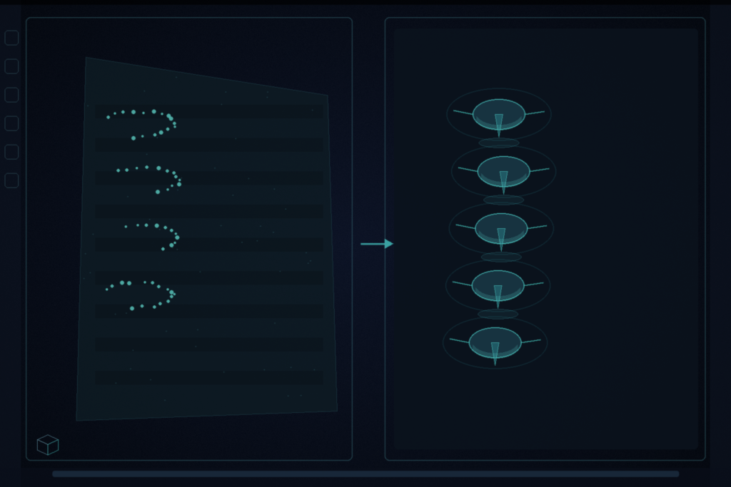
Hang an intraoperative lumbar ultrasound and you never see the whole spine. Bone casts deep shadows. The probe FOV is small. The surface you get from segmentation is a partial point cloud with holes and noise. ZODIAC takes that partial bone surface and returns a watertight 3D lumbar mesh in about eight seconds. Miruna-Alexandra Gafencu and Vlad Bratulescu (equal contribution), with Yordanka Velikova, Mohammad Farid Azampour, and Nassir Navab (Chair for Computer Aided Medical Procedures, Technical University of Munich; Munich Center for Machine Learning; Gafencu also Konrad Zuse School of Excellence in Reliable AI) posted arXiv:2608.24422 on 25 August 2026. The paper is ZODIAC: Zero-shot Octree-based Diffusion for Anatomical Completion. The claimed code URL github.com/miruna20/ZODIAC returned 404 when checked on 31 August 2026.
Prior ultrasound shape-completion nets learn a conditional map from synthetic incomplete-complete pairs. Real intraoperative holes do not follow that simulated occlusion distribution. Those methods also finish vertebrae one at a time, so global curvature and relative pose can drift. ZODIAC learns only from complete CT spines, then conditions at inference without paired training.
Octree diffusion prior, then blended completion
Complete lumbar meshes are stored as adaptive geometric octrees that spend resolution near bone surfaces. A VAE compresses each octree into a latent octree and decodes a continuous signed-distance field. Marching cubes extracts the mesh. A two-stage denoising diffusion model learns the prior over complete shapes. A low-resolution UNet denoises base-depth split signals for global structure and inter-vertebral layout. A high-resolution graph UNet then denoises per-node latents for surface detail inside that envelope.
At test time the partial ultrasound point cloud is rasterized into the same base-depth split grid with a one-sided mask: only positively observed geometry is locked. Unobserved entries stay free, because acoustic shadow is not the same as empty space. Blended completion initializes the reverse process from a noised version of that partial grid. At every low-resolution reverse step, observed patches are re-noised to the current noise level and blended back in, while the prior fills the rest. Blending runs only on the low-resolution stage. The high-resolution stage refines the whole surface. Re-noising matters: pinning the clean observation at every step (ZODIAC-clean) worsens Chamfer, HD95, and F1 across phantom and volunteer splits.
Complete CT spines for training, Balgrist US for stress tests
Training uses 91 VerSe20 lumbar meshes with deformation augmentation plus 322 TotalSegmentator lumbar meshes. The paper reports 504 spines in the training run. Representation is a 2563 SDF. VAE then LR diffusion then HR diffusion, batch size 1, on an RTX 4080. Inference uses 200 diffusion steps and about eight seconds per spine.
Evaluation covers two anthropomorphic lumbar phantoms and six Balgrist volunteer scans (three subjects, handheld and robotic each), the public paired US-CT set. A difficult subset of three acquisitions leaves one or more vertebrae entirely absent from the partial input. Against the paired supervised twin TP-ODIAC on those difficult whole-spine cases, zero-shot ZODIAC reports HD95 8.02 mm versus 10.26 mm (a 22% cut) and Chamfer down about 19%. F1 still trails the supervised twin by about 11 to 15% depending on the split. The per-vertebra supervised baseline Shape Completion in the Dark is more brittle when coverage is noisy or incomplete.
How this lands in a viewer
If you already hang intraoperative ultrasound next to planning CT in a DICOM viewer, treat this as a whole-spine mesh rail: load the partial bone surface from US segmentation, run completion, and hang the watertight lumbar mesh for orientation during epidural or pedicle work. Fail closed when the US segmentation is empty or the vertebral levels cannot be identified. Do not treat volunteer HD95 of 8.02 mm on three difficult cases as a device claim. Rebuild once the authors restore the repo; as of 31 August 2026 the GitHub URL in the abstract still 404s.
AnatoProto is blind fetal US sweep in, AC plane flags out. FARR3D is CBCT volume in, liver overlay without CBCT labels. SLIP is interactive 3D segmentation with latency and undo. This paper is partial intraoperative lumbar US surface in, complete lumbar mesh out.
Rebuild from arXiv:2608.24422 when code lands. The preprint is the source of record until a camera-ready version exists.
Sources
- Gafencu, M.-A., Bratulescu, V., Velikova, Y., Azampour, M.F., Navab, N. ZODIAC: Zero-shot Octree-based Diffusion for Anatomical Completion. arXiv:2608.24422, posted 25 August 2026. https://arxiv.org/abs/2608.24422. Claimed code: github.com/miruna20/ZODIAC (HTTP 404 on 31 August 2026).
- Sekuboyina, A., et al. VerSe: A Vertebrae labelling and segmentation benchmark for multi-detector CT images. Med. Image Anal. 2021. DOI: 10.1016/j.media.2021.102166
- Wasserthal, J., et al. TotalSegmentator: Robust segmentation of 104 anatomic structures in CT images. Radiol. Artif. Intell. 2023. DOI: 10.1148/ryai.230024
- Cavalcanti, N.A., et al. A large, paired dataset of robotic and handheld lumbar spine ultrasound with ground-truth CT benchmarking. Sci. Data 2025.
- Gafencu, M.A., et al. Shape completion in the dark: completing vertebrae morphology from 3D ultrasound. Int. J. Comput. Assist. Radiol. Surg. 2024.
- Xiong, B., et al. OctFusion: Octree-based diffusion models for 3D shape generation. Computer Graphics Forum 2025.
- Ho, J., Jain, A., Abbeel, P. Denoising diffusion probabilistic models. NeurIPS 2020.
- Avrahami, O., Lischinski, D., Fried, O. Blended diffusion for text-driven editing of natural images. CVPR 2022.
- Lugmayr, A., et al. RePaint: Inpainting using denoising diffusion probabilistic models. CVPR 2022.