
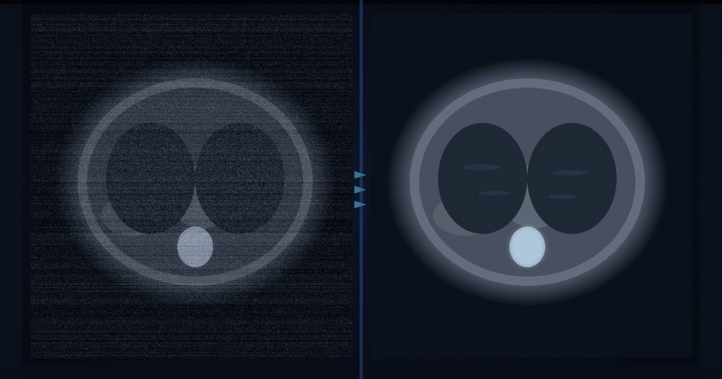
You hang a low-dose CT stack and wish the grain were quieter, without a paired normal-dose series sitting in the archive for training. PIPG-LDCT takes one low-dose measurement, builds two approximately independent noisy training branches from that single acquisition by modeling mixed Poisson-Gaussian projection noise, trains an image-domain Noise2Noise-style U-Net on those branches, then iterates by pushing restored images back into a sinogram prior. At inference you get a cleaner CT stack out. Xianlei Han, Shaoyu Wang, Jiancheng Fang, Weiwen Wu, and Qiegen Liu (Nanchang University / Sun Yat-sen) posted arXiv:2609.02654 around 3 September 2026. The paper is Physics-Driven Independent Pair Generation for Iterative Self-Supervised Low-Dose CT Denoising. Code is public at github.com/yqx7150/PIPG-LDCT.
The training trick is physics, not a second scan. A learned sinogram prior plus the LDCT noise model infer latent photon counts so the Poisson and Gaussian parts can be separated. Binomial thinning acts on the Poisson part; Gaussian thinning acts on the readout part; residual scaling variance-matches each branch to the observation. Both branches are reconstructed, then used as a self-supervised pair. No paired normal-dose CT is required to build those training pairs.
One measurement becomes two training branches
Most self-supervised denoisers lean on generic image statistics. PIPG-LDCT starts in projection space. Posterior photon-count inference under the mixed Poisson-Gaussian model matters more than a plain Poisson split. Variance matching keeps the branch noise levels honest relative to the observed low-dose measurement. The image-domain U-Net is the workhorse once the pair exists. Cross-domain iteration closes the loop: forward-project the restored images, refresh the sinogram prior, and regenerate cleaner training pairs for the next round.
At inference the pipeline is sinogram prior plus residual fusion, then image-domain refine. That is the path you would hang beside a low-dose series if you were wiring this into a viewer rail.
Where the numbers landed
Evaluation covers simulated AAPM Mayo, LIDC-IDRI, and LoDoPaB-CT at 0.5% and 1.0% dose (I0 of 5e3 and 1e4), plus a real GE clinical cardiac CT case and a real mouse chest CT. Among the self-supervised baselines the authors evaluated, PIPG-LDCT ranked best. It is comparable to, and often beats, supervised RED-CNN on PSNR, SSIM, and RMSE. On AAPM at 0.5% dose, theirs reports PSNR 38.39 against RED-CNN at 37.77. On LIDC at 0.5% dose, 40.89 against 39.10. Ablations put the largest share of the gain on the image-domain network, with clear help from the posterior split, variance matching, and the cross-domain iteration.
How this lands in a viewer
If you already hang low-dose CT in a DICOM viewer, treat PIPG-LDCT as a self-supervised denoise rail that does not wait for paired normal-dose labels. Load the noisy stack, run the prior-plus-residual fusion path, then the image-domain refine, and hang the cleaner series for review. Fail closed when scatter or beam-hardening leave residual correlation between the generated branches, when posterior or prior error breaks the independence assumption the training pair needs, or when you need true 3D or multi-slice behavior (the current implementation is 2D). Do not treat the RED-CNN comparisons as a guarantee on your scanner’s noise model until you re-validate on local data.
CT-DeltaBench scores prior-versus-current CT difference reports. Axial CT organ boxes hang left/right spatial calls from a single slice. This paper is low-dose CT in, cleaner stack out, without paired normal-dose training pairs.
Rebuild from arXiv:2609.02654. As of 3 September 2026 the abstract and PDF respond (HTTP 200). Code: https://github.com/yqx7150/PIPG-LDCT (HTTP 200 on 3 September 2026). The preprint is the source of record until a camera-ready version exists.
Sources
- Han, X., Wang, S., Fang, J., Wu, W., Liu, Q. Physics-Driven Independent Pair Generation for Iterative Self-Supervised Low-Dose CT Denoising. arXiv:2609.02654, posted ~3 September 2026. https://arxiv.org/abs/2609.02654 (HTTP 200 on 3 September 2026). PDF: https://arxiv.org/pdf/2609.02654 (HTTP 200 on 3 September 2026). Code: https://github.com/yqx7150/PIPG-LDCT (HTTP 200 on 3 September 2026).