
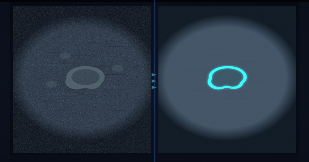
Hang a lesion frame with cluttered background: hair on dermatoscopy, speckle on ultrasound, folds on endoscopy. FreNet injects a SAM visual prior into the raw pixels before encoding, reconfigures features in frequency and space during encoding, and returns a cleaner lesion overlay. Yinan Liu, Jiankang Hong, Zhen Gao, and Ye Luo (Tongji University) posted arXiv:2609.03535 around 3 September 2026. The paper is FreNet: Feature Reconfiguration With Visual Prior for Medical Lesion Segmentation. No public code repository is linked in the preprint (checked 4 September 2026).
Complex background and diverse lesion morphology still break a lot of overlay rails. FreNet attacks that in two places. First, an Implicit Prior Neural Network (IPNN) models a continuous spatial field and applies pixel-wise reconfiguration weights so the SAM prior quiets background early, before the backbone sees the frame. Second, Dual-domain Feature Reconfiguration (DFR) sits between PVTv2 stages: a Frequency Decoupling Module (FDM) separates bands, and a Spatial Localization Module (SLM) relocates and sharpens lesion response. The same dual branch can wrap TransUNet or SAM instead of PVTv2.
Prior in the pixels, then frequency and space
IPNN takes coordinates plus the prior mask, builds dense reconfiguration weights through an implicit decoder, and writes a reconfigured input that the encoder actually consumes. That is early lesion-aware activation, not a late decoder hint. DFR then works stage by stage. FDM uses a ratio-based frequency split so high-frequency clutter and low-frequency structure get different treatment. SLM keeps spatial continuity while pulling lesion evidence back together. Residual fusion feeds the next stage. Grad-CAM and t-SNE in the paper show tighter lesion clusters and quieter background after both modules are on.
Evaluation covers nine 2D benchmarks across three modalities: dermoscopy (ISIC2018, PH2), ultrasound (BUSI, STU), and endoscopy polyps (CVC-ColonDB, CVC-ClinicDB, Kvasir, ETIS, CVC-300).
Where the numbers landed
On ETIS, Ours(SAM) reports Dice 82.0, which is +7.2 against SAM on that set. Across the nine datasets the authors report Dice gains of 0.7 to 5.0 against SOTA and 0.7 to 7.2 against SAM, with the lowest false-positive rate among the compared methods. The full trainable stack is about 38.31M parameters (SAM 4.07M, IPNN 0.04M, DFR 2.97M, backbone 25.40M). Ablations on ISIC2018, BUSI, and ETIS need SAM plus IPNN plus DFR for the best scores; dropping either IPNN or DFR hurts. Fig. 6 bins SAM mask quality into low, medium, and high and still shows gains, including a large lift on ETIS where SAM masks are often weak.
How this lands in a viewer
If you already hang dermoscopy, breast ultrasound, or polyp endoscopy in a DICOM viewer, treat FreNet as a prior-then-reconfigure overlay rail. Load the 2D frame, run IPNN so the SAM prior reweights pixels before encode, let DFR clean frequency and space features on the way through the backbone, and hang the returned lesion contour for review. Fail closed when the SAM prior is garbage for that frame (the paper still reports gains across quality bins, but a collapsed prior is still a stop condition), when you need volumetric CT or MRI stacks rather than 2D lesion frames, or when compute budget cannot absorb the extra cost the authors flag (they point to knowledge distillation as future work). Do not invent a GitHub link; none appears in the preprint.
MedPixel hangs a phrase or loose box into an overlay plus a short sentence. GazeRefine turns clinician gaze into a training-free overlay. This paper is lesion frame in, background quieted before encoding, cleaner overlay out.
Rebuild from arXiv:2609.03535. As of 4 September 2026 the abstract and PDF respond (HTTP 200). No public code link appears in the preprint. The preprint is the source of record until a camera-ready version exists.
Sources
- Liu, Y., Hong, J., Gao, Z., Luo, Y. FreNet: Feature Reconfiguration With Visual Prior for Medical Lesion Segmentation. arXiv:2609.03535, posted ~3 September 2026. https://arxiv.org/abs/2609.03535 (HTTP 200 on 4 September 2026). PDF: https://arxiv.org/pdf/2609.03535 (HTTP 200 on 4 September 2026). No public code repository linked in the preprint.