
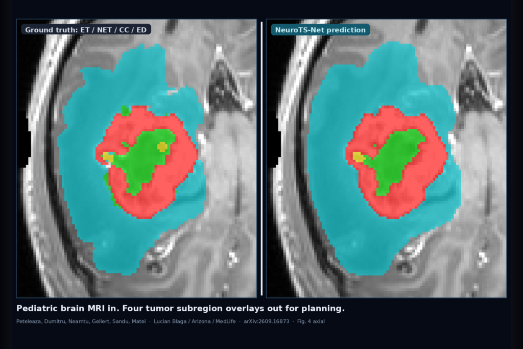
Hang a pediatric brain mpMRI study and ask for one overlay language that separates enhancing tumor, non-enhancing tumor, cystic component, and peritumoral edema. The structures are small, rare, and often low contrast. Manual delineation eats planning time and varies between readers. Training separate networks per subregion is wasteful when the four sequences already sit in one hanging. Darius Peteleaza, Razvan-Gabriel Dumitru, Bogdan Neamtu, Arpad Gellert, Mariana Sandu, and Claudiu Matei (Lucian Blaga University of Sibiu with University of Arizona, Pediatric Clinical Hospital of Sibiu, Johns Hopkins, and MedLife Polisano) posted arXiv:2609.16873 on 15 September 2026. The paper is NeuroTS-Net: Multi-Class Semantic Segmentation of Pediatric Brain Tumors in Multi-Modal MRI.
They train one compact 3D encoder-decoder on BraTS 2026 pediatric Task 2 mpMRI (T1N, T1C, T2W, T2F) with no external data and no pretrained weights. On the internal validation set, whole-tumor and tumor-core Dice reach 0.938 and 0.937. On the official challenge validation set those composites stay at 0.927 and 0.926 while still beating matched nnU-Net and MedNeXt runs under the same protocol.
What hangs on the viewer
Upstream, four co-registered sequences enter a five-resolution NeuroTS encoder-decoder. A dual-scale raw-detail stream keeps fine intensity and boundary cues that usual aggressive downsampling throws away. Adaptive low-resolution context selection picks which coarse features matter for the current tumor layout. Multipath downsampling preserves detail while the network still sees broader anatomy. Labels are voxel-wise enhancing tumor (ET), non-enhancing tumor (NET), cystic component (CC), and edema (ED). Tumor core and whole tumor are composites for scoring, not separate heads.
Downstream for a DICOM shop is a single multi-class SEG overlay you can hang next to the source series. Fig. 4 shows the qualitative check readers care about: axial, coronal, and sagittal panels with T1N/T1C/T2W/T2F on the left, ground truth in the middle, and the NeuroTS-Net prediction on the right. Red, green, yellow, and cyan mark ET, NET, CC, and ED. The predicted whole-tumor contour tracks the reference closely, with the usual local boundary wobble on cyst and edema edges. The model is compact (about 18.7M parameters) and the paper reports mean inference around 33 seconds per case on their hardware, faster than the larger nnU-Net and MedNeXt baselines they matched.
Why four overlays from one pass matter
Pediatric planning and response assessment need more than a binary blob. Enhancing rind, non-enhancing core, cyst, and edema drive different decisions, and they live on different contrasts in the same study. If your viewer already supports multi-label SEG and a four-sequence hanging, this paper is a concrete blueprint for one overlay vocabulary instead of four modality-locked or class-locked models. Whole tumor and tumor core give you quick QC numbers. The four tissue classes are what a reader audits against T1C and T2F.
Use the published composites as deployment checks, not as a product claim. Internal WT/TC near 0.94 tells you the overlay is in a usable band for assistive display on their split. Official validation WT/TC near 0.93 under the same protocol is the number closer to challenge distribution. Enhancing tumor and cyst remain harder than the composites, which is exactly where a reader should zoom and edit.
Where it still fails
Edema and cystic component are the weak classes. On the official validation set every compared method, including NeuroTS-Net, scored zero Dice for ED under the lesion-wise matching rules when predicted and reference components do not match. The authors flag rare-class overrepresentation in the internal split and cohort differences as likely causes. Connected-component post-processing is applied only to ET and CC predictions, so edema false positives or misses still hit the score hard. Evaluation used a single internal split and training seed, so patient and seed variability are not fully mapped. Fail closed when the series is not pediatric BraTS-style mpMRI, when edema floods into healthy white matter with no T2F support, when enhancing islands appear without T1C correlate, or when the whole-tumor contour fragments across slices.
For a viewer or clinic AI shop
Treat this as a pediatric brain-tumor rail: four-sequence mpMRI in, one multi-class overlay out with ET, NET, CC, and ED layers (plus optional WT/TC composites for QC). Hang the source T1C and T2F first. Then hang the four-color mask so a reader can audit enhancing rim versus cyst versus edema against the contrasts that define them. Fail closed when whole-tumor overlap against a quick QC contour is poor, when ED or CC lights up on an ED/CC-absent case, when the study mixes adult glioma protocols the BraTS-PEDs training never saw, or when someone wants edema metrics published from a model that still collapses under the official lesion-wise ED rule. If your platform already supports multi-label SEG and multi-sequence hangings, this paper is a practical pattern for shipping one compact pediatric subregion model instead of four separate class nets.
Rebuild from arXiv:2609.16873. As of 17 September 2026 the abstract and PDF respond. Code is linked at github.com/maenstru56/NeuroTS.
Sources
- Peteleaza, D., Dumitru, R.-G., Neamtu, B., Gellert, A., Sandu, M., Matei, C. NeuroTS-Net: Multi-Class Semantic Segmentation of Pediatric Brain Tumors in Multi-Modal MRI. arXiv:2609.16873, posted 15 September 2026. https://arxiv.org/abs/2609.16873. PDF: https://arxiv.org/pdf/2609.16873.
- BraTS 2026 pediatric Task 2 / BraTS-PEDs (cited challenge data and scoring).
- Isensee et al. nnU-Net; Roy et al. MedNeXt (cited baselines under matched protocol).
- Code: https://github.com/maenstru56/NeuroTS.