
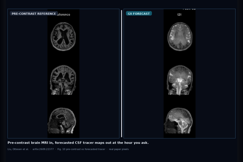
Hang a predicted CSF tracer enhancement volume and a change map next to the baseline T1w, at a requested hour after injection, without waiting for the delayed repeat scan. That is the clinic ask Liu, Ottesen, Nguyen, Ringstad, Mossige, Svensson, Bjørnerud, Eide, and Emblem take on in arXiv:2609.22377. From a pre-contrast T1-weighted brain MRI and an elapsed time ∆t, their networks forecast the follow-up intrathecal-tracer T1w, not a same-time virtual-contrast trick. The work targets glymphatic clearance, idiopathic normal pressure hydrocephalus (INPH), and CNS drug-delivery study workflows where patients otherwise return for scans over one or more days.
Repeated post-injection imaging is hard on elderly subjects and on scheduling. A model that fills the missing mid-course volume from anatomy already on the console would let a viewer or research rail keep reading while the true follow-up is still hours away. The paper keeps that claim research-grade: same 3D U-Net backbone, two training targets, fixed patient-level splits, and an external INPH late-time set.
What hangs on the viewer
Input is a co-registered pre-contrast T1w volume plus the hour you care about. Output is a forecasted follow-up T1w at that ∆t, plus the implied change relative to the reference. In a DICOM viewer that means a predicted enhancement series you can hang beside the baseline, a diverging change overlay for sulcal and ventricular signal, and a time scrubber that asks for 5 h, 24 h, or 48 h without requiring those acquisitions yet.
The authors compare two heads on the same backbone. Direct image-to-image regression (I2I) maps reference plus ∆t to the follow-up in one forward pass. Conditional flow matching (CFM) integrates a learned velocity field over ten Euler steps (ten network evaluations). Both stay inside a brain mask for the primary score.
How it works in plain words
Main-cohort patients are split 104 / 22 / 23 into train / validation / test. Training uses every earlier-to-later within-patient pair so the network sees forecasting, not same-time synthesis. Volumes are co-registered within patient, resampled to 1.5 mm isotropic, and intensity-normalized. The network never sees the true follow-up at inference; ∆t is injected as a conditioning signal.
I2I is trained with a brain-weighted L1 loss on the follow-up image. CFM starts the flow at the reference (with light noise in training) and learns the change velocity, again with a brain-weighted L1 on velocity. At test time CFM runs ten NFEs; I2I runs once. Checkpoints are picked by lowest validation masked MAE on one 12 GB RTX A2000 training setup.
Evaluation is built around the change signal. The identity baseline returns the pre-contrast scan unchanged, so its masked MAE equals the mean absolute true change. A voxel-wise temporal polynomial is a second weak baseline. Metrics are averaged within patient first, then across patients with bootstrap intervals, so densely sampled subjects do not dominate.
What the numbers say
Internal test is 23 patients and 180 pairs. Identity masked MAE is 0.089. CFM reaches 0.058. I2I reaches 0.036, about a 60% cut versus copying the pre-contrast scan and 38% lower than CFM. Enhancement Dice at threshold 0.05 is 0.647 for I2I versus 0.513 for CFM. I2I wins on 22 of 23 internal patients at the patient-level mean.
External INPH evaluation uses 51 held-out patients and 67 pairs, mostly at 20-30 h and beyond 40 h, with ventriculomegaly and strong ventricular signal. Identity MAE is 0.075, CFM 0.063, I2I 0.056. Patient-level I2I wins in 34 of 51 external subjects. The hardest internal window is 20-30 h, where identity peaks and both models still beat it; residuals stay largest where enhancement is strongest.
Where it fails and what not to trust
Late INPH morphology differs from the main cohort, and the external time mix does not match the internal distribution. CFM sample standard-deviation maps rank likely error voxels, yet a ten-sample ensemble still trails single-pass I2I on MAE. Visual hard cases at 20-30 h keep gross anatomy but leave residual error in the brightest enhancement zones.
This is not a cleared medical device. Treat forecasts as research overlays for glymphatic, INPH, and intrathecal drug-delivery studies, with a human gate before any clinical decision. Rebuild thresholds, masks, and timing bins on your own protocol before you hang them in a production viewer.
For a viewer or clinic AI shop
Wire pre-contrast T1w plus a requested ∆t in; hang the I2I follow-up volume and a change map out. Prefer the single-pass regression head when latency matters; keep CFM uncertainty maps as an optional second rail if you want a spatial error hint. Surface the hour on the overlay so readers never confuse a forecast with an acquired series.
If you already ship a brain MRI viewer for research cohorts, the natural hang points are a forecast series in the same frame of reference as the baseline, a masked change LUT, and a provenance tag naming model, ∆t, and arXiv:2609.22377. Audit on your own INPH and delayed-tracer studies before you advertise hand-off. Rebuild from the paper rather than trusting the published MAE alone.
Rebuild from arXiv:2609.22377. PDF: https://arxiv.org/pdf/2609.22377.
Sources
- Liu, Q., Ottesen, J. A., Nguyen, T., Ringstad, G., Mossige, I., Svensson, S. F., Bjørnerud, A., Eide, P. K., Emblem, K. E. Forecasting Intrathecal Tracer Enhancement from Pre-Contrast Brain MRI: Direct Regression versus Flow Matching. arXiv:2609.22377, 2026. https://arxiv.org/abs/2609.22377. PDF: https://arxiv.org/pdf/2609.22377.
- Task framing: pre-contrast T1w + elapsed hours ∆t → forecasted follow-up intrathecal-tracer T1w enhancement (not same-time virtual contrast). Primary metric: masked MAE versus identity and CFM; enhancement Dice at threshold 0.05.