
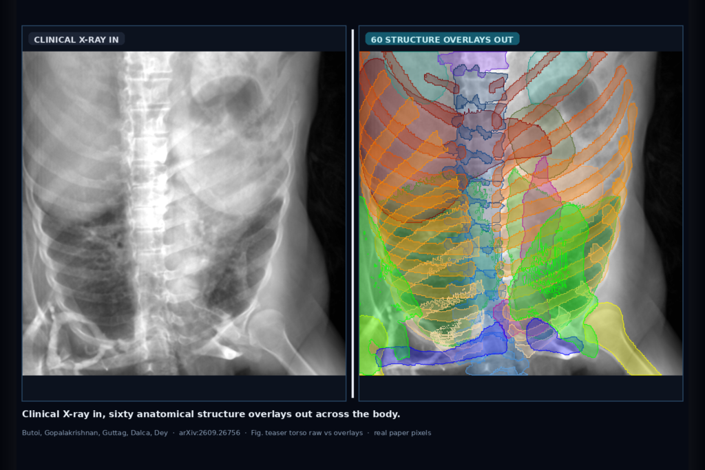
Hang a clinical X-ray into a viewer and get overlapping structure overlays across the body, not one organ model at a time. That is the clinic ask Butoi, Gopalakrishnan, Guttag, Dalca, and Dey take on in arXiv:2609.26756, posted around late September 2026. The paper is FleXray: Universal Clinical X-ray Segmentation, from MIT CSAIL with Massachusetts General Hospital and Harvard Medical School. Instead of hand-labeling dense multi-organ X-ray masks, the team builds a physics-based generative data engine from labeled whole-body CTs, then trains a generalist segmentor that predicts 60 anatomical structures on real radiographs.
X-ray is still the highest-volume modality worldwide, yet most quantitative anatomy tools live on CT and MRI. Projection overlap makes dense pixel labels hard even for experts, so public X-ray datasets usually cover a few chest or limb organs under narrow views. FleXray treats that annotation gap as a data-engineering problem: render digitally reconstructed radiographs (DRRs) with projected CT labels, randomize geometry and organ attenuation, close the appearance gap with generative editing, and mix in a little real peripheral X-ray supervision where CT coverage is thin.
What hangs on the viewer
Upstream is a routine clinical radiograph or fluoro frame: chest, spine, pelvis, limbs, pediatric or adult, frontal, lateral, or oblique. No prompts and no acquisition metadata are required at inference. Downstream outputs are independent probability maps for up to 60 structures (skull, ribs 1 through 12, vertebrae C1 through L5, long bones, hands and feet, lungs, heart, liver, kidneys, spleen, and related skeletal anatomy). Because X-ray rays can hit several organs in one pixel, the maps are multi-label and overlapping rather than a single exclusive label map.
For a DICOM viewer or clinic AI rail, the hangable pieces are structure overlays on the projection, structure chips for morphometry (Cobb angle from vertebra masks, lung fields, hip landmarks), and optional reuse of those masks as semantic priors for 2D/3D registration during fluoro-guided navigation. The authors also ship a local browser tool at flexray.csail.mit.edu so teams can try overlays on arbitrary X-rays without standing up training infra first.
How it works in plain words
The data engine aggregates densely labeled CT sources (notably MOOSE whole-body segmentations plus targeted head-neck, pediatric, fracture, shoulder, and elbow sets). At training time it samples projection geometry, field of view, magnification, and per-structure attenuation, then projects each 3D mask independently so overlaps stay honest. Offline, a pretrained image editor turns analytic DRRs into more clinical-looking radiographs while an automatic filter drops grossly hallucinated edits (about 4% rejected). Real hand, foot, and MURA arm annotations fill peripheral gaps that whole-body CT rarely images.
FleXray is an ensemble of five 2D U-Nets trained on different mixes of online DRRs versus generatively enhanced DRRs, with 16-sample test-time augmentation at inference. Partial-label losses ignore structures a source never annotated instead of treating them as absent, and targeted negatives block anatomically impossible classes (for example, hand classes on foot X-rays). The released recipe also includes a harmonized 60-structure vocabulary and a Hugging Face dataset of enhanced DRRs plus splits for fair follow-on work.
What the numbers say
On eight held-out real X-ray datasets covering chest, ribs, fluoro pelvis, elbow, hip, lower limb, wrist, and pediatric torso, FleXray reaches an equal-dataset mean Dice of 0.875. It beats every supported baseline (PAXray, TotalSegmentator2D, FluoroSAM with oracle prompts) on seven of eight sets, and trails dedicated per-dataset nnU-Nets by only about 0.039 Dice on average (0.875 versus 0.914). On DeepFluoro it even exceeds the dataset-trained nnU-Net (0.915 versus 0.866), which matters when training data is scarce and angles vary.
Downstream reuse is the clinic angle. From vertebra masks on AASCE spine X-rays, FleXray recovers major Cobb angle with mean absolute error 5.13 degrees and zero unmeasurable failures across 218 images, inside typical inter-observer ranges (about 6 to 7 degrees). Injecting FleXray distance transforms into an iterative 2D/3D registration pipeline (xvr) cuts median mean target registration error from hundreds of millimeters down to about 1.2 mm from a rough frontal init, and clears catastrophic failures that image similarity alone leaves behind. Fine-tuning the same backbone on scarce labels also helps new pelvic anatomy and bone-tumor detection budgets where random-init nnU-Net lags.
Where it fails and what not to trust
Evaluation datasets with dense multi-structure ground truth are still sparse, so many quantitative scores sit on curated research sets rather than messy hospital archives full of burned-in text. Severe trauma, unusual implants, and strongly out-of-distribution hardware can break overlays because pathologies and devices are not systematically synthesized beyond what source CTs already contain. Soft-tissue contrast on plain film remains limited; overlapping ribs and abdominal organs can still confuse lower-rib predictions even when generative enhancement helps.
The model is research software with released weights, code, data, and a browser demo. It is not a cleared medical device. Treat overlays as assistive maps that need human review before measurement export or navigation, and rebuild thresholds on your own scanners, pediatric mix, and implant load.
For a viewer or clinic AI shop
Wire clinical X-ray or fluoro in; hang multi-structure overlays out, with per-channel opacity controls so readers can isolate ribs, vertebrae, lungs, or pelvis without drowning the radiograph. Prefer exposing the overlapping multi-label nature instead of forcing a winner-take-all color map that erases true projection overlap. Natural next products are Cobb or landmark tools derived from vertebra and pelvic masks, and a registration assist that uses the same overlays as a Chamfer prior when C-arm pose search gets stuck.
If you already ship a radiography or OR viewer, start with a side panel of structure chips, a one-click full overlay, and a gate that requires human confirm before chart or navigation export. Validate on your fluoro angles and text-heavy CR/DR archives before you advertise hand-off. Rebuild from arXiv:2609.26756. PDF: https://arxiv.org/pdf/2609.26756. Demo: https://flexray.csail.mit.edu. Data: VictorButoi/flexray-data.
Sources
- Butoi, V. I., Gopalakrishnan, V., Guttag, J. V., Dalca, A. V., Dey, N. FleXray: Universal Clinical X-ray Segmentation. arXiv:2609.26756, 2026. https://arxiv.org/abs/2609.26756. PDF: https://arxiv.org/pdf/2609.26756. Demo: https://flexray.csail.mit.edu. Dataset: https://huggingface.co/datasets/VictorButoi/flexray-data.
- Key held-out segmentation result from the paper: equal-dataset mean Dice 0.875 across eight real X-ray sets; average gap to per-dataset nnU-Nets about 0.039 Dice.
- Downstream signals from the paper: Cobb MAE 5.13 degrees on AASCE (218 images, no failures); Chamfer-augmented 2D/3D registration median mTRE about 1.2 mm from frontal init on DeepFluoro held-out frames. Treat as research workflow evidence, not a device claim.