
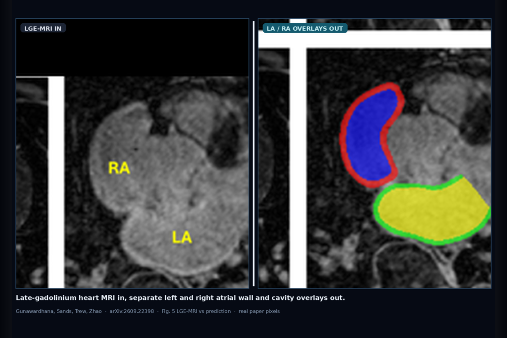
Hang a late-gadolinium heart MRI into a cardiac or EP viewer and you want separate left and right atrial wall masks plus cavity masks, not one merged bi-atrial blob. That is the clinic ask Gunawardhana, Sands, Trew, and Zhao take on in arXiv:2609.22398, posted around mid-September 2026 from the Auckland Bioengineering Institute. The paper is Multicentre Bi-atrial Segmentation from LGE-MRI for Atrial Fibrillation with a 2D and 3D Framework, and the authors name their pipeline TASSNet. They train on Utah LGE-MRI, then measure transfer to Waikato and Kobe without fine-tuning, so you can see what survives a site shift before you wire overlays into fibrosis or ablation planning tools.
Fibrosis quantification on LGE sits on thin atrial wall borders next to the blood pool. A few voxels of wall error can swing scar burden. Manual slice-by-slice labels are slow and hard to scale. Prior challenges often score a combined bi-atrial wall or only cavities, which hides chamber-specific wall thickness and septal errors. TASSNet keeps four labels: LA wall, RA wall, LA cavity, and RA cavity.
What hangs on the viewer
Upstream is a 3D late-gadolinium enhancement cardiac MRI volume from an AF workup. Downstream are four class masks that pad back to the original grid: left and right atrial walls, left and right atrial cavities. Stage 1 first localises an atrial ROI with a 3D network (100% crop success on the 41 held-out test scans in the paper). Stage 2 runs fine segmentation on that crop, then restores the masks to full volume so a DICOM viewer can hang them beside the source series.
For a viewer or clinic AI rail, the hangable pieces are LA/RA wall overlays for fibrosis ROIs, cavity overlays for chamber volumes, and a clear split between wall and cavity so scar metrics do not leak into the blood pool. The authors also show that cavity overlays transfer across centres more cleanly than wall overlays, which is the practical warning for any multi-site EP deployment.
How it works in plain words
TASSNet is a two-stage U-Net stack with ResNeXt encoder blocks and instance normalisation. After the ROI crop, a 2D U-Net works slice by slice for in-plane edge detail, while a 3D U-Net keeps through-plane continuity on the same crop. Softmax maps from both streams are averaged voxel-wise and argmaxed into the final four-class mask. The paper compares that ensemble against nnU-Net variants, SegResNet, Swin UNETR, SAMed, and U-Mamba setups under the same train-on-Utah, test-elsewhere protocol.
Training used DiceFocal loss on patient-level Utah splits (80 train, 20 held-out test). Waikato (11) and Kobe (10) stayed fully external. No target-site fine-tuning. Labels came from Amira slice-by-slice delineation of the four structures. That setup is closer to a real multi-vendor import path than a same-hospital holdout.
What the numbers say
On Utah, the ResNeXt ensemble is the balanced option across the four structures: RA wall Dice 0.753, LA wall 0.620, RA cavity 0.920, LA cavity 0.923. Cavities sit near 0.92 while walls lag, which matches how hard thin LGE borders are even in-domain.
On Waikato without fine-tuning, cavities still hold (RA 0.867, LA 0.870) while walls drop (RA 0.681, LA 0.561). On Kobe, the harder shift, LA cavity stays at 0.902 and RA cavity at 0.832, but RA wall falls to 0.511 and LA wall to 0.521. Average Dice across structures in the paper’s 3D error maps lands near 84.6% Utah, 79.4% Waikato, and 74.9% Kobe. The takeaway for a clinic shop is not a leaderboard spike. It is that cavity overlays are the safer first hang across sites, and wall overlays need local review or adaptation before you trust fibrosis numbers.
Where it fails and what not to trust
Wall segmentation is the main failure mode under domain shift, especially on Kobe, where intensity and through-plane sampling diverge from Utah. The paper’s qualitative failures show broken thin-wall borders and local leaks near pulmonary veins and valvular planes. Inter- and intra-observer variability on the labels was not quantified, so absolute Dice should be read as method ranking evidence, not a cleared measurement claim.
TASSNet is research software and a multicentre benchmark, not a medical device. Treat wall overlays as assistive contours that need EP or radiology review before scar export or ablation planning. Rebuild thresholds on your own LGE protocol, vendor mix, and AF subtype load. Prefer shipping cavity and wall as separate channels so a reader can hide a noisy wall layer without dropping chamber volumes.
For a viewer or clinic AI shop
Wire LGE cardiac MRI in; hang four independent LA/RA wall and cavity overlays out, with per-channel opacity and a site-shift banner when the series comes from a scanner family you have not validated. Keep fibrosis tools gated on wall-mask quality checks (surface distance or thinness heuristics) instead of auto-exporting scar burden from a soft wall Dice near 0.5.
If you already ship a cardiac or EP viewer, start with cavity overlays for volume panels, then add wall overlays behind a confirm step. Validate on at least one external LGE protocol before you advertise multi-site hang. Rebuild from arXiv:2609.22398. PDF: https://arxiv.org/pdf/2609.22398.
Sources
- Gunawardhana, M., Sands, G. B., Trew, M. L., Zhao, J. Multicentre Bi-atrial Segmentation from LGE-MRI for Atrial Fibrillation with a 2D and 3D Framework. arXiv:2609.22398, 2026. https://arxiv.org/abs/2609.22398. PDF: https://arxiv.org/pdf/2609.22398.
- In-domain (Utah) ResNeXt ensemble Dice from the paper: RA wall 0.753, LA wall 0.620, RA cavity 0.920, LA cavity 0.923.
- External transfer without fine-tuning (same ensemble): Waikato cavities ~0.87 with walls 0.681 / 0.561; Kobe LA cavity 0.902 / RA cavity 0.832 with walls 0.511 / 0.521. Treat as research transfer evidence, not a device claim.