
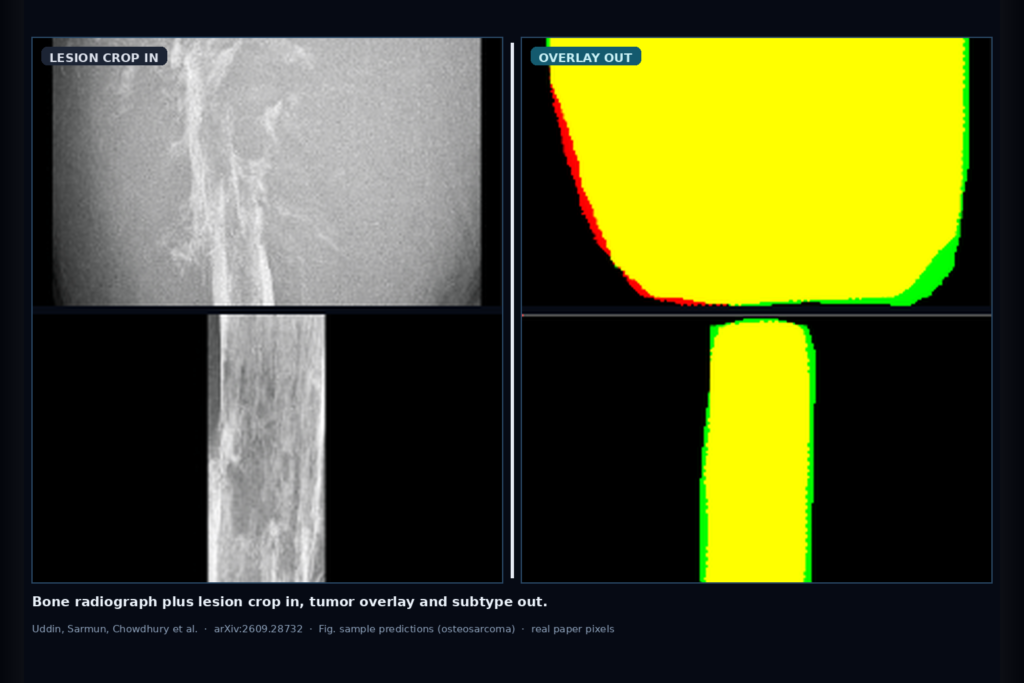
Hang a bone radiograph into a musculoskeletal viewer and you want two things next to the series: a lesion overlay you can scrub, and a subtype call that separates osteosarcoma from common benign look-alikes without sending every case to a second modality first. That is the clinic ask S.M. Nasif Uddin, Rusab Sarmun, Muhammad E. H. Chowdhury, Adam Mushtak, Israa Al-Hashimi, and Sohaib Bassam Zoghoul take on in arXiv:2609.28732, posted late September 2026 from Ahsanullah University of Science and Technology, the University of Dhaka, Qatar University, and Hamad Medical Corporation. The paper is Integrating Local Detail and Global Context: A Dual-Input Multi-Task Learning Framework for Bone Tumor Diagnosis. The authors name the network OsteoHiFuse-Net: a dual-stream DenseNet121 with bidirectional cross-modal attention between a YOLO lesion crop and the full radiograph, branching to lesion segmentation and three-class subtype classification on the multi-institutional BTXRD set (n=3,746).
Single-crop classifiers see margin texture but miss periosteal reaction and cortical breach outside the box. Full-image segmenters see anatomy but dilute small lesions. OsteoHiFuse-Net keeps both views in parallel, fuses them with cross-modal attention and hierarchical multi-scale fusion, then trains segmentation and classification together so the shared features serve surgical margin drawing and triage at once.
What hangs on the viewer
Upstream is a standard bone radiograph. A YOLOv11 detector proposes a lesion box, expands it 10%, squares it, and resamples a 256×256 crop. That crop pairs with the full radiograph as the dual input. Downstream is a predicted lesion mask (overlay on the crop or mapped back to the full frame), a three-class subtype label among osteosarcoma, osteochondroma, and other benign, and optional confidence or AUC-style scores for QC.
For a DICOM or MSK viewer rail, the hangable pieces are: the YOLO box as a first ROI suggestion, the segmentation overlay as a toggle layer, the subtype chip next to the study (with an explicit research/assistive flag), and a residual or disagreement view when the predicted mask and a human edit diverge. Detection stays a preprocessing stage in this paper, not a joint head, so plan a confidence gate (the authors reviewed crops below 0.5) before you trust the dual-input pass.
How it works in plain words
BTXRD supplies radiographs with boxes, masks, and clinical metadata across normal, benign, and malignant cases. The authors consolidate labels into three management-facing classes: osteosarcoma (malignant), osteochondroma (common benign), and other benign (giant cell tumour and simple bone cyst rolled in; rare subtypes dropped). Patient-level splits keep all studies from one patient in the same fold (80/10/10 train/val/test spirit with a held-out patient-level test).
YOLOv11x is trained only on the training split to avoid leakage, then used to crop ROIs for every split. Each ROI is paired with its full radiograph. Two DenseNet121 encoders run in parallel: one on the crop for local margin and matrix detail, one on the full image for global skeletal context. Bidirectional cross-modal attention lets each stream query the other. Hierarchical multi-scale feature fusion and CBAM-style attention sharpen the fused maps. A CA-UNet-style decoder produces the segmentation; a classification head reads the fused representation for the three-class call. Training mixes Dice and focal loss on the mask with a weighted classification loss that ups the osteosarcoma weight so missed malignancy costs more than a benign mix-up.
At inference the path is detector → dual-input OsteoHiFuse-Net → mask + subtype. The detector’s validation mAP@0.5 was 0.755 with precision 0.764 and recall 0.725, which is good enough to feed crops but still the first failure point for subtle low-contrast lesions. Multifocal detections each produce their own crop-and-full pair, so a viewer can page ROIs without rewriting the dual-input graph. Augmentation during training covered affine jitter, brightness/contrast and gamma shifts, mild blur and noise, which is the usual radiograph acquisition scatter rather than fancy synthetic pathology.
What the numbers say
On the held-out patient-level test split, OsteoHiFuse-Net reports an overall (sample-weighted) Dice of 0.896 and IoU of 0.821, with sensitivity 0.919 and specificity 0.924. Per-class Dice sits at 0.923 for osteosarcoma, 0.882 for osteochondroma, and 0.947 for other benign. Irregular permeative osteosarcoma margins are harder than smooth benign cortices, which matches the ordering.
Classification reaches a macro F1 of 0.928 and a macro AUC of 0.993. Per-class F1 is 0.936 (osteosarcoma), 0.976 (osteochondroma), and 0.871 (other benign). Osteosarcoma AUC is 0.999 with recall 0.917 and precision 0.957; osteochondroma AUC 0.993; other benign AUC 0.987. Overall accuracy is 0.955. The confusion matrix concentrates mistakes between osteosarcoma and other benign when early malignant and matrix-producing benign cases share mixed radiodensity. That is the band to watch in a triage UI: high malignant AUC does not erase borderline benign look-alikes.
Ablations in the paper favour the full dual-input plus cross-modal attention stack over single-stream baselines on both Dice and macro F1 (full stack lands at the abstract’s 0.896 / 0.928). Qualitative overlays show most errors as thin green (miss) or red (overcall) rings at the boundary, with yellow true-positive cores filling the lesion bulk, which is what you want for a scrubbable viewer layer.
Where it fails and what not to trust
This is research software on a public radiograph cohort, not a cleared medical device. Rebuild and validate on your vendor mix, age mix, and local subtype prior before you hang subtype next to a reportable impression.
Detection is the cascade bottleneck. Small, low-contrast, or subtly cortically disrupted lesions can be missed upstream; the paper treated detection as preprocessing, not a joint multi-task head. Class consolidation hides fine benign subtypes, so do not claim full WHO-style taxonomy. Per-location, per-subtype counts are thin, so flat-bone and rare-site generalization is unproven. Dual streams cost memory and latency versus a single crop CNN, which matters on thin edge boxes. Radiographs alone miss soft-tissue and intramedullary detail that MRI or CT would show; the authors flag hybrid extensions as future work.
Do not advertise the osteosarcoma AUC as a standalone screening claim without a local prevalence study. Do not skip the low-confidence YOLO review step. Do not hide that “other benign” is a consolidated bucket.
For a viewer or clinic AI shop
Wire bone radiograph in; hang YOLO ROI, dual-input mask overlay, and a three-class subtype chip out. Keep detection confidence and a human confirm gate before the dual-input pass. Prefer overlay-first UX (lesion scrubber) with subtype as a secondary assistive panel labeled research until you correlate against your MSK reads on a tagged cohort.
Log detector score, crop coordinates, predicted Dice-proxy vs any human edit, subtype probabilities, and which stream dominated attention if you expose that debug view. Keep osteosarcoma probability and the raw three-class softmax so a radiologist can see when “other benign” and osteosarcoma are close. Validate at least one internal holdout that mirrors BTXRD’s three-class taxonomy and one site where your case mix differs (pediatric long-bone heavy vs adult flat-bone heavy). If you already ship an MSK hanging protocol, start with ROI + overlay on the primary radiograph viewport, then add the subtype chip in a side panel that never overwrites the report impression field. Rebuild from arXiv:2609.28732. PDF: https://arxiv.org/pdf/2609.28732.
Sources
- Uddin, S.M.N., Sarmun, R., Chowdhury, M.E.H., Mushtak, A., Al-Hashimi, I., Zoghoul, S.B. Integrating Local Detail and Global Context: A Dual-Input Multi-Task Learning Framework for Bone Tumor Diagnosis. arXiv:2609.28732, 2026. https://arxiv.org/abs/2609.28732. PDF: https://arxiv.org/pdf/2609.28732.
- BTXRD (Yao et al., 2025): multi-institutional bone tumor radiographs, n=3,746, with boxes, masks, and metadata used as the training resource in this paper.
- Segmentation (Table 4, held-out test): overall Dice 0.896, IoU 0.821; osteosarcoma Dice 0.923; osteochondroma 0.882; other benign 0.947.
- Classification (Table 5): macro F1 0.928, macro AUC 0.993; osteosarcoma AUC 0.999, F1 0.936, recall 0.917; overall accuracy 0.955.
- YOLOv11x ROI stage (Table 3, validation): precision 0.764, recall 0.725, mAP@0.5 0.755, mAP@0.5:0.95 0.412.