
Hang a chest X-ray or a CT slice into a pulmonary viewer and a short clinical phrase that names the finding, and the useful hang is an infection overlay that follows that phrase, not a generic mask that paints every plausible opacity the same way. Image-only segmenters struggle when lesions are diffuse, low contrast, or look like neighboring tissue. Text helps, but many text-guided models still push every case through one shared update pathway. Md Maklachur Rahman, Md Hasan Al Banna, Assame Arnob, and Tracy Hammond (Texas A&M), with Saraf Anjum, take that gap seriously in arXiv:2609.28860, posted 24 September 2026. They call the method MRSeg: Multimodal Routing and Region Refinement for Language-Guided Medical Image Segmentation. The shop-facing idea is simple. Keep the vision and language backbones frozen, let each image-text pair choose a sparse adapter route, refine evidence at a region level, then decode a dense infection mask. This is a paper walkthrough for practitioners, not a product claim.
Public code is at https://github.com/maklachur/MRSeg. The training recipe stays parameter-efficient: about 7.11 million trainable parameters and 7.60 GFLOPs on the reported setup, with ConvNeXt-Tiny and PubMedBERT held frozen while LoRA, routing, region bridges, and the decoder learn.
What hangs on the viewer
Upstream is a chest image resized to 224×224 plus a clinical description padded or truncated to 24 tokens. On QaTa-COV19 that image is a chest X-ray; on MosMedData+ it is a CT slice. Downstream is a binary infection mask from a sigmoid at 0.5. For a DICOM viewer rail, the hangable pieces are: a text-conditioned infection overlay scrubber, a path that accepts a short clinical phrase with the series, and a model card that lists frozen encoders plus a small set of trainable adapters rather than a full retrain of both towers.
The practical claim for a clinic AI shop is not a brand-new backbone. MRSeg hands you a routed adapter stack and two Region Bridge modules that sit on frozen visual and biomedical language features. Inference still ends in a multiscale decoder that fuses shallow image detail with the refined semantic maps. You do not need to fine-tune the entire ConvNeXt and PubMedBERT stacks to try the hang.
How it works in plain words
Three moves matter. First, a Medical Image Adapter lightly corrects grayscale chest intensities into the three-channel path the vision backbone expects, without wiping the pretrained prior. Frozen ConvNeXt-Tiny then emits four feature scales; frozen PubMedBERT turns the clinical phrase into token features. Lightweight LoRA and normalization updates sit on later vision stages and on the last four language layers, but the heavy weights stay fixed.
Second, Pair Adapter makes the update case-specific. A joint router reads a global pool of the deepest visual feature and a mean pool of the text tokens, then picks a sparse mixture over four low-rank adapter bases, keeping the top two. That same route controls separate adapter banks for mid-level vision, deep vision, and text. The route is shared so the modalities stay coordinated; the adapter weights stay feature-specific so text and vision are not forced through one projection. Up-projections start at zero, so the module begins as an identity map.
Third, Region Bridge closes the gap between phrase-level language and pixel-level masks. Text-derived queries gather dense visual tokens into a handful of latent regions (12 at the mid scale, 6 at the deep scale). Those regions talk to each other and to the adapted text, then send a residual correction back onto the dense feature map. Two bridges run in parallel on the two semantic scales. The decoder upsamples the refined deep map, fuses the mid map, brings in shallower skips through a gated residual, and adds a detail branch from the adapted image. Training uses equal BCE-with-logits and soft Dice.
In shop language: the phrase and the scan decide which adapter bases fire, regions organize infection evidence before pixels are painted, and frozen towers keep the trainable budget small.
What the numbers say
On QaTa-COV19, MRSeg reports 90.90 Dice and 83.32 mIoU. On MosMedData+, it reports 81.53 Dice and 68.82 mIoU (Table 1). Those sit above the listed text-guided priors in the paper’s comparison, including TGCAM on QaTa-COV19 and MAdapter on MosMedData+, at 7.11M trainable parameters and 7.60 GFLOPs. Ablations in Table 2 put the largest drop on removing text, and consistent drops when Pair Adapter or the Region Bridges are removed. Independent image and text routers underperform the shared pair-conditioned route. With only 30% of the training split, the model still reaches 89.60 Dice on QaTa-COV19 and 77.83 on MosMedData+ (Table 3), which is useful context for shops that cannot label every case.
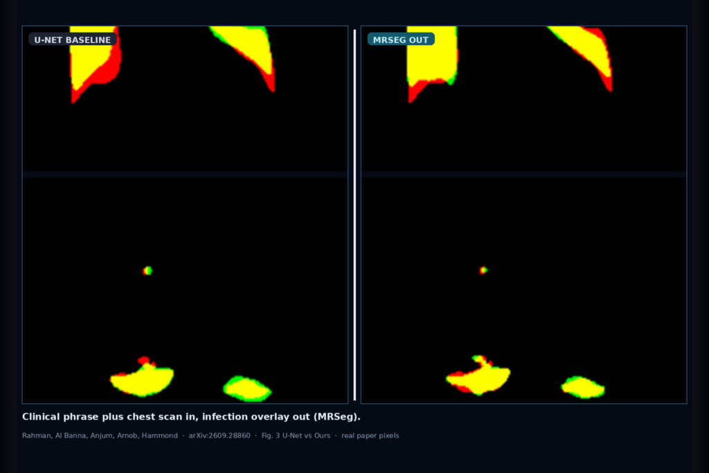
Figure 3 overlays true positives in yellow, false negatives in red, and false positives in green. Against U-Net and several text-guided baselines, MRSeg shows more complete lesion coverage and fewer missed or spurious patches on both the X-ray and CT examples. Treat those panels as qualitative support, not as a promise on your local protocol.
Where it fails and what not to trust
This is research software on two public pulmonary infection image-text-mask benchmarks, not a cleared medical device. Rebuild and validate on your own acquisition protocol, phrase style, and lesion mix before you hang overlays next to reportable findings. The latent regions are learned only through the segmentation loss; do not read them as supervised anatomy. Prompts are capped at 24 tokens in the paper’s setup, so longer report prose is not what was tested. The paper itself flags evaluation beyond these pulmonary sets and sensitivity to text variation.
Do not treat 90.90 / 81.53 Dice as a guarantee on a different vendor stack or on non-infection targets. Do not skip a local holdout with clinical review of false positives near vessels, fissures, or motion artifact. And do not hide that the method still needs paired image-text-mask supervision on the target domain even though the backbone weights stay frozen.
For a viewer or clinic AI shop
Wire chest image plus clinical phrase in; hang an infection overlay out. Prefer MRSeg when you want text-conditioned lesion hang without a full encoder retrain: frozen ConvNeXt-Tiny and PubMedBERT, routed Pair Adapter, Region Bridge refinement, multiscale decode. Keep a QC scrubber in the viewer so support can spot over-paint (green-class errors) and missed patches (red-class errors) against a senior read. Log which phrase template was used, whether the Medical Image Adapter saw X-ray or CT intensities, and the exact checkpoint.
Validate on at least one internal cohort before you claim language-guided infection hang in production. Start from the public repo and the preprint. Rebuild from arXiv:2609.28860. PDF: https://arxiv.org/pdf/2609.28860. Code: https://github.com/maklachur/MRSeg.
Sources
- Rahman, M.M., Al Banna, M.H., Anjum, S., Arnob, A., Hammond, T. Multimodal Routing and Region Refinement for Language-Guided Medical Image Segmentation. arXiv:2609.28860, 2026. https://arxiv.org/abs/2609.28860. PDF: https://arxiv.org/pdf/2609.28860. Code: https://github.com/maklachur/MRSeg.
- Abstract headline: 90.90 / 83.32 Dice/mIoU on QaTa-COV19 and 81.53 / 68.82 on MosMedData+, with 7.11M trainable parameters and 7.60 GFLOPs.
- Table 1: MRSeg tops the listed image-only and text-guided comparisons on both datasets in the paper’s reporting.
- Fig. 3 qualitative: yellow = true positive, red = false negative, green = false positive; Ours cleaner than U-Net and several text-guided baselines on QaTa-COV19 and MosMedData+ examples.